
Chapter 21 • Chest Wall Resection 357
Lung
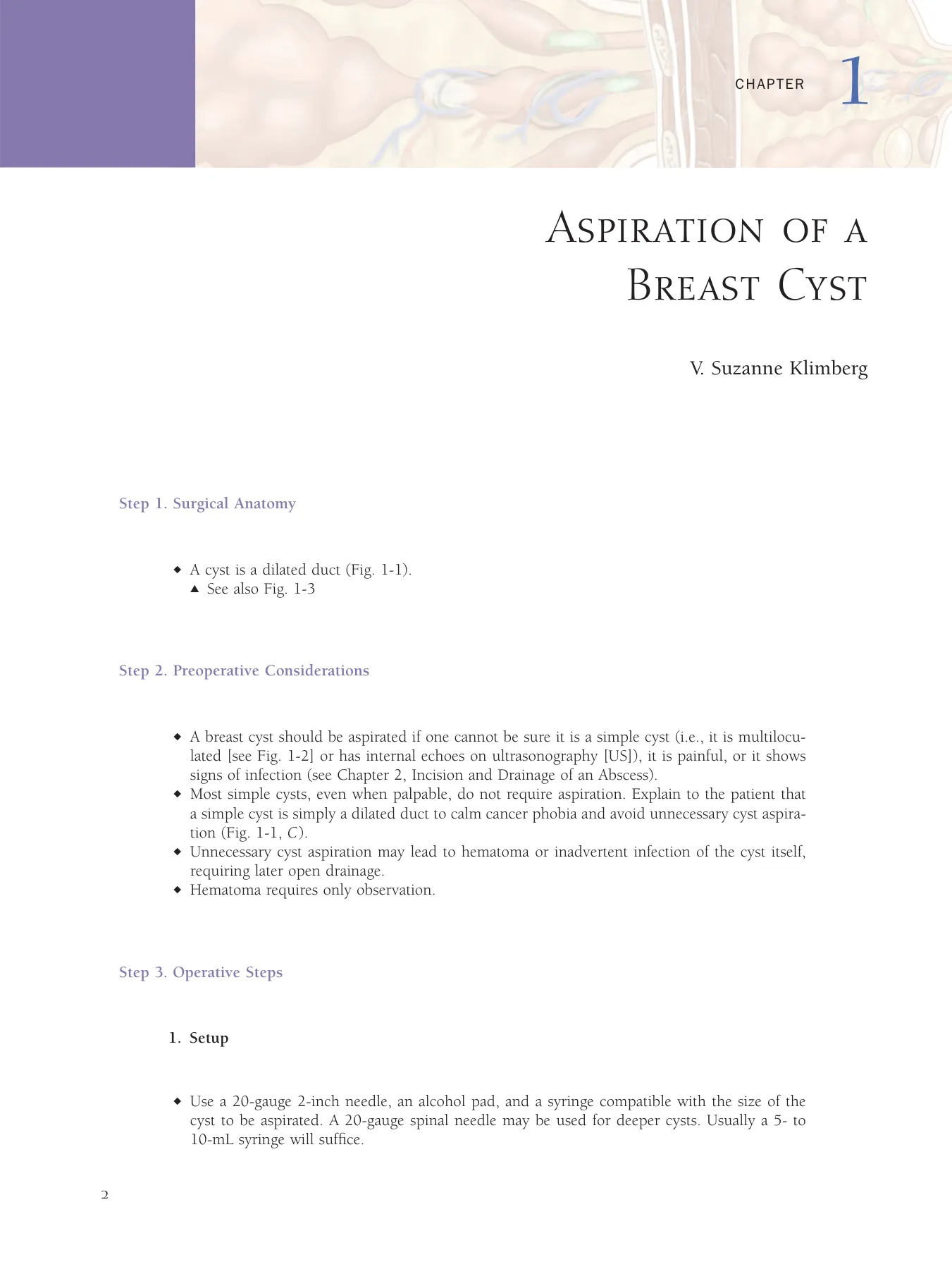
Rib
Pleural adhesions
dissected
Figure 21-7
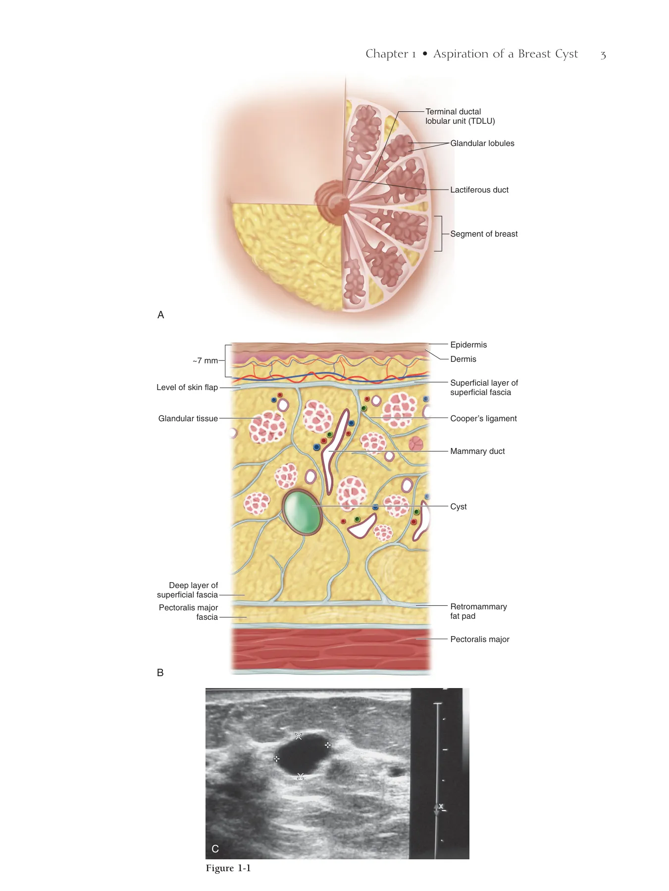
Incision through
intercostal muscle
Rib
Pectoralis muscle
(cut)
Neurovascular bundle
double ligated
Pericardium
Figure 21-8
358 Section V • Extensive Resections
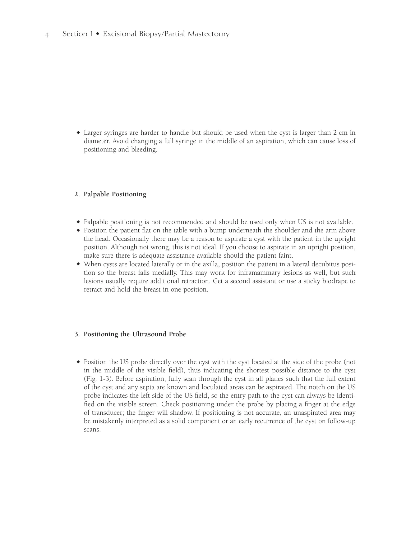
◆ As the incision is extended around the margins of the resection, the periosteum of the ribs
should be elevated with a Doyen rib raspatory before using a rib cutter (Fig. 21-9).
◆ If the margin extends medially to the sternum, a sternal saw may be used for partial or total
sternotomy. The incision should be extended to the sternocostal margin where the IMA may
be palpated, ligated, and divided. The sternum may then be divided while protecting the
pericardium.
Chapter 21 • Chest Wall Resection 359
Periosteum
elevated
with Doyen
Rib
Ligated neurovascular
bundle
Rib cutter
Ligated neurovascular
bundle
A
B
C
Figure 21-9
360 Section V • Extensive Resections
◆ The specimen should be removed en bloc, leaving the chest defect and exposed lung
(Fig. 21-10, A). It is important to inspect the underlying lung for evidence of invasion. If inva-
sive disease is detected grossly or by biopsy, a wedge resection of the involved lung should be
performed (Fig. 21-10, B). The defect should be oversewn and tested for air leaks before closing
the chest.
◆ Before closing the chest wall defect, two chest tubes should be placed. An angled chest tube is
inserted inferior to the defect along the anterior axillary line and positioned anteromedially over
the diaphragm. The second chest tube is inserted more posteriorly along the mid-axillary line
and positioned toward the lung apex. Small-caliber tubes are usually suffi cient to clear the
resulting pneumothorax.
3. Reconstruction
◆ After measuring the defect, a methyl methacrylate and mesh sandwich should be shaped to fi t
the chest wall defect. The sandwich is layered as methyl methacrylate between two layers of
mesh (or one large piece folded in half).
◆ The methyl methacrylate should be prepared and mixed on a back table. The methyl metha-
crylate should be shaped to be 1 cm thick and that circumferentially leaves a 1-cm margin
short of the edges of the defect. This will avoid the potential for uncomfortable rubbing and
clicking of the hardened methyl methacrylate against the ribs. As the methyl methacrylate is
hardening, the mesh should be applied to both sides to form the sandwich. The hardening
process is an exothermic reaction that has the potential to cause thermal injury to the lung.
The sandwich should be allowed to harden and cool before being sewn into the chest wall
defect.
◆ The methyl methacrylate and mesh sandwich is secured to the edges of the wound using either
bone anchors or nonabsorbable 0 or no. 1 polypropylene sutures.
Chapter 21 • Chest Wall Resection 361
Lung
Pericardium
Rib (cut)
Pectoralis
muscle (cut)
Intercostal
muscle (cut)
A
B
Figure 21-10
362 Section V • Extensive Resections
◆ If sutures are used, the sandwich should be secured with horizontal mattress stitches placed in
the intercostal spaces to avoid injury to the intercostal neurovascular bundles (Fig. 21-11).
Sutures can alternatively encircle the ribs along the edges of resection. This method has a higher
incidence of entrapping or injuring the intercostal neurovascular bundles while securing the
mesh. Surgeon’s knots should be tied on the anterior side of the mesh for either closure.
Chapter 21 • Chest Wall Resection 363
Mattress
suture
Methyl methacrylate
“sandwich”
A
B
Methyl methacrylate “sandwich”
Mesh
Mesh
Methyl
methacrylate
Intercostal
muscle (cut)
Figure 21-11
364 Section V • Extensive Resections
◆ After the methyl methacrylate and mesh sandwich is secured, reconstruction of the soft tissue
defect can begin. A pedicled latissimus fl ap with a skin paddle is the most versatile fl ap for this
area. Figure 21-12, A highlights the planned latissimus fl ap. Figure 21-12, B illustrates exposure
of the latissimus muscle with the skin paddle. Figure 21-12, C shows the rotated fl ap covering
the defect and sewn in place.
Chapter 21 • Chest Wall Resection 365
Incision for
latissimus flap
Latissimus
dorsi muscle
Skin paddle
Scapula
Iliac crest
Flap covering defect
and sewn in place
A
C
B
Figure 21-12
366 Section V • Extensive Resections
◆ An omental fl ap based on the right gastroepiploic artery may also be used to cover the defect.
An upper midline laparotomy incision is used to approach the omentum. The omentum is
transected from the splenocolic ligament to the base of the right gastroepiploic artery. Division
and ligation of the vasa recta of the anterior and posterior leaves of the omentum are performed
using 3-0 absorbable ties (Fig. 21-13).
◆ After exiting the midline incision, the omentum may be delivered to the chest by a retrosternal,
transdiaphragmatic, or subcutaneous tunnel to the defect (Fig. 21-14). Care should be taken
not to strangulate or twist the omental pedicle. Figure 21-15, A shows the omentum exposed.
Figure 21-15, B shows the tunneled omentum in place. Planning of the defect coverage before
the omentum is tunneled is optimal. Figure 21-15, C shows the fl ap covered with a split-
thickness skin graft 3 weeks later.
Chapter 21 • Chest Wall Resection 367
Retrosternal
tunnel
Transdiaphragmatic
tunnel
Subcutaneous
tunnel
Esophagus
Stomach
Vasa recta
Spleen
Right gastroepiploic
artery
Left gastro-
epiploic
artery
Omentum
Omental arterial
arch
Figure 21-13
Figure 21-14
Flap covered with
split-thickness skin graft
Omentum exposed
“Tunneled” omentum
in place
A
C
B
Figure 21-15
368 Section V • Extensive Resections
◆ Pedicled or free fl aps using transverse or vertical rectus abdominis muscle fl aps with skin
paddles are possible alternatives for covering the defect. Free fl aps may be anastomosed to the
ipsilateral IMA as infl ow (Fig. 21-16).
◆ Flaps should be closed in two layers. The deep dermal layer is closed with 3-0 absorbable
sutures. A running subcuticular closure using absorbable 4-0 sutures is used for skin.
◆ Subcutaneous drains are also needed in the soft tissue reconstruction to prevent hematoma
(Fig. 21-17). Internal Doppler probes may be placed for monitoring fl ap arterial supply.
Step 4. Postoperative Care
◆ Immediate postoperative extubation should be attempted in the operating room if possible.
Minimized exposure to positive-pressure ventilation, especially in the setting of a lung resection,
is optimal.
◆ Patients will require intensive care unit admission and may need ventilator management after
surgery. Ventilator management should include preventive care for ventilator-associated pneu-
monia. Aggressive pulmonary toilet after extubation should be encouraged to prevent atelectasis
and nosocomial pneumonia.
◆ Because of the extreme pain usually experienced with this procedure, postoperative epidural
pain control should be considered.
◆ Wound care and fl ap management are necessary to monitor for fl ap ischemia or necrosis. Arte-
rial supply can be evaluated by an internal Doppler ultrasound probe placed during surgery, a
hand-held external Doppler probe, and capillary refi ll. Standard criteria can be used for remov-
ing chest tubes and subcutaneous drains.
◆ Complication rates for this procedure have been reported as high as 27% to 41%. Wound and
pulmonary complications are the most common. Wound complications include infection,
delayed healing, partial fl ap or skin graft necrosis, or complete fl ap loss. Pulmonary complica-
tions include reintubation, fl ail chest, or pleural infection. Despite the morbidity risk, the
mortality rate for this procedure is low (<5%).
◆ A low threshold should be used for treating suspected wound or pulmonary infections.
Chapter 21 • Chest Wall Resection 369
Inferior
epigastric artery
Free flap using transverse
rectus abdominis muscle
and skin paddle
Internal
mammary artery
Lung
Sternum
Jackson-Pratt
drain
Chest tubes
Internal
Doppler probe
Figure 21-16
Figure 21-17
370 Section V • Extensive Resections
Step 5. Pearls and Pitfalls
◆ Appropriate patient selection for chest wall resection is crucial. Curative intent requires ruling
out systemic and multifocal disease.
◆ Preoperative evaluation of the patient’s pulmonary function and reserve is important to mini-
mize operative morbidity, especially if lung resection is anticipated.
◆ The complexity of these resections is often high because of previous operations (including past
reconstructions), radiation, and patient comorbidities. Orchestrating preoperative collaboration
between oncologic and reconstructive surgeons is critical for designing the best procedure for
the patient.
◆ Preoperative skin biopsies around the planned margins of resection should be sent for frozen-
section analysis before the initial incision because failure of the resection is usually from lym-
phovascular invasion of the skin. Frozen sections should also be sent from the deep resection
margins before reconstruction begins.
◆ Ideally, margins should be at least 2 cm and should extend one intercostal space above and
below the defect. Anatomic location may sometimes limit margins.
◆ Change the chest tube from suction to water seal as soon as possible to avoid venous conges-
tion in chest wall fl aps.
Chapter 21 • Chest Wall Resection 371
Bibliography
Beahm E, Hunt K, Pollock R: Surgical procedures for advanced local and regional malignancies of the breast. In Bland K, Copeland E
(eds): The Breast, vol 2, 3rd ed. St. Louis, Saunders, 2004, pp 1235-1254.
Chang RR, Mehrara BJ, Hu QY, et al: Reconstruction of complex oncologic chest wall defects: A 10-year experience. Ann Plast Surg
2004;52:471-479; discussion 479.
Faneyte IF, Rutgers EJ, Zoetmulder FA: Chest wall resection in the treatment of locally recurrent breast carcinoma: Indications and outcome
for 44 patients. Cancer 1997;80:886-891.
Fix R, Vasconez L: Use of omentum in chest-wall reconstruction. Surg Clin North Am 1989;69:1029-1046.
McCraw J, Arnold P: Latissimus dorsi. In McCraw and Arnold’s Atlas of muscle and musculocutaneous fl aps. Norfolk, Va, Hampton Press,
1986, pp 157-226.
Pameijer CR, Smith D, McCahill LE, et al: Full-thickness chest wall resection for recurrent breast carcinoma: An institutional review and
meta-analysis. Am Surg 2005;71:711-715.
Step 1. Surgical Anatomy
◆ Table 22-1 and Figure 22-1 detail the anatomic structures that must be identifi ed to perform
a forequarter amputation.
Forequarter Amputation
Jennifer B. Manders, Jaime D. Lewis,
and Elizabeth A. Shaughnessy
CHAPTER 22
372
TABLE 22-1 Important Anatomic Structures in Forequarter Amputation
ANTERIOR CHEST (SEE FIG. 22-1, A)
POSTERIOR CHEST
Pectoralis major and minor muscles
Superfi cial (see Fig. 22-1, B)
Deltoid muscle
Serratus anterior muscle
Brachial plexus with axillary vein and artery
Deltoid muscle
Biceps brachii muscle
Trapezius muscle
Coracobrachialis muscle
Scapula
Coracoid process of the scapula
Infraspinatus muscle
Clavicle
Supraspinatus muscle
Mid-depth (see Fig. 22-1, C)
Levator scapulae muscle
Rhomboid major muscle
Rhomboid minor muscle
Deep (see Fig. 22-1, C)
Teres major muscle*
Teres minor muscle*
Deep and arm (see Fig. 22-1, C)
Triceps muscle*
*Muscles constituting the rotator cuff.
Step 2. Preoperative Considerations
◆ Review of the anatomy of the shoulder girdle, including musculature, ligament and tendon
insertions, and neurovascular structures, is essential.
◆ Because the normal anatomy may be obscured by or involved with a tumor, preoperative
imaging with computed tomography (CT) or magnetic resonance imaging (MRI) may help with
the identifi cation of anatomic structures of interest. MRI of the brachial plexus is extremely
useful to identify the proximal extent of tumor involvement. These imaging studies may then
be used to develop an operative plan.
Chapter 22 • Forequarter Amputation 373
Figure 22-1
Clavicle
Anterior View
Y-shaped incision
Biceps brachii
muscle
Serratus anterior
muscle
Pectoralis major muscle
Pectoralis
minor muscle
A
Brachial plexus and
axillary vessels
Deltoid muscle
Posterior View
B
Posterior incision
Deltoid muscle
Teres minor muscle
Teres major muscle
Infraspinatus
muscle
Trapezius
muscle
Supraspinatus
muscle
Coracoid process
Scapula
Rhomboid minor
muscle
Rhomboid major
muscle
Supraspinatus
muscle
Infraspinatus
muscle
Levator scapulae
Scapula
Deltoid muscle
Teres minor muscle
Teres major muscle
Latissimus dorsi
muscle
Trapezius muscle (cut)
C
374 Section V • Extensive Resections
◆ On physical examination and imaging, attention should be paid to the surgical margins of
resection. If the tumor extends beyond the clavicle, into the posterior triangle of the neck, or
invades the chest wall, then residual disease after resection is likely.
◆ Patients should be counseled extensively about the risks and benefi ts associated with this pro-
cedure, and that anticipated survival and long-term prognosis are likely unaffected by forequar-
ter amputation as a palliative or curative procedure.
◆ Phantom limb pain is often mild to moderate and usually controlled well with low-dose narco-
tics. Up to 60% to 90% of patients experience phantom limb pain, which may be exacerbated
with previous exposure to chemotherapy or prolonged, uncontrollable preoperative pain.
◆ Metastatic work-up should include a bone scan and CT scans of the chest, abdomen, and
pelvis. Thoracoscopy may be used to evaluate for the presence of pleural or parenchymal
involvement.
Step 3. Operative Steps
1. Indications
◆ Forequarter amputation has been described historically as a useful procedure for locally
advanced, high-grade bone and soft tissue sarcomas involving the shoulder girdle. More recently,
its utility has been limited because of the increased use of neoadjuvant chemotherapy and
radiation.
◆ Forequarter amputation may be of benefi t in the case of locally advanced or recurrent metastatic
carcinomas that affect the shoulder and are otherwise nonresectable by more common
operations.
◆ Indications for the procedure may include the following:
▲ Local invasion along the rotator cuff and the scapula, including invasion into or ulceration
of the axillary neurovascular bundle (brachial plexus or axillary vessels)
▲ Lymphangiosarcoma of the upper extremity (to include Stewart-Treves syndrome—resulting
from long-term lymphedema after modifi ed radical mastectomy with radiation)
▲ Salvage after failure of conservative management
▲ Severe, intractable pain with loss of limb function
▲ Local tumor-related complications: paralysis, tumor fungation, hemorrhage, sepsis, severe
lymphedema, venous stasis with resultant limb necrosis, radiation-induced complications
(severe brachial plexopathy)
2. Setup and Positioning
◆ The patient should be intubated with a double-lumen endotracheal tube.
◆ The patient should be placed in the lateral decubitus position, on a beanbag, with the affected
side up. A pillow should be placed between the knees and, potentially, a roll placed under the
dependent axilla (Fig. 22-2, A). Alternatively, if the hip is fl exed, a bean bag can be skipped
and the hip secured with tape to the table (Fig. 22-2, B). The affected extremity should be pre-
pared in the fi eld and covered with a stockinette per the surgeon’s preference. The upper
extremity is held by an assistant during the resection because dissection is both anterior and
posterior. The extremity can then be maneuvered as necessary by resting the patient’s bent
elbow over the assistant’s arm.
Chapter 22 • Forequarter Amputation 375
Posterior View
A
Bean bag
Arm in stockinette
Pillow
Tape
B
Figure 22-2
376 Section V • Extensive Resections
◆ Skin preparation of the upper extremity is performed circumferentially for its entire length,
extending to the midline of the chest and back, and including the lateral thorax.
3. Operative Procedure
◆ Anterior exposure is performed to facilitate exploration of the infraclavicular portion of the
brachial plexus and axillary vessels. This is begun with a Y-shaped incision below the clavicle,
with one limb placed posteriorly across the clavicle onto the back (see Fig. 22-1, A) and one
limb carried inferiorly along the deltopectoral groove, onto the chest wall, meeting in a loop
under the arm (Fig. 22-3; see Figs. 22-1, A and B).
◆ The pectoralis major muscle is released from the humeral insertion, exposing the neurovascular
bundle. The conjoined tendon (short head of biceps and the coracobrachialis), the coracobra-
chialis muscle, short head of the biceps brachii muscle, and pectoralis minor muscle are all
subsequently released from the coracoid process, completing exposure of the neurovascular
bundle (Fig. 22-4).
Chapter 22 • Forequarter Amputation 377
Right upper
extremity
Incision over
shoulder
Incision over
clavicle
Head
Planned
incision
Anterior view
Tendon of long
head of biceps
brachii muscle
Tendon of short
head of biceps
brachii muscle
Musculocutaneous
nerve
Pectoralis major
muscle (cut)
Pectoralis minor
muscle
Neurovascular
bundle (brachial
plexus with
axillary blood
vessels)
Coracobrachialis
muscle
Deltoid
muscle
Coracoid
process
Figure 22-3
Figure 22-4
378 Section V • Extensive Resections
◆ Neurovascular encasement by tumor is then assessed. The subclavian artery and vein and the
brachial plexus are individually doubly ligated and transected proximal to tumor and proximal
to the planned clavicle resection (Fig. 22-5).
◆ The incision is extended posterolaterally over the shoulder, curving medially at the scapular
tip, resulting in the construction of a medially based posterior fasciocutaneous fl ap. All muscles
anchoring the scapula to the chest wall medial to the scapula (trapezius, and the deeper rhom-
boid major and minor muscles as well as the levator scapulae muscle) are released, after the
arm is rotated anteriorly to place these muscles on tension. The serratus anterior muscle now
anchors the upper extremity (Fig. 22-6).
Chapter 22 • Forequarter Amputation 379
Anterior view
Subclavian artery
and vein, ligated
Brachial plexus, ligated
Coracobrachialis muscle
Pectoralis major
muscle (cut)
Pectoralis minor
muscle
Clavicular extension
of incision
Skin flap
folded back
Intercostal
muscle
Exposed
rib
Serratus
anterior
muscle
Trapezius muscle (cut)
Figure 22-5
Figure 22-6
380 Section V • Extensive Resections
◆ The two skin incisions are connected under the axilla. The clavicle is osteotomized back one-
third the distance from the acromion to the sternum (Fig. 22-7). The serratus anterior muscle
is released from the scapula, usually along the rib insertion, and all remaining attachments to
the humerus are divided. The arm is removed.
◆ The pectoralis major muscle is transected close to its origin or sutured to the chest wall (Fig.
22-8). Closed-suction drains are placed, and an epineural catheter can be inserted into the
brachial plexus sheath, with delivery of a 0.25% bupivacaine bolus. A catheter delivering con-
tinuous bupivacaine can also be placed in the subcutaneous space. The fl ap is closed over the
defect (Fig. 22-9).
Step 4. Postoperative Care
◆ The epineural catheter is kept in place until the patient is converted to oral pain medications.
Neuropathic pain related to a brachial plexopathy or phantom limb pain not responsive to
narcotics can be treated with γ-aminobutyric acid (GABA) receptor antagonists.
◆ Once incisions are healed, the patient can be fi tted for a shoulder or arm prosthesis, or both.
Physical and occupational therapy may be of great benefi t for the patient, enabling her to resume
activities of daily living.
Chapter 22 • Forequarter Amputation 381
Subclavian vein
and artery
Clavicle
Anterior
Pectoralis major
muscle
Pectoralis muscle
Clavicle
Posterior
Figure 22-7
Figure 22-8
Figure 22-9
382 Section V • Extensive Resections
Step 5. Pearls and Pitfalls
◆ Large defects caused by surgical resection can make the fi nal wound edges diffi cult to reap-
proximate and may require the use of a split-thickness skin graft. If a posterior myocutaneous
fl ap can be maintained without sacrifi cing surgical margins, this can aid in primary closure.
Preoperative counseling and preparation of a potential harvest site should be included in surgi-
cal planning.
◆ Prostheses offered after forequarter amputation lack proprioception and precise movement.
Shoulder pads help maintain normal contour, which is signifi cantly affected by this radical
procedure.
◆ Intervention with psychological counseling is extremely benefi cial, especially in the preoperative
setting, to help the patient prepare herself for the signifi cant life change she will experience.
Physical and occupational therapy should be instituted early in the patient’s postoperative
course. If available, contact with support groups or with other patients who have undergone
forequarter amputation should be sought.
Bibliography
Bhagia SM, Elek EM, Grimer RJ, et al: Forequarter amputation for high-grade malignant tumours of the shoulder girdle. J Bone Joint Surg
Br 1997;79:924-926.
Goodman MD, McIntyre B, Shaughnessy EA, et al: Forequarter amputation for recurrent breast cancer: A case report and review of the
literature. J Surg Oncol 2005;92:134-141.
Moore KL: Clinically Oriented Anatomy, 3rd ed. Baltimore, Williams & Wilkins, 1992.
Sugarbaker PH: Forequarter amputation. In Sugarbaker PH, & Malawer MM (eds.), Musculoskeletal surgery for cancer: principles and
techniques, pp. 306-316. New York, Thieme Medical Publishers, Inc., 1992.
Volpe CM, Peterson S, Doerr RJ, et al: Forequarter amputation with fasciocutaneous deltoid fl ap reconstruction for malignant tumors of
the upper extremity. Ann Surg Oncol 2007;4:298-302.
Wittig JC, Bickels J, Kollender Y, et al: Palliative forequarter amputation for metastatic carcinoma to the shoulder girdle region: Indications,
preoperative evaluation, surgical technique, and results. J Surg Oncol 2001;77:105-113.
Step 1. Surgical Anatomy
◆ The placement of a MammoSite (Cytyc Surgical Products, Marlborough, MA) balloon requires
a basic understanding of breast anatomy as well as a minimum level of ultrasonographic knowl-
edge of the breast (Fig. 23-1).
◆ Cavity evaluation with ultrasonography is required to be able to measure skin-to-cavity distance
(see Fig. 23-1) and to assess the entire lumpectomy cavity (Fig. 23-2) so the proper device and
size can be selected. Skin-to-cavity distance is excellent at 18 mm with a fairly regular cavity
of 36 mm × 25 mm (the cavity will conform to the expanded balloon).
Step 2. Preoperative Considerations
◆ The decision to place a MammoSite balloon or other brachytherapy device begins at the time
of the initial evaluation of the patient with breast cancer.
◆ The size of the cancer itself as well as the tumor-to-breast size ratio is evaluated (a small breast
may not be able to accommodate a MammoSite balloon, although the newer devices allow more
fl exibility). For MammoSite placement, a minimum distance of 5 mm from skin to lumpectomy
cavity will be necessary after the lumpectomy. Other devices require less distance (potentially
down to a 2-mm skin-to-cavity distance).
◆ The following are the generally accepted patient criteria for accelerated partial breast
irradiation:
▲ Age 50 years or older
▲ Tumor size less than 3 cm
▲ Negative margins
MammoSite Balloon
Catheter Placement and
Other Brachytherapy
Delivery Devices
Peter D. Beitsch
CHAPTER 23
384
Chapter 23 • MammoSite Balloon Catheter Placement 385
Retromammary fat
Cooper’s ligament
Parenchyma
Pectoralis muscle
Skin
B
A
Figure 23-1
Skin to cavity distance - 18 mm
Cavity size 36 mm x 25 mm
Skin to cavity distance - 18 mm
Cavity size 36 mm x 25 mm
Figure 23-2
386 Section VI • Surgical Techniques to Assist Irradiation
▲ Negative sentinel lymph node
▲ Ductal histology: infi ltrating or ductal carcinoma in situ
◆ Discussing the possibility of accelerated partial breast irradiation before surgery, including a
radiation oncology consult, will allow the patient to make a proper informed decision.
Step 3. Operative Steps
1. Intraoperative Placement
◆ Although most MammoSite balloons (and other brachytherapy devices) are placed postopera-
tively in the offi ce, selected patients will have a balloon placed in the operating room. Reasons
for intraoperative placement could include the following:
▲ Patient unable to tolerate minor procedures in the offi ce because of fears or the need for very
close monitoring
▲ Patient convenience during a re-lumpectomy for a close or positive margin
◆ The MammoSite placement begins after the lumpectomy and sentinel node biopsy (with intra-
operative margin and sentinel node evaluation).
◆ A new lateral incision (or potentially medial or other incision) is made remote from the lumpec-
tomy incision (Fig. 23-3).
◆ The trocar provided in the MammoSite kit (or a Kelly clamp or hemostat) is used to create a
track into the lumpectomy cavity (Fig. 23-4).
◆ The defl ated MammoSite balloon is placed through the incision into the track and advanced
into the lumpectomy cavity. After the balloon is in the cavity, it is infl ated to assess its position
in the cavity (Fig. 23-5).
Chapter 23 • MammoSite Balloon Catheter Placement 387
Figure 23-3
Trocar
A
B
Deflated
mammosite
Figure 23-4
Figure 23-5
388 Section VI • Surgical Techniques to Assist Irradiation
◆ The MammoSite balloon is defl ated and the lumpectomy cavity is closed in layers to ensure a
minimum of 5 mm and, ideally, greater than a full centimeter of tissue between the cavity and
the skin (Fig. 23-6).
◆ Once the skin is closed, the balloon is reinfl ated (Fig. 23-7) and the skin-to-balloon distance
is evaluated by ultrasonography (Fig. 23-8). The skin-to-balloon distance in Figure 23-8 is
16 mm, and the balloon diameter is 37 mm.
◆ The skin is closed and the usual sterile dressings are applied (Fig. 23-9).
Chapter 23 • MammoSite Balloon Catheter Placement 389
US probe
Figure 23-7
Skin to balloon distance 16 mm
Balloon size 37 mm
Skin to balloon distance 16 mm
Balloon size 37 mm
Figure 23-8
Figure 23-9
Figure 23-6
390 Section VI • Surgical Techniques to Assist Irradiation
2. Postoperative Placement
Lateral Placement
◆ Most MammoSite balloons (and other brachytherapy devices) are placed in the offi ce once the
pathologic examination is complete. This ensures that the margins are negative and the sentinel
lymph nodes are without metastases.
◆ Once the pathology requirements have been met, the fi rst step is to evaluate the cavity and the
skin-to-cavity distance (minimum 5 mm, and preferably >7 mm) to ensure the patient is still
a candidate for MammoSite balloon placement. However, other devices require less skin-to-
cavity distance and could be considered if spacing appears to be an issue (Fig. 23-10, A).
◆ The patient is then sterilely prepared and draped. A lateral stab incision (or occasionally medial
or other locations) is made with a no. 11 blade (Fig. 23-10, B).
◆ The trocar provided in the MammoSite kit (or a Kelly clamp or hemostat) is used to create
a track into the lumpectomy cavity. This is done under ultrasonographic guidance
(Fig. 23-10, C).
◆ The uninfl ated balloon is placed through the skin nick, down the track, and into the cavity
under ultrasonographic guidance (Figs. 23-10, D and E).
◆ The MammoSite balloon is infl ated and a sterilely draped ultrasound probe is used to evaluate
the skin-to-cavity distance and the balloon-to-cavity conformance (Fig. 23-10, F).
◆ A sterile dressing is then applied.
Chapter 23 • MammoSite Balloon Catheter Placement 391
A
B
C
D
E
F
US probe
Figure 23-10
392 Section VI • Surgical Techniques to Assist Irradiation
Scar Entry Technique
◆ To ensure adequate skin spacing, the cavity is evaluated by ultrasonography before deciding
the patient is a MammoSite candidate (Fig. 23-11, A).
◆ The patient is then sterilely prepared and draped. A hemostat or Kelly clamp is used to slightly
open the incision and then plunged into the cavity under direct ultrasonographic guidance
(Fig. 23-11, B).
◆ The uninfl ated MammoSite balloon is placed through the incision into the cavity
(Fig. 23-11, C).
◆ The balloon is infl ated and the incision is sutured on either side to ensure the wound stays
closed (Fig. 23-11, D).
◆ The balloon is evaluated by ultrasonography to ensure adequate skin spacing and good balloon-
to-cavity conformance (Fig. 23-11, E).
Chapter 23 • MammoSite Balloon Catheter Placement 393
A
C
E
B
D
Figure 23-11
394 Section VI • Surgical Techniques to Assist Irradiation
3. New Multicatheter Brachytherapy Delivery Devices
◆ All these devices allow treatment plans that reduce radiation dose to the skin and therefore
allow a narrower skin-to-cavity distance.
▲ ClearPath (Renata Medical, Irvine, CA) is a single-entry, multicatheter device with a set of
outer, nonfunctional struts to displace the breast tissue (similar to the balloon) and inner
treatment struts (Fig. 23-12).
▲ Contura (SenoRx, Inc., Aliso Viejo, CA) is a single-insertion, multicatheter balloon device
that has a stiff polyurethane balloon (which helps with cavity conformance) and fi ve catheters
contained within the balloon. There are also suction ports at the distal and proximal ends
of the balloon to allow for aspiration of fl uid around the balloon (Fig. 23-13).
▲ SAVI (Cianna Medical, Inc., Aliso Viejo, CA) is a single-entry, multicatheter device with 6,
8, or 10 struts that are in contact with the breast tissue (Fig. 23-14).
Chapter 23 • MammoSite Balloon Catheter Placement 395
A
B
Figure 23-12
©Renata Medical, Irvine, CA.
Figure 23-13
©SenoRx, Inc., Aliso Viejo, CA.
Figure 23-14
©Cianna Medical, Inc., Aliso Viejo, CA.
396 Section VI • Surgical Techniques to Assist Irradiation
4. Electronic Brachytherapy System
◆ Current brachytherapy techniques all involve a radioactive source. Xoft (Sunnyvale, CA) has
developed the Axxent, a miniaturized x-ray source that delivers a dose of radiation to the
lumpectomy cavity similar to an iridium source (Fig. 23-15). The miniaturized x-ray source is
delivered through a balloon device similar to a MammoSite balloon. The Xoft balloon has
suction ports at the proximal and distal ends to allow for aspiration of fl uid from around the
balloon (Fig. 23-16).
Step 4. Postoperative Care
◆ After the last treatment (typically on Friday after the afternoon fraction), the balloon and
multicatheter devices are removed by defl ating the balloon or collapsing the multicatheters
and pulling them out through the entry track. Steri-Strips are used to close the entry
stab incision.
◆ Patients are usually seen in 1 to 2 weeks to evaluate the small incision and to assess the radia-
tion effects.
Chapter 23 • MammoSite Balloon Catheter Placement 397
Multiple balloon
shapes and sizes
Drainage port valve
Multilumen extrusion
Drainage holes x 7
Balloon inflation
valve
Radiation probe
lumen with
inserted stylet
Figure 23-15
©Xoft, Inc., Sunnyvale, CA.
Figure 23-16
398 Section VI • Surgical Techniques to Assist Irradiation
Step 5. Pearls and Pitfalls
◆ When placing a MammoSite (or any other device), there may be a signifi cant amount of drain-
age from the small lateral incision. Discussing this with the patient will often save a midnight
phone call.
◆ After placing a MammoSite balloon, fl uid may be trapped between the balloon and the lumpec-
tomy cavity wall. Gentle breast massage can be used to evacuate this fl uid through the track
and out the lateral stab incision.
◆ If the MammoSite is being placed in the operating room, sometimes the skin-to-balloon distance
is inadequate. This can be solved by opening the wound, removing the thin skin fl aps, and
reapproximating more subcutaneous tissue before closing the skin.
◆ There is a fi nite amount of time between the lumpectomy surgery and the placement of the
MammoSite or other brachytherapy device (usually less than 4 weeks). As the lumpectomy
cavity heals, it will become progressively more fi brotic and less distensible, eventually disap-
pearing entirely. This healing fi brosis may make symmetric deployment of the MammoSite
balloon diffi cult (this may be less of an issue with the stiffer Contura polyurethane balloon or
the multicatheter devices). Therefore, earlier placement of the MammoSite or other device is
advisable.
◆ One of the advantages of brachytherapy is that all the radiation therapy is completed before the
initiation of systemic therapy. However, to avoid radiation recall, chemotherapy should be
delayed at least 3 weeks after completion of the brachytherapy.
Chapter 23 • MammoSite Balloon Catheter Placement 399
Bibliography
Arthur DW, Vicini FA: Accelerated partial breast irradiation as a part of breast conservation therapy. J Clin Oncol 2005;23:1726-1735.
Chen PY, Vicini FA, Benitez P, et al: Long-term cosmetic results and toxicity after accelerated partial-breast irradiation. Cancer
2006;106:991-999.
Keisch M, Vicini F, Kuske RR, et al: Initial clinical experience with the MammoSite breast brachytherapy applicator in women with early-
stage breast cancer treated with breast-conserving therapy. Int J Radiat Oncol Biol Phys 2003;55:289-293.
Kuske RR, Winter K, Arthur DW, et al: Phase II trial of brachytherapy alone after lumpectomy for select breast cancer: Toxicity analysis
of RTOG 95-17. Int J Radiat Oncol Biol Phys 2006;65:45-51.
NSABP B-39, RTOG 0413: A randomized phase III study of conventional whole breast irradiation versus partial breast irradiation for
women with stage 0, I, or II breast cancer. Clin Adv Hematol Oncol 2006;4:719-721.
Vicini FA, Antonucci JV, Wallace M, et al: Long-term effi cacy and patterns of failure after accelerated partial breast irradiation: A molecular
assay-based clonality evaluation. Int J Radiat Oncol Biol Phys 2007;68:341-346.
Vicini FA, Beitsch PD, Quiet CA, et al: First analysis of patient demographics, technical reproducibility, cosmesis, and early toxicity: Results
of the American Society of Breast Surgeons MammoSite breast brachytherapy trial. Cancer 2005;104:1138-1148.
Zannis V, Beitsch P, Vicini F, et al: Descriptions and outcomes of insertion techniques of a breast brachytherapy balloon catheter in
1403 patients enrolled in the American Society of Breast Surgeons MammoSite breast brachytherapy registry trial. Am J Surg
2005;190:530-538.
Step 1. Surgical Anatomy
◆ The adult breast lies between the second and sixth ribs and between the sternal edge and the
mid-axillary line.
◆ Figure 24-1 demonstrates the major anatomic structures of the chest wall from an external
view.
Step 2. Preoperative Considerations
◆ An increasing number of studies are attempting to decrease the overall treatment time for radia-
tion after lumpectomy. To be considered for intraoperative radiation therapy (IORT) of the
breast, the patient should be a breast-conservation candidate with an invasive ductal carcinoma
with no extensive intraductal component. The tumor size should be 3 cm or less based on
physical examination, mammography, and ultrasonography. The patient must be at least 48
years of age, and the tumor must be able to be visualized on ultrasonography.
◆ Exclusion criteria for breast IORT are invasive lobular carcinoma, ductal carcinoma in situ,
extensive intraductal component, and pregnancy.
◆ The tumor is localized with a wire, and technetium sulfur colloid is injected for the sentinel
lymph node biopsy.
◆ Breast ultrasonography is performed to defi ne the target volume. Three distances are measured
along the axis of the planned IORT delivery, usually the shortest distance from skin to tumor:
(1) from the skin to the middle of the breast cancer, (2) from the skin to the posterior edge of
the tumor, and (3) from the skin to the anterior surface of the pleural surface. These measure-
ments will then be used by the treating radiation oncologist to determine dosimetry curves and
the electron energy and applicator size required to deliver 15 Gy to the 90% isodose line cover-
ing the tumor with a 1-cm anteroposterior margin and a 2-cm lateral margin.
In Vivo Intraoperative
Radiation Therapy for
Breast Cancer
Nancy Klauber-DeMore, Carolyn I. Sartor, and David W. Ollila
CHAPTER 24
400
Chapter 24 • In Vivo Intraoperative Radiation Therapy for Breast Cancer 401
Lymph node
Milk duct
Lacteriferous
duct
Papilloma
Milk gland
Fat
Subcutaneous
abscess
Subarealor
abscess
Cyst
Fascia of
pectoralis major
Retromammary
fat plain
Pectoralis major
Pectoralis minor
Rib
DCIS
TDLU (Terminal
Ductal Lobular
Unit)
Invasive mammary
carcinoma
A
Common carotid artery
Pectoralis
minor muscle
Clavicle
Pectoralis
major muscle
Axillary artery
and vein
Internal mammary
neurovascular bundle
Intercostal
neurovascular bundle
1
2
3
4
5
Sternum
Deltoid muscle
Lattisimus
dorsi muscle
Serratus
anterior muscle
Rectus abdominus
muscle
Internal jugular vein
B
Figure 24-1
402 Section VI • Surgical Techniques to Assist Irradiation
◆ The operating room table is turned 180 degrees so it can be moved under the Mobetron
(IntraOp Medical Corporation, Sunnyvale, CA) machine. The patient is placed on the operating
table in the supine position and general anesthesia is administered. A secure seatbelt is tightly
fastened. Isosulfan blue (Lymphazurin) or methylene blue dye is injected peritumorally. The
patient is prepared and draped in standard fashion.
Step 3. Operative Steps
1. Incision
◆ The incision needs to be large enough to encompass the diameter of the applicator used for the
IORT. For example, if a 6-cm-diameter applicator is used, the incision must be at least 6 cm
(Fig. 24-2).
2. Tumor Exposure
◆ The sentinel node biopsy is performed before IORT in standard fashion (see Chapter 10).
◆ An incision is made in the breast and very wide, thin skin fl aps are raised (Fig. 24-3).
Chapter 24 • In Vivo Intraoperative Radiation Therapy for Breast Cancer 403
A
B
A
B
Figure 24-2
Figure 24-3
404 Section VI • Surgical Techniques to Assist Irradiation
◆ A silk stitch is placed in the tumor and the stitch is kept long (Fig. 24-4). The stitch will be
used later to position the tumor in the center of the applicator.
3. Delivery of Intraoperative Radiation Therapy
◆ IORT is delivered using a Mobetron device, which is a self-shielded, magnetron-driven, X-band
linear accelerator specifi cally developed for IORT. This machine produces a megavoltage elec-
tron beam of energies ranging from 4 to 12 MeV. The radiation is delivered from the Mobetron
to the tumor bed through an attached applicator.
◆ The sizes of applicators available for the Mobetron range from 3 to 10 cm in diameter. An
applicator is selected that encompasses the tumor along with a 2-cm radial margin. The appli-
cator is placed into the incision (Fig. 24-5).
Chapter 24 • In Vivo Intraoperative Radiation Therapy for Breast Cancer 405
A
B
Figure 24-4
A
B
Figure 24-5
406 Section VI • Surgical Techniques to Assist Irradiation
◆ The stitch is manipulated to place the tumor in the center of the applicator (Fig. 24-6).
◆ A moist, sterile gauze is placed between the skin and the radiation applicator to introduce a
further tissue-equivalent barrier to absorb the low-energy electrons scattered by the applicator
itself.
◆ The applicator is positioned such that there is a minimal air gap between the end of the appli-
cator and the tumor. At the discretion of the radiation oncologist, a surface bolus (5 or 10 mm)
may be attached to the end of the applicator before stabilization to improve surface dose.
◆ The applicator is then secured by a modifi ed Bookwalter retraction system (Fig. 24-7). Visual
inspection with the aid of a Surch-Lite lighted stylet (Aaron Medical, St. Petersburg, FL) is used
Chapter 24 • In Vivo Intraoperative Radiation Therapy for Breast Cancer 407
A
B
Figure 24-6
A
B
Figure 24-7