
52 Section I • Excisional Biopsy/Partial Mastectomy
Step 3. Operative Steps
1. Duct Selection and Cannulation
◆ Cleanse the nipple papilla (exfoliate using nonabrasive to remove keratin plugs).
◆ Have patient well hydrated and relaxed.
◆ Use sedation.
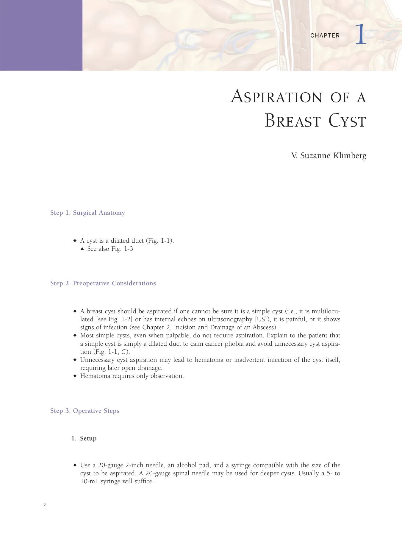
◆ Massage as if expressing at lactation using a thin, slippery lotion (e.g., Udderly Smooth Udder
Cream; Redex Industries, Inc., Salem, OH; Fig. 5-3).
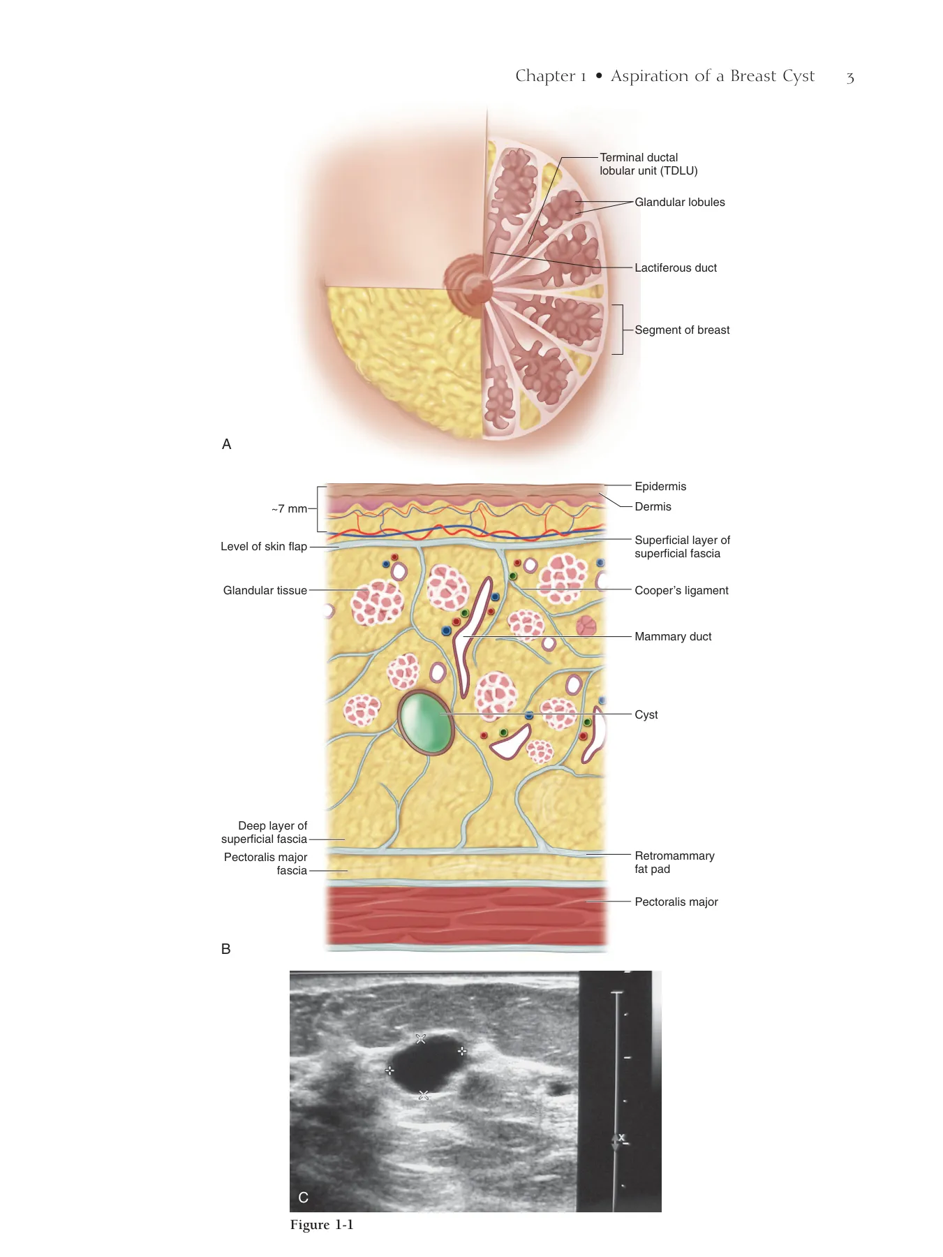
◆ Cannulate the fl uid-producing duct (Fig. 5-4; the skin markings outline the area of abnormal
fi ndings). In case of pathologic discharge, use a 2-0 Prolene suture cut to a gentle taper; for
breast cancer cases, the orifi ce will be smaller and produce much less fl uid. In these cases, fi rst
inject Lymphazurin (Tyco Healthcare Group LP, Norwalk, CT) and local anesthetic in the region
around the tumor to assist in selecting the correct low-volume duct. Cannulate with the 2-0
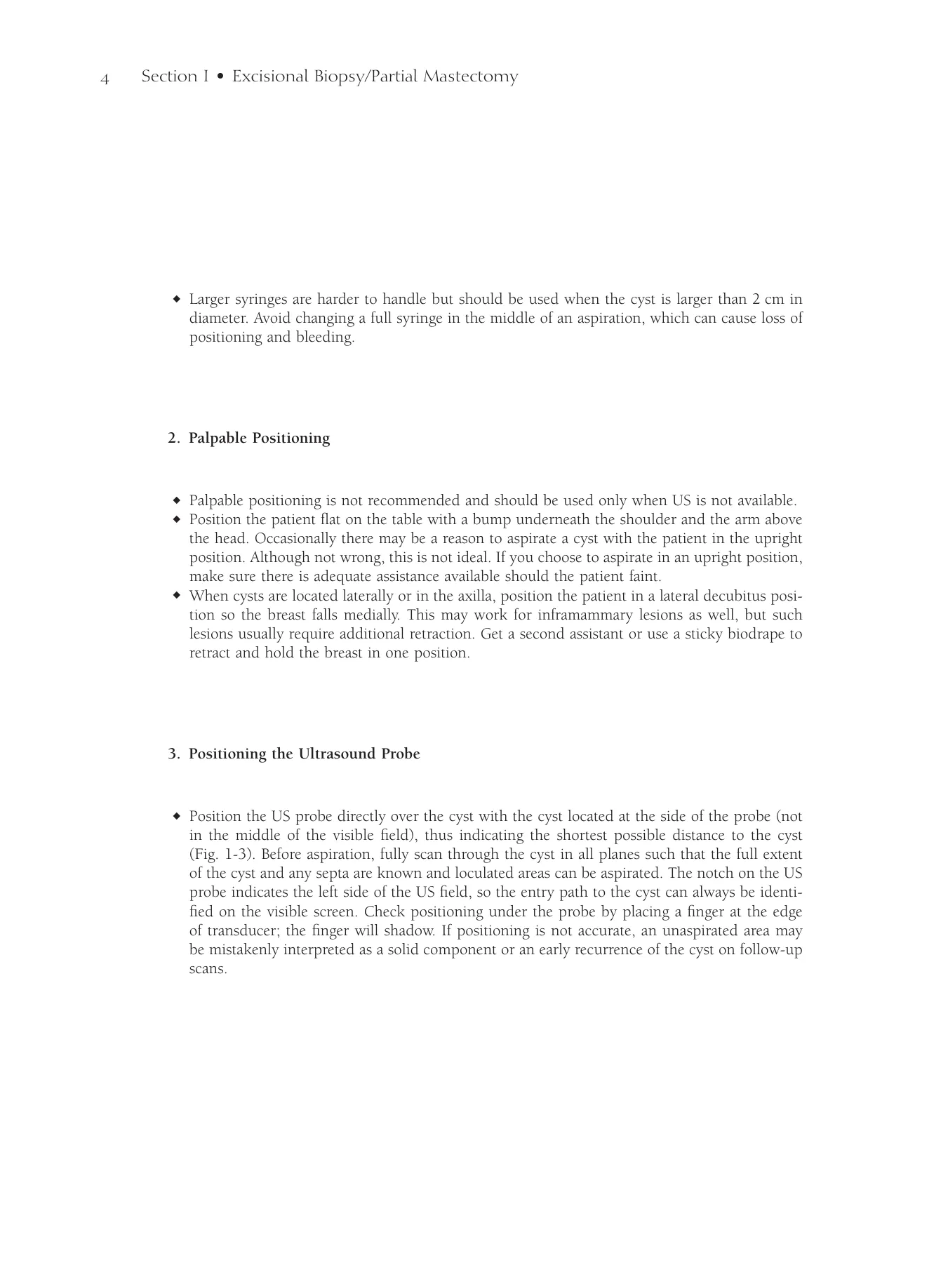
Prolene if possible or, if necessary, use more rigid introducers; the best current controllable tip
is the EntreeTM dilating trocar (ConMed EndoSurgery, Utica, NY; Fig. 5-5).
◆ Progressively dilate in Seldinger fashion over the 2-0 Prolene using 24-, 22-, and 20-gauge
Angiocaths (Becton Dickinson Medical Surgical Systems, Sandy, UT; Fig. 5-6). With each
Angiocath, inject 1 to 5 mL of local anesthetic into the duct for distention.
Chapter 5 • Breast Ductoscopy 53
Centripetal Massage Methods
Figure 5-3
Figure 5-4
Entree™ Dilating trocar
Figure 5-5
Figure 5-6
54 Section I • Excisional Biopsy/Partial Mastectomy
2. Ductoscopy—Diagnostic
◆ The endoscope sheath is introduced in Seldinger fashion, removing the hollow introducer and
replacing it with the optical fi bers of the scope. Ensure that the endoscope is well focused and
white balanced before introducing it into the breast ducts (Fig. 5-7).
◆ The duct then can be distended using saline or a local anesthetic while preventing leakage by
compressing the nipple papilla against the endoscope with the thumb and index fi nger.
◆ The endoscope is advanced as distention is obtained. Distraction of the nipple and gentle motion
of the underlying breast mound can be used to negotiate the varying branches. The scope
sheaths all come with markings to help keep track of depth.
◆ When you come to multiple branches, the largest-diameter branch usually leads to the most
proliferative lesion.
◆ To fi nd the position of the scope tip if an abnormality that needs excisional biopsy is seen, turn
off all the overhead lights in the operating room and use the transillumination from the scope
tip to identify the site for biopsy.
3. Ductoscopy—Therapeutic
◆ When directing breast cancer procedures, remember that the volume of expressed fl uid may
be small, making ductal cannulation more challenging. Do not perforate the ductal system, or
efforts to distend the ducts will fail and ductoscopy will not be useful. Be gentle and expect
that distention may take longer.
◆ The ductal orifi ce chosen should usually lie in the radial sector of the nipple papilla 100 to
110 degrees in either direction from the known lesion position in the breast (see Fig. 5-4).
For example, if a known cancer is at 2:00 (i.e., 2 o’clock) and 5 cm out from the nipple papilla,
the discharging orifi ce connecting to that cancer will be on the nipple papilla at 2:00 ± 100 to
110 degrees, or roughly between 9:45 to 4:45.
◆ Always direct the endoscope down the largest branches fi rst because these are more likely to
connect to the tumor.
◆ Once the obstructing tumor is found, mark the position by transillumination.
◆ Work your way backward, directing the endoscope down each smaller branch and marking the
positions of intraductal luminal defects by transillumination on the breast surface.
◆ Leave the endoscope at the most proximal extent of nipple-ward progression of intraluminal
disease.
◆ Design the excision of the subsegmental quadrant to include from scope tip to a pie-shaped
wedge to the periphery of the breast from the known tumor. The width of this wedge is deter-
mined by the intraluminal disease foci identifi ed in side branches.
Chapter 5 • Breast Ductoscopy 55
Micro-endoscope
Figure 5-7
56 Section I • Excisional Biopsy/Partial Mastectomy
Step 4. Postoperative Care
◆ These patients require no different care than that provided after any surgical breast-tissue exci-
sion. It is normal for bloody fl uid to drain from the nipple for several days after ductoscopy,
but rarely longer than 3 weeks.
Step 5. Pearls and Pitfalls
◆ Papillomas in the nipple papilla may make it hard for the surgeon to decide what he or she is
initially looking at because the duct is full of material. Usually the duct is very dilated below
the papilloma, so advance the endoscope until the normal duct is seen below the papilloma. If
the papilloma is within 12 mm of the nipple papilla surface, it usually can be excised by remov-
ing the scope and, using a 3-mm skin punch biopsy, removing the ductal opening and pushing
down in the direction in which the scope most easily advances. Usually a 15-mm or larger duct
can be removed in this way. The hole does not need to be sutured because the circular muscle
of the nipple papilla will pursestring the opening as soon as the local anesthetic wears off. It
will bleed, however, much like a fi ngertip paper cut. Figure 5-8 shows a range of normal,
benign, and malignant fi ndings (ductal carcinoma in situ [DCIS]).
◆ Massaging the breast deeply in a centripetal fashion, using a smooth hydrating lotion, is the
best way to express fl uid (see Fig. 5-3).
Chapter 5 • Breast Ductoscopy 57
Clean duct
Bifurcation
Papilloma
Papillomas
Papillomas
DCIS
Low-grade DCIS
Low-grade DCIS
DCIS
High-grade DCIS
High-grade DCIS
DCIS
Figure 5-8
58 Section I • Excisional Biopsy/Partial Mastectomy
◆ Dekeratinizing agents should not contain shards of particulate material or be sticky and diffi cult
to wash off because both can interfere with ductal orifi ce identifi cation. A nipple aspirator may
be used in diffi cult cases (Fig. 5-9).
◆ When the image dims, usually this means more proteinaceous secretions or blood may be
present. It may be necessary to fi ll the ductal system with saline and empty it several times to
get a clear image for complete ductoscopy.
◆ Cytologic study will identify cancer in less than one third of cases in which it has been seen
endoscopically. Remember that excisional biopsy must still be the standard for correctly diag-
nosing an endoscopically visible lesion.
◆ Small, widely spaced lesions (<0.2 mm) are rarely malignant.
◆ Multiple proliferative lesions in the ductal wall are commonly associated with widespread DCIS
or atypical ductal hyperplasia.
Bibliography
Dietz JR, Crowe JP, Grundfest S, et al: Directed duct excision by using mammary ductoscopy in patients with pathologic nipple discharge.
Surgery 2002;132:582-587.
Dooley WC: Routine operative breast endoscopy for bloody nipple discharge. Ann Surg Oncol 2002;9:920-923.
Dooley WC: Routine operative breast endoscopy during lumpectomy. Ann Surg Oncol 2003;10:38-42.
Dooley WC, Francescatti D, Clark L, Webber G: Offi ce-based breast ductoscopy for diagnosis. Am J Surg 2004;188:415-418.
Jacobs VR, Kiechle M, Plattner B, et al: Breast ductoscopy with a 0.55-mm mini-endoscope for direct visualization of intraductal lesions.
J Minim Invasive Gynecol 2005;12:359-364.
Kim JA, Crowe JP, Woletz J, et al: Prospective study of intraoperative mammary ductoscopy in patients undergoing partial mastectomy for
breast cancer. Am J Surg 2004;188:411-414.
Matsunaga T, Kawakami Y, Namba K, Fujii M: Intraductal biopsy for diagnosis and treatment of intraductal lesions of the breast. Cancer
2004;101:2164-2169.
Matsunaga T, Ohta D, Misaka T, et al: Mammary ductoscopy for diagnosis and treatment of intraductal lesions of the breast. Breast Cancer
2001;8:213-221.
Moncrief RM, Nayar R, Diaz LK, et al: A comparison of ductoscopy-guided and conventional surgical excision in women with spontaneous
nipple discharge. Ann Surg 2005;241:575-581.
Okazaki A, Okazaki M, Asaishi K, et al: Fiberoptic ductoscopy of the breast: A new diagnostic procedure for nipple discharge. JPN J Clin
Oncol 1991;21:188-193.
Sauter ER, Ehya H, Klein-Szanto AJ, et al: Fiberoptic ductoscopy fi ndings in women with and without spontaneous nipple discharge.
Cancer 2005;103:914-921.
Sauter ER, Ehya H, Schlatter L, MacGibbon B: Ductoscopic cytology to detect breast cancer. Cancer J 2004;10:33-41.
Sauter ER, Klein-Szanto A, Ehya H, MacGibbon B: Ductoscopic cytology and image analysis to detect breast carcinoma. Cancer
2004;101:1283-1292.
Sharma R, Dietz J, Wright H, et al: Comparative analysis of minimally invasive microductectomy versus major duct excision in patients
with pathologic nipple discharge. Surgery 2005;138:591-596.
Yamamoto D, Senzaki H, Nakagawa H, et al: Detection of chromosomal aneusomy by fl uorescence in situ hybridization for patients with
nipple discharge. Cancer 2004;97:690-694.
Chapter 5 • Breast Ductoscopy 59
Aspiration cup
Figure 5-9
Step 1. Surgical Anatomy
◆ Figure 6-1 demonstrates the anatomic positions of various breast abnormalities.
◆ Figure 6-2 shows the subcutaneous anatomy of the breast.
Step 2. Preoperative Considerations
◆ It is preferable to obtain a defi nitive diagnosis through needle core biopsy before performing
excisional breast biopsy (EBB).
◆ The so-called triple-negative test was developed to identify patients in whom EBB could be
obviated and refers to a combination of results from physical examination, imaging (mammo-
graphy or ultrasonography [US]), and pathology in palpable lesions less than 1 cm in diameter.
When all three modalities indicate a benign lesion, the probability of a diagnostic error is less
than 1%.
◆ Obtaining a preoperative diagnosis of cancer allows for a single defi nitive cancer operation
(including axillary staging) in 75% to 100% of cases, compared with 25% to 40% of patients
in whom the diagnosis has not been established before EBB.
◆ The indications for EBB include the following:
▲ Any suspect new fi nding or change in follow-up clinical examination or imaging
▲ A lesion that is almost certainly benign but is greater than 2 cm, painful, or recurring, or
where the mass represents growing cysts or fi broadenomas
▲ Solid masses with equivocal clinical or radiologic features, high-risk lesions, or discordant
lesions
▲ Patient preference
◆ Lesions diagnosed on core biopsy that have a signifi cant risk of underestimating cancer char-
acterized as high-risk, including atypical ductal or lobular hyperplasia, lobular carcinoma in
situ, papilloma, and radial scar, should undergo EBB. Lesions diagnosed as malignant should
undergo EBB or segmental mastectomy.
Excisional Breast
Biopsy of Palpable or
Nonpalpable Lesions
V. Suzanne Klimberg
CHAPTER 6
60
Chapter 6 • Excisional Breast Biopsy of Palpable or Nonpalpable Lesions 61
Lymph node
Milk duct
Lactiferous
duct
Papilloma
Milk gland
Fat
Subcutaneous
abscess
Subareolar
abscess
Cyst
Fascia of
pectoralis major
Retromammary
fat plain
Pectoralis major
Pectoralis minor
Rib
DCIS
TDLU (Terminal
Ductal Lobular
Unit)
Invasive mammary
carcinoma
Figure 6-1
Epidermis
Terminal ductal
lobular unit (TDLU)
Glandular lobules
Lactiferous duct
Segment of breast
Dermis
Superficial layer of
superficial fascia
Cooper’s ligament
Pectoralis major
Mammary duct
Mass
Retromammary
fat pad
Glandular tissue
Level of skin flap
~7 mm
Pectoralis major
fascia
A
B
Deep layer of
superficial fascia
Figure 6-2
62 Section I • Excisional Biopsy/Partial Mastectomy
Step 3. Operative Steps
1. Setup
◆ Excision of palpable breast lesions can be performed under local anesthesia and sedation or
general anesthesia. Local anesthesia using a mixture of short- and long-acting anesthetics with
epinephrine is ideal. Supplemental sedation relaxes the patient during the procedure. Some
have suggested that better margin clearance is obtained with general anesthesia.
◆ The patient’s body should be juxtaposed to the edge of the table and the arm ipsilateral to the
index lesion should be positioned at 90 degrees, such that the surgeon and assistant can stand
close to the patient above or below the arm (Fig. 6-3).
◆ Padding under the upper arm should be level with the table and the wrist should be partially
cocked back with padding. In a patient with a large, pendulous breast or lesion in the lower
outer quadrant, a biodrape instead of an assistant can be used to hold the breast in an optimal
position for EBB.
2. Intraoperative Localization
◆ Intraoperative localization can be accomplished simply by palpating the location and extent of
the index lesion.
◆ US with a 7.5- or 10-MHz probe can be used to increase the accuracy of palpable lesion local-
ization, or to localize the nonpalpable lesion (Fig. 6-4).
3. Incision
◆ Although it is desirable to place the EBB incision to obtain the best cosmetic result, consider-
ation should be given to subsequent surgical procedures if the lesion is suspected to be malig-
nant (Fig 6-5).
◆ Incisions along Langer’s lines usually provide the best cosmesis in the upper pole of the
breast.
◆ For large masses in the lower half of the breast, an incision along a Langer’s line may produce
a signifi cant cosmetic deformity, pulling the nipple–areolar complex toward the inframammary
crease on closure. Radial incisions are preferred in this circumstance. Others prefer radial inci-
sions because cancer grows in a radial fashion (see Fig. 6-1). The edge of the dermis is then
freed to facilitate retractor placement as well as optimal closure.
Chapter 6 • Excisional Breast Biopsy of Palpable or Nonpalpable Lesions 63
Incision lines
Figure 6-3
Figure 6-4
Figure 6-5
64 Section I • Excisional Biopsy/Partial Mastectomy
4. Margin Estimation
◆ The surgeon should aim for a 1-cm margin around a mass as determined by palpation and US
guidance (Fig. 6-6).
◆ Figure 6-7 shows the US probe being used to determine how deep the EBB needs to be by
placing it laterally in the wound.
◆ Intraoperative US can be used to visualize approximately 50% of nonpalpable lesions and can
improve margin negativity with both palpable and nonpalpable lesions by a line-of-sight
technique (see Fig. 6-6).
Chapter 6 • Excisional Breast Biopsy of Palpable or Nonpalpable Lesions 65
1 cm
A
C
D
B
1 cm
1 cm
1 cm
1 cm
1 cm
1 cm
1 cm
1 cm
1 cm
Ultrasound
probe
Lesion
E
Figure 6-6
66 Section I • Excisional Biopsy/Partial Mastectomy
5. Specimen Imaging
◆ Once a lesion has been removed, inspect the mass to detect positive or close margins. This can
be done by visual inspection or by specimen US (Fig. 6-8).
◆ Specimen US, using water on the specimen or submersion of the specimen in a bowl of water,
can determine close or positive margins on all six sides of the specimen. Reexcision, if neces-
sary, can then be directed to a specifi c margin.
6. Pathology Marking
◆ The specimen should be marked either while still in the patient or immediately after delivery
from the wound in order not to confuse orientation.
◆ It is good practice to mark specimen orientation in the same way every time—for example,
“superior” with a short stitch and “lateral” with a long stitch.
◆ In addition, ink from the marking pen can be used to mark the anterior surface (Fig. 6-9). It
is not permanent and allows further orientation and ink marking during pathologic examination
with six different colors (one for each side).
7. Palpation of Cavity
◆ Palpation of the cavity after removal of the primary lesion, even when margins appear negative,
can sometimes fi nd nodules or disease that was not initially apparent.
Chapter 6 • Excisional Breast Biopsy of Palpable or Nonpalpable Lesions 67
A
B
Hematoma
Posterior margin
Figure 6-7
Figure 6-8
Figure 6-9
68 Section I • Excisional Biopsy/Partial Mastectomy
8. Shaved Margins
◆ If a particular margin is close, instead of performing a whole-site reexcision, the intraoperative
pathology or permanent inked margins can be used to determine the specifi c involved margin
(or margins), and the problem can be addressed using a shave technique (Fig. 6-10).
◆ Two clamps are placed approximately 1 cm above and on either side of the index lesion site.
◆ A fl at, approximately 1-cm-thick slice of tissue is shaved from the involved margin (e.g., lateral),
the new margin is marked with ink, and the specimen is sent for pathologic examination.
9. Hemostasis
◆ The breast is extremely vascular, so it is important to obtain excellent hemostasis using elec-
trocautery and ties. A mixture of short- and long-acting local anesthesia containing epinephrine
facilitates hemostasis and postoperative pain control.
10. Closure
◆ For the best cosmetic results, the subcutaneous tissue of the cavity just below the incision is
closed. It is critical that the tissue thickness there be at least 7 mm (Fig. 6-11) in case partial
breast irradiation is used later.
◆ Either a running or interrupted absorbable suture can be used to close the dermis. A subcuticu-
lar running stitch is best to close the skin.
◆ A drain is unnecessary and may adversely affect cosmesis.
Chapter 6 • Excisional Breast Biopsy of Palpable or Nonpalpable Lesions 69
Knife cutting
shaved margin
Nipple
Lump removed
≥ 7 mm
Figure 6-10
Figure 6-11
70 Section I • Excisional Biopsy/Partial Mastectomy
Step 4. Postoperative Care
◆ A pressure dressing is not necessary. Glue or Steri-Strips oriented horizontally over the wound
is suffi cient.
◆ A sports bra is suffi cient to stabilize the breast for at least the fi rst 48 hours.
Step 5. Pearls and Pitfalls
◆ Too little padding under the upper arm can cause neuropraxia or neuralgia of the axillary nerve.
Too little under the wrist can cause radial nerve praxis.
◆ Hematomas can be avoided by meticulous attention to hemostasis. Stick ties should be used
for any major vessels or oozing, not simply electrocautery. A drain should be placed only
if generalized oozing continues. Make sure to take patients off of any nonsteroidal anti-
infl ammatory drugs (for 7 to 10 days) because of their antiplatelet effects. Hematomas can occur
for up to 10 days after surgery.
◆ Blisters from Steri-Strips not placed parallel to the plane of the incision or under tension can
make scars more unsightly than the incision itself. Incision glue is a good alternative.
◆ Palpation followed by US of the cavity can sometimes help locate a missed lesion. Reexcision
of the cavity can also be attempted if, based on reexamination of the mammogram, the operator
is confi dent of the location of the missed lesion. If the lesion still cannot be located, the incision
should be closed and a repeat mammogram taken; if the lesion is confi rmed still to be present,
it should be localized using needle localization and the patient taken directly back to the oper-
ating room.
Chapter 6 • Excisional Breast Biopsy of Palpable or Nonpalpable Lesions 71
Bibliography
Chalas E, Valea F: The gynecologist and surgical procedures for the breast disease. Clin Obstet Gynecol 1994;37:948-953.
Crow JP, Rim A, Patrick R, et al: A prospective review of the decline of excisional breast biopsy. Am J Surg 2002;184:353-355.
Kerlikowske K, Smith-Bindman R, Ljung BM, Grady D: Evaluation of abnormal mammography results and palpable breast abnormalities.
Ann Intern Med 2004;139:274-284.
Schwartz GF, Veronesi U, Clough KB, et al: Consensus Conference on Breast Conservation. J Am Coll Surg 2006;203:198-207.
Vetto J, Pommier R, Schmidt W, et al: Diagnosis of palpable breast lesions in younger women by the modifi ed triple test is accurate and
cost-effective. Arch Surg 1996;131:967-974.
Step 1. Surgical Anatomy
◆ Figure 7-1 demonstrates the anatomic positions of various breast abnormalities.
◆ Figure 7-2 shows the subcutaneous anatomy of the breast.
Step 2. Preoperative Considerations
◆ It is preferable to obtain a defi nitive diagnosis through needle core biopsy before performing
excisional breast biopsy (EBB). Stereotactic biopsy guided by mammography is the preferred
method of diagnosis if the lesion is not seen by ultrasonography (US).
◆ The indications for needle localization breast biopsy (NLBB) include the following:
▲ Any suspect new fi nding or change in follow-up clinical examination or mammography not
seen by US
▲ Suspect calcifi cation (fi ve or more clustered calcifi cations), solid masses with equivocal clini-
cal or mammographic features, high-risk lesions, or discordant lesions; or because of patient
preference
▲ Lesions diagnosed on core biopsy that have a signifi cant risk of underestimating cancer
characterized as high-risk, including atypical ductal or lobular hyperplasia, lobular carcinoma
in situ, papilloma, and radial scar, should undergo NLBB if not palbable or can be excised
by hematoma-directed ultrasound guidance (HUG) if visible on US. Lesions diagnosed as
malignant should undergo segmental mastectomy.
◆ The needle for NLBB is placed while the patient is in the sitting position with the breast com-
pressed by a plate that is numbered on one side and lettered on the other. The lesion is local-
ized using a needle and wire, that, if possible, is placed behind the target lesion to facilitate
excision (Fig. 7-3). Various needles are available to localize lesions, including the Kopans
needle (checkmark wire), the Hawkins needle (multiple barbs and the only retractable wire
for repositioning), and the Homer needle (J wire), which is the only virtually nontransectable
needle.
Needle Localization
Breast Biopsy
V. Suzanne Klimberg, Keiva Bland, and Kent C. Westbrook
CHAPTER 7
72
Chapter 7 • Needle Localization Breast Biopsy 73
Lymph node
Milk duct
Lactiferous
duct
Papilloma
Milk gland
Fat
Subcutaneous
abscess
Subareolar
abscess
Cyst
Fascia of
pectoralis major
Retromammary
fat plain
Pectoralis major
Pectoralis minor
Rib
DCIS
TDLU (Terminal
Ductal Lobular
Unit)
Invasive mammary
carcinoma
Figure 7-1
Epidermis
Terminal ductal
lobular unit (TDLU)
Glandular lobules
Lactiferous duct
Segment of breast
Dermis
Superficial layer of
superficial fascia
Cooper’s ligament
Pectoralis major
Mammary duct
Mass
Retromammary
fat pad
Glandular tissue
Level of skin flap
~7 mm
Pectoralis major
fascia
A
B
Deep layer of
superficial fascia
Figure 7-2
Cranial
caudal
view
Medical
lateral
view
Needle wire
Needle wire
Index lesions circled
Figure 7-3
74 Section I • Excisional Biopsy/Partial Mastectomy
◆ It is advisable to leave the needle over the wire so that the wire is strengthened and is more
easily palpated and seen on US.
◆ The patient should be scheduled for excision as soon as feasible after placement because the
wire and needle placement and the retained needle itself can be uncomfortable.
Step 3. Operative Steps
1. Setup
◆ As with the excision of palpable breast lesions, NLBB can be performed under local anesthesia
with sedation or general anesthesia.
◆ The patient’s body should be juxtaposed to the edge of the table and the arm ipsilateral to the
index lesion should be positioned at 90 degrees, such that the surgeon and assistant can stand
close to the patient above or below the arm (Fig. 7-4). Appropriate padding of the arm should
be used.
◆ Care should be taken when removing bandages covering the extraneous portion of the wire
and needle because it is easily dislodged.
2. Intraoperative Localization
◆ Intraoperative localization can be performed simply by palpating the needle (Fig. 7-5).
◆ US with a 7.5- or 10-MHz probe provides additional accuracy; the tip of the needle wire can
be diffi cult to palpate; if necessary, US can be used to fi nd the tip and excise the needle and
wire.
Chapter 7 • Needle Localization Breast Biopsy 75
A
B
Figure 7-4
Figure 7-5
76 Section I • Excisional Biopsy/Partial Mastectomy
3. Incision
◆ Although it is desirable to place the EBB incision to obtain the best cosmetic result, such as
through an incision in the inframammary fold and following the needle wire down to the tip
of the needle (Fig. 7-6), most often the lesion is accessed by making an incision directly over
the tip of the palpable wire or next to the needle and wire and following it down to the tip to
fi nd the target lesion (Fig. 7-7).
◆ For large masses in the lower half of the breast, incision along a Langer’s line may produce a
signifi cant cosmetic deformity, pulling the nipple–areolar complex toward the inframammary
crease on closure. Radial incisions are preferred in this circumstance. Others prefer radial inci-
sions because cancer grows in a radial fashion along the ductal system (see Fig. 7-1). The edge
of the dermis is then freed to facilitate retractor placement as well as optimal closure.
Chapter 7 • Needle Localization Breast Biopsy 77
B
C
D
E
Figure 7-6
Figure 7-7
A
78 Section I • Excisional Biopsy/Partial Mastectomy
4. Excision
◆ After making the incision, a clamp should be used to secure the hub of the needle wire construct
proximal to the targeted lesion (Figs. 7-8 and 7-9).
◆ Rakes can then be used to open up the wound over the lesion (Fig. 7-9), with the goal of
removing a cylinder of tissue around the wire (Fig. 7-10).
◆ Ideally, the wire should be placed behind the index lesion so that the surgeon can cut along
the needle wire posteriorly, in effect marking the posterior extent of the resection.
◆ A clamp is used to secure the tissue distal to the tip of the needle wire (Fig. 7-11).
◆ The tissue surrounding the needle–wire is then lifted out of the wound, with dissection con-
tinuing to take a cylinder of tissue around the needle wire (Fig. 7-12).
Chapter 7 • Needle Localization Breast Biopsy 79
Nipple
Skin
Glandular
tissue
Avascular
fatty plain
Wire
Clamp
on wire
Lesion
Figure 7-8
Figure 7-9
Figure 7-10
Figure 7-11
A
B
Figure 7-12
80 Section I • Excisional Biopsy/Partial Mastectomy
◆ Once the dissection has reached the proximal clamp, the lock on the wire is released
(Fig. 7-13) and the needle removed, leaving the wire in place with the proximal clamp on it
(Fig. 7-14).
◆ The clamp is then loosened and the wire is exited through the wound (Fig. 7-15).
Chapter 7 • Needle Localization Breast Biopsy 81
A
B
Figure 7-13
Figure 7-14
Figure 7-15
82 Section I • Excisional Biopsy/Partial Mastectomy
◆ The dissection is then completed, delivering the specimen with wire intact through the wound
(Fig. 7-16).
5. Margin Estimation
◆ The surgeon should aim for a cylindrical 1-cm margin around the mass and wire (see
Fig. 7-10).
6. Specimen Imaging
◆ Once the lesion has been removed, inspect the mass to detect positive or close margins. This
can be done by visual inspection.
Chapter 7 • Needle Localization Breast Biopsy 83
A
B
C
Figure 7-16
84 Section I • Excisional Biopsy/Partial Mastectomy
◆ Plain radiography of the specimen confi rms that the targeted lesion and calcifi cations were
removed and shows how close they were to a margin (Fig. 7-17). Although specimen radio-
graphy cannot confi rm completely negative margins because it is taken in only one plane, it
does confi rm a positive one.
7. Pathology Marking
◆ The specimen should be marked as described in Chapter 6 (Fig. 7-18), with a superior short
stitch, a lateral long stitch, and ink anteriorly.
8. Palpation of Cavity
◆ Palpation of the cavity after removal of the primary lesion, even when margins appear negative,
can sometimes fi nd nodules or disease that was not initially apparent.
9. Shaved Margins
◆ If a particular margin is close, instead of performing a whole-site reexcision, the intraoperative
pathology or permanent inked margins can be used to determine the specifi c involved margin
(or margins), and the problem can be addressed using a shave technique in which a
1-cm thickness from each wall of the cavity is removed sequentially (Fig. 7-19).
10. Hemostasis
◆ The breast is extremely vascular, so it is important to obtain excellent hemostasis using elec-
trocautery and ties. A mixture of short- and long-acting local anesthesia containing epinephrine
facilitates hemostasis and postoperative pain control.
Chapter 7 • Needle Localization Breast Biopsy 85
Knife cutting
shaved margin
Nipple
Figure 7-17
Figure 7-18
Figure 7-19
86 Section I • Excisional Biopsy/Partial Mastectomy
11. Closure
◆ For the best cosmetic results, the subcutaneous tissue of the cavity just below the incision is
closed. It is critical that the tissue thickness left there be at least 7 mm (Fig. 7-20) in case partial
breast irradiation is used later.
◆ Either a running or interrupted absorbable suture can be used to close the dermis. A subcuticu-
lar running stitch is best to close the skin.
◆ A drain is unnecessary and may adversely affect cosmesis.
Step 4. Postoperative Care
◆ A pressure dressing is not necessary. Glue or Steri-Strips over the wound is suffi cient.
◆ A sports bra is suffi cient to stabilize the breast for at least the fi rst 48 hours.
Step 5. Pearls and Pitfalls
◆ Four to 9% of lesions can be missed with NLBB. When removing a lesion, the surgeon should
always try to visualize where he or she would reexcise if the lesion were missed the fi rst time.
Palpation and then US of the cavity can sometimes help locate a missed lesion. Reexcision of
the cavity can also be attempted if, based on reexamination of the mammogram, the operator
is confi dent of the location of the missed lesion. If the lesion still cannot be located, the patient
should be closed and a repeat mammogram taken; if the lesion is confi rmed still to be present,
it should be relocalized using needle localization and the patient taken directly back to the
operating room.
Bibliography
Chalas, Valea F: The gynecologist and surgical procedures for the breast disease. Clin Obstet Gynecol 1994;37:948-953.
Crow JP, Rim A, Patrick R, et al: A prospective review of the decline of excisional breast biopsy. Am J Surg 2002;184:353-355.
Kerlikowske K, Smith-Bindman R, Ljung BM, Grady D: Evaluation of abnormal mammography results and palpable breast abnormalities.
Ann Intern Med 2004;139:274-284.
Schwartz GF, Veronesi U, Clough KB, et al: Consensus Conference on Breast Conservation. J Am Coll Surg 2006;203:198-207.
Vetto J, Pommier R, Schmidt W, et al: Diagnosis of palpable breast lesions in younger women by the modifi ed triple test is accurate and
cost-effective. Arch Surg 1996;131:967-974.
Chapter 7 • Needle Localization Breast Biopsy 87
Lump removed
≥ 7 mm
Figure 7-20
Step 1. Surgical Anatomy
◆ Familiarity with the ultrasonographic characteristics of the normal breast as well as those of
benign and malignant lesions is essential (see also Chapter 3).
◆ Figure 8-1 demonstrates the normal ultrasonographic anatomy of the breast.
◆ Figure 8-2 illustrates the more common characteristics that distinguish benign from malignant
lesions.
Step 2. Preoperative Considerations
◆ Indications for radioisotope occult lesion localization (ROLL) include the following:
▲ Microcalcifi cations
▲ Parenchymal distortions (i.e., radial scars, atypical hyperplasia)
▲ Nonpalpable, suspect soft tissue masses
▲ Impalpable cancers after neoadjuvant chemotherapy
▲ Foreign bodies in soft tissue
◆ ROLL is performed for the diagnostic excision biopsy of suspect lesions as well as the thera-
peutic excision of a proven cancer. The aim is to remove cancerous tissue with clear resection
margins within the smallest possible amount of glandular tissue, to achieve excellent cosmetic
results. ROLL is frequently performed under general anesthesia as a day case. Initial diagnostic
imaging (i.e., mammography, ultrasonography) should be available at the time of localization
and in the operating room to plan the surgical approach and to compare with the specimen
radiograph to confi rm excision of the target lesion.
Radioisotope Occult
Lesion Localization
Hodigere S.J. Ramesh, Matilde M. Audisio,
and Riccardo A. Audisio
CHAPTER 8
88
Chapter 8 • Radioisotope Occult Lesion Localization 89
Skin
Subcutaneous
Glandular tissue
Retromammary fat
Pectoral muscle
Pleura
Age - 28
Skin
Subcutaneous
Glandular tissue
Retromammary fat
Pectoral muscle
Pleura
Age - 28
Figure 8-1
Smooth / sharp
Indistinct / jagged
Grows within tissue planes
Disrupted
Transverse > Longitudinal
Longitudinal > Transverse
Posterior enhancement
Posterior shadowing
Benign
Malignant
Margins
Adjacent
architecture
Shape
Retrotumoral
effect
Figure 8-2
90 Section I • Excisional Biopsy/Partial Mastectomy
◆ The nonpalpable lesion can be targeted mammographically or ultrasonographically, such as the
lesion shown in Figure 8-3.
◆ Technetium-99m–labeled (half-life of 6 hours) macroaggregate is freshly prepared by radio-
pharmacy and delivered a few hours before localization. The localization fl uid (0.2 mL) is
delivered in a preloaded, sterile-packed syringe in a protective lead case (Fig. 8-4). Care should
be taken to shake the preloaded syringe gently to mix the macroaggregates before injection.
◆ The absorbed radiation dose to hospital personnel is low and requires neither radiation protec-
tion control nor classifi cation of exposed workers as class A or B. Special containers for radio-
active wastes are necessary in the administration room but not in the operating room, where
possible contamination is negligible. In cases when a surgeon performs 100 procedures per
year, a fi nger dose (FD) dose of approximately 1 mSv is received, which is well within the
annual dose limit of 150 mSv. Annual whole-body dosage of assisting staff may reach 0.04 mSv,
compared with an annual limit of 6 mSv. These low doses indicate that no additional radiation
protection measures are necessary, although they may be required by law.
◆ Localization is performed by a trained radiologist in the radiology department. After reviewing
initial diagnostic imaging and case notes, the radiologist injects 0.2 mL 99mTc-labeled nanocol-
loid into the core of the lesion under ultrasonographic or stereotactic guidance 1 to 4 hours
before surgery. The radiation dosage delivered is 1 MBq, equivalent to 0.02 mSv (=1 chest
radiograph). The lesion is most frequently localized by ultrasonography (∼75%; Fig. 8-5; also
see Fig. 8-3); stereotaxis may be used when the lesion is not visible on ultrasonography
(∼25%).
◆ A fi nal check for radioactive signal is made in the anesthesia room while the patient is in a
sitting as well as a supine position; this not only ensures the working status of the system
(gamma camera and radioisotope) but provides a fi nal opportunity to plan, discuss, and mark
the most appropriate surgical approach to the lesion while the patient is awake (Fig. 8-6).
◆ General anesthesia is recommended, although local anesthesia may be considered in a suitable
candidate.
Chapter 8 • Radioisotope Occult Lesion Localization 91
1
2
2
1
Lesion
Figure 8-3
Figure 8-4
Figure 8-5
Figure 8-6
92 Section I • Excisional Biopsy/Partial Mastectomy
Step 3. Operative Steps
1. Patient Position
◆ The patient is positioned supine on the operating table with the arm ipsilateral to the lesion
abducted to expose the outer breast quadrants and fully extended at the elbow, resting on a
well-padded arm board (Fig. 8-7).
Chapter 8 • Radioisotope Occult Lesion Localization 93
A
B
Figure 8-7
94 Section I • Excisional Biopsy/Partial Mastectomy
2. Incisions
◆ The appropriate skin incision is chosen to facilitate surgical removal in accordance with onco-
logic principles while providing the best cosmetic results. ROLL allows great fl exibility in
approaching the lesion, bearing in mind that the incision line should fall within the boundaries
of a subsequent mastectomy or lumpectomy, should such a procedure become necessary.
Whenever possible, it is our preference to use an inframammary incision for lesions in the lower
quadrants (Fig. 8-8), a periareolar incision for lesions in the central quadrant (Fig. 8-9),
Chapter 8 • Radioisotope Occult Lesion Localization 95
A
B
Submammary
approach
Pectoralis major muscle
Lactiferous
duct
Lobule of
gland
Tumor
Figure 8-8
A
Periareolar
approach
B
C
Pectoralis
major muscle
Tumor
Lobule of
gland
Figure 8-9
96 Section I • Excisional Biopsy/Partial Mastectomy
or an axillary incision for lesions in the upper-outer quadrant (Fig. 8-10). These incision lines
provide excellent cosmetic results.
◆ Skin incisions paralleling Langer’s lines result in thin, cosmetically acceptable scars for peri-
pheral lesions requiring a direct anterior approach (Fig. 8-11).
Chapter 8 • Radioisotope Occult Lesion Localization 97
Axillary
approach
C
Left nipple
Anterior
axillary fold
Posterior axillary fold
Anterior
axillary fold
A
Anterior axillary fold
Posterior axillary fold
Left nipple
B
Figure 8-10
Lines of Langer
Figure 8-11
98 Section I • Excisional Biopsy/Partial Mastectomy
3. Targeting the Lesion
◆ The gamma camera with a mounted collimator is wrapped in a sterile polythene cover (Fig. 8-
12). The collimator fi lters the stream of gamma rays so that only those from the injection site
traveling parallel to the probe tip are scanned. Without a collimator, rays from all directions
are recorded and localization may be more diffi cult.
◆ The most intense area of radioactivity (i.e., center of targeted lesion) is identifi ed with the
gamma probe by listening for audible signal at the injection site (Fig. 8-13).
◆ After the skin is incised, a check for gamma signal facilitates choosing the most direct approach
to the lesion.
4. Excision and Marking of Index Lesion
◆ Excision biopsy is carried out following audible gamma signals and using fi nger palpation of
the tissue being excised. The wound is deepened to include the pectoral fascia, aiming to achieve
5 to 10 mm of healthy margins encasing the neoplastic lesion. The localized tissue is checked
for signal before being dissected from the glandular tissue (Fig. 8-14).
◆ Excision of the target lesion is confi rmed by the absence of residual signal in the cavity
(Fig. 8-15).
Chapter 8 • Radioisotope Occult Lesion Localization 99
B
Figure 8-12
Figure 8-13
Figure 8-14
Figure 8-15
100 Section I • Excisional Biopsy/Partial Mastectomy
◆ The specimen is then marked with standard orientation stitches for pathologic examination,
and checked once again for presence of radioactive signal (Fig. 8-16).
5. Specimen Radiography
◆ Specimen radiography ensures that the lesion is present within the excised tissue; the presence
of healthy tissue margins may provide reassurance as to the oncologic adequacy of the removal,
although this must be confi rmed by pathology (Fig. 8-17).
6. Closure
◆ Meticulous hemostasis is ensured. Drainage of the breast cavity is not necessary.
◆ The excisional cavity is obliterated by approximating the breast plate overlying the pectoral
fascia with 2-0 Vicryl absorbable stitches. Stitches in the fat are avoided (see Fig. 8-9, C).
◆ Skin edges are approximated with continuous subcuticular absorbable 3-0 Vicryl Rapide stitches
(Fig. 8-18).
◆ The wound edges may be reinforced with ¼-inch Steri-Strips (Fig. 8-19). Breathable dressings
or pressure dressings are avoided.
◆ See Chapter 6 for a detailed explanation of excisional breast biopsy.
Step 4. Postoperative Care
◆ Patients are encouraged to resume normal activity on the evening of surgery. Postoperative
assessment should include evaluation of the wound for hematoma formation.
Chapter 8 • Radioisotope Occult Lesion Localization 101
Figure 8-16
Figure 8-17
Figure 8-18
Figure 8-19