
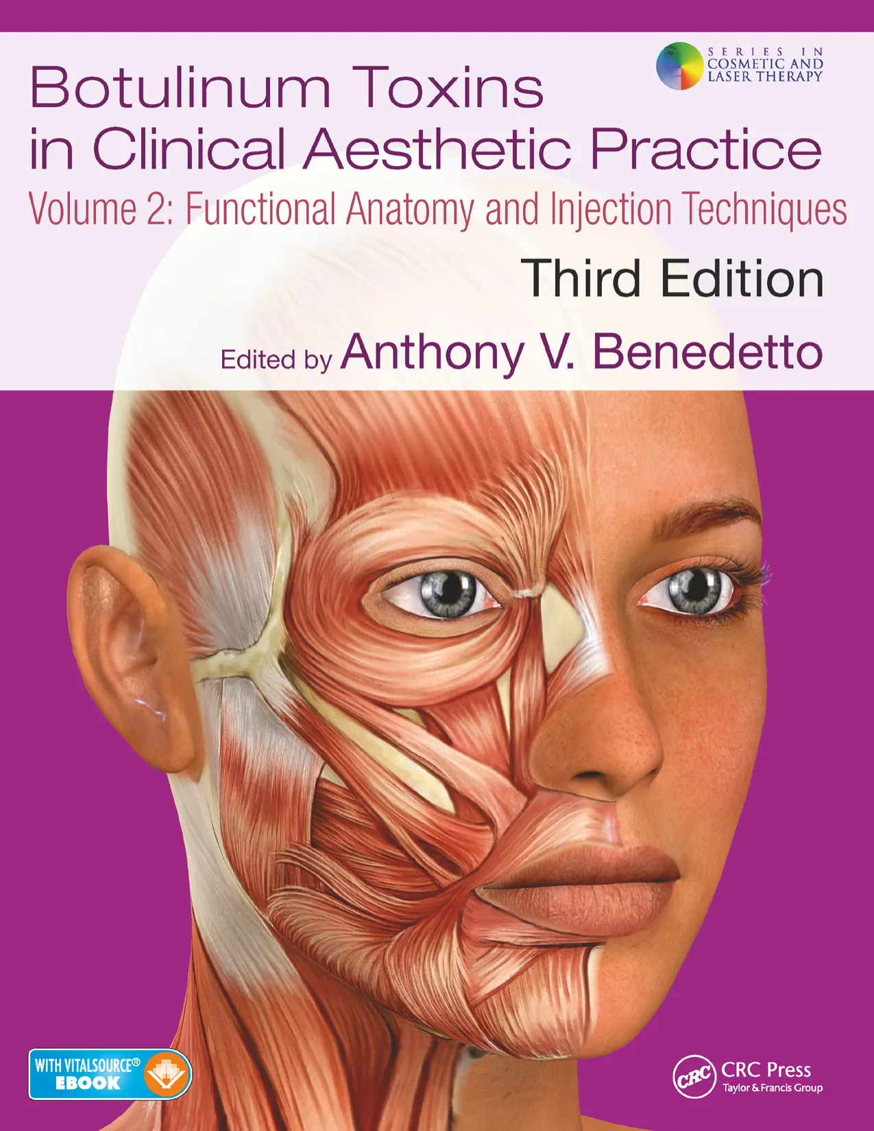

Botulinum Toxins in Clinical Aesthetic Practice
142
point must be at least 2 cm above the eyebrows. Thirdly, the upper
row of treatable horizontal wrinkles can be found midway between
the superior margin of active frontalis and the lower row of treatable
wrinkles. Five injections of 4 units of OnaBTX-A are approved for a
total of 20 units placed at the intersection of the upper and lower rows
of treatable horizontal wrinkles with the following vertical land
marks (see Figure 13.55c):
1. On the lower row of treatable horizontal wrinkles first inject 4
units of OnaBTX-A at the midline of the face.
2. On the same lower row of treatable wrinkles, inject another 4
units of OnaBTX-A on the right and 4 units on the left side of
the face, approximately 0.5–1.5 cm medial to the anterior tem
poral fusion line or temporal crest (see Figure 13.69).
3. On the upper row of horizontal wrinkles, midway between
the central and lateral injection points of the lower row, inject
another 4 units of OnaBTX-A on the right and 4 units on the
left side of the face (see Figure 13.55c).
However, as one develops a more refined injection technique,
improved and more natural appearing results can be realized when
one injects BoNT starting just lateral to the midline, approximately
2–4 cm above the medial aspect of the eyebrows. From this start
ing point, injections are applied across the forehead at three or four
points in an upward and downward pattern following the curvature
of the arc of the eyebrows and maintaining the same distance of
2–3 cm above and parallel to the upper border of the eyebrows. The
highest point of injection should be directly over the highest point of
an arched eyebrow (Figures 13.60 and 13.61). This pattern will keep
the OnaBTX-A high enough above the eyebrows and allow the lower
muscle fibers along the transverse length of the frontalis to gently lift
X
X
X
X
X
X
X
X
X
X
X
X
Figure 13.57 Random pattern injections into a forehead that is high and wide with
multiple parallel wrinkles. This patient had 2 U of OnaBTX-A injected at each site.
2
2
2
2
2
2
2
2
2
(a)
(b)
Figure 13.58 (a) This 68-year-old patient with an average sized forehead and low-set eyebrows was unaware that her right eyebrow was higher than her left. (b) After
OnaBTX-A treatment they were symmetrical. Note the higher dosing on the right versus the left.
2
2
2
2
2
(a)
(b)
Figure 13.59 This 50-year-old patient has a high and narrow forehead and multiple rows of forehead wrinkles. Note the left eyebrow is higher than the right one (a) before
and (b) after treatment with OnaBTX-A.
13. cosmetic uses of botulinum toxin a in the upper face
143
and deliberately produce a naturally peaked and aesthetically pleas
ing arched eyebrow (Figures 13.2, 13.64, and 13.65). Injections that
are too high above the lateral tail of the eyebrows may cause unaf
fected lower fibers of the lateral frontalis to excessively elevate the
lateral aspect of the eyebrows, resulting in the so-called “Mephisto”
or “Mr. Spock” look (Figures 13.62, 13.66, and 13.67). This occurred
more frequently when a “V” pattern of injection was performed in the
forehead (as discussed in the first edition of this text).
The simplest approach to treating the forehead in men is to inject
as much as 25 U of OnaBTX-A or even more intramuscularly in a
horizontal, linear fashion across the forehead, maintaining the same
distance of approximately 3-4 cm above and parallel to the eyebrows
and wrinkles (Figures 13.62 and 13.63). This pattern of injection min
imizes the curvature of the eyebrows, resulting in a more masculine
appearing eyebrow (Figure 13.63).
Another technique is to inject subcutaneously approximately
2–4 U of OnaBTX-A at sites approximately 2 cm apart and across the
entire forehead horizontally at a point midway between the brow and
the hairline also maintaining a constant distance above the eyebrows
(Figures 13.58 and 13.63). This is advisable if the hairline is set low
and if there are only one or two rows of horizontal wrinkles across
the forehead (Figure 13.68).
In most patients, muscle fibers of the frontalis do not extend any
more laterally than the anterior temporal fusion lines. If the width
of the forehead is narrow, that is, less than 12 cm between the right
and left anterior temporal fusion lines, or temporal crests, then four
or five subcutaneous injections of 2–4 U of OnaBTX-A at each injec
tion site across the forehead are sufficient (Figure 13.59). One can feel
the anterior temporal fusion line by first identifying the zygomatic
process of the frontal bone, which is the superior aspect of the upper
lateral wall of the bony orbit (Figure 13.69). Its posterior edge con
tinues upward, as a palpable protruding ridge along the lateral edge
of the frontal bone, and arches upward and backward, delineating
the anterior and superior boundary of the temporal fossa. If an indi
vidual has a wider brow, that is, more than 12 cm between the right
and left anterior temporal fusion lines, then five, six, or possibly more
injection points across the forehead are probably necessary, with
2–4 U of OnaBTX-A injected at each site subcutaneously in women
(Figures 13.57, 13.65 and 13.66). The stronger the frontalis, the more
units of OnaBTX-A will be required to produce the desired effect
(Figures 13.66 and 13.68).
Gentle massage upward and laterally at the injection sites for a few
seconds helps relieve the acute and transient pain of a transcutaneous
injection and can help disperse the toxin locally. Prolonged or heavy-
handed massage, especially in a downward direction, can disperse the
liquid OnaBTX-A beyond the intended area of injection, weakening
adjacent lower muscle fibers and producing an unwanted brow ptosis.
OUTCOMES (RESULTS) WHEN TREATING HORIZONTAL
FOREHEAD LINES (SEE APPENDIX 5)
An adequate result when treating the frontalis is to completely
eliminate the horizontal lines of the forehead when the patient is at rest,
and to provide the ability for some movement and minimal wrinkling
when the patient is animated or actively expressing an emotion. Ideally,
weakening of the frontalis should last at least 3 full months when a
sufficient dose of OnaBTX-A is injected. Frequently, after repeat treat
ments and occasionally even after the first treatment session in some
patients, the effects of OnaBTX-A weakening can last as long as 4–6
months after it is injected.41 Overgenerous intramuscular injections
of the frontalis with high doses of OnaBTX-A will eliminate totally
all movement of the muscle, even with forced contraction, creating a
flat mask-like and motionless forehead and even some degree of brow
ptosis especially in patients (young or old) with inelastic skin. There
usually is never a good aesthetic reason for such total myorelaxation of
the frontalis or any other muscle of facial expression even if the effect
is only temporary. In addition, the overall duration of results usually
2
2
2
2
2
2
2
(a)
(b)
Figure 13.60 (a) A 40-year-old patient with a low and wide forehead with the right eyebrow higher than the left before treatment with OnaBTX-A. Note the position of the
injection points: lower on the right lateral brow and higher over the left brow. (b) Note the position of the right eyebrow 1 week after treatment with OnaBTX-A.
5
4
5
4
4
2
2
(a)
(b)
Figure 13.61 (a) This 36-year-old patient has an average low and narrow forehead with the right eyebrow higher than the left before a treatment of OnaBTX-A. (b) Two
weeks after OnaBTX-A, the eyebrows are symmetrical.
Botulinum Toxins in Clinical Aesthetic Practice
144
is not extended in any muscle when OnaBTX-A is injected at a higher
dose than that which is recommended or normally sufficient for the
individual’s present problem.79
Occasionally and especially with the initial treatment of forehead
wrinkles, the effect of OnaBTX-A weakening may not occur symmet
rically, and there may be wrinkling on one side of the forehead and
not on the other, even at rest. It is imperative that the physician warns
the patient of this before treatment and requires the patient to return
2–3 weeks after a treatment session so that any minor asymmetries
can be corrected. This is easily accomplished by injecting 1–2 U of
OnaBTX-A in the vicinity of the persistent asymmetric wrinkling
and muscle hyperactivity (Figures 13.62 and 13.67).60 Remember, this
should always be done at least 2–3 cm above the bony orbital rim,
so as not to produce brow ptosis inadvertently. This is particularly
important for those patients who have some degree of dermatochala
sis and multiple rows of low-lying horizontal forehead lines (Figure
13.70a). By allowing the lower fibers of the frontalis to remain active,
there typically may be a wrinkle or two directly above the eyebrow
and a degree of asymmetric brow elevation that might persist. Total
elimination of these particularly persistent wrinkles directly above
the lateral aspect of the eyebrows usually cannot be reduced with
out causing some amount of brow ptosis (Figure 13.70b,c). Most of
the time, these narrow horizontal lines immediately adjacent to, if
not within, the upper lateral border of the eyebrows can be identi
fied during pretreatment physical examination and management
planning, and the patient should be made aware of them (Figures
13.63, 13.67, 13.70, and 13.71). The patient also should be given the
option to eliminate these lines by other cosmetic procedures (e.g.,
fillers, energy-based therapy, or both), before initiating injections of
OnaBTX-A. These minor horizontal forehead lines usually are the
manifestation of excessively lax skin and a hyperactive frontalis being
recruited in a compensatory manner to elevate a weighty brow to pre
vent brow hooding and visual field obstruction. The patient is bet
ter off totally ignoring these lines. If they remain after the injections
of OnaBTX-A have completely taken effect and still are extremely
bothersome to the patient, they can be treated by injecting intrader
mally a minimal amount of highly diluted OnaBTX-A adjacent to the
wrinkle. However, no matter how dilute and how little the amount of
OnaBTX-A is injected, brow ptosis still may result. Otherwise, injec
tions of a soft tissue filler, preferably a hyaluronic acid (e.g. a cohesive
polydensified matrix hyaluronic acid to minimize the Tyndall effect)
product 2–4 weeks later, can diminish these wrinkles while bolster
ing the lax, excessive brow skin and creating an overall additional
aesthetic enhancement to the lateral brow and periorbital area.
Complications (Adverse Sequelae) When Treating Horizontal
Forehead Lines (See Appendix 6)
When treating the frontalis with OnaBTX-A, appropriate injection
patterns and meticulous injection techniques are extremely impor
tant in avoiding brow ptosis.54,57,83 This is best accomplished by
remaining at least 3–4 cm above the supraorbital bony margin or
2–3 cm above the eyebrow depending on the idiosyncratic anatomy of
the individual patient being treated. This will enable the muscle fibers
of the frontalis to remain functional with an adequate resting tone in
the area directly above the brow so that the eyebrows will not drop
and produce hooding over the upper eyelids (Figures 13.67 and 13.72).
In most patients, horizontal forehead lines are present in
conjunction with glabellar frown lines. In these patients, the current
preferred practice is to treat the upper third of the face during the
6
6
6
6
6
3
3
3
3
3
3
3
3
3
3
3
3
3
3
(a)
(b)
(c)
(d)
Figure 13.62 (a) Typical injection points and amount of units for a man who works outdoors and has strong depressors of the brow and a strong levator of the forehead
causing very deep furrows. Before treatment with OnaBTX-A injections. (b) Same patient 2 weeks after the initial OnaBTX-A treatment and before an additional 6 U of
OnaBTX-A. Note the elevated “Mephisto” or “Mr. Spock” lateral eyebrows and where an additional 3 U of OnaBTX-A were injected on each side to lower the lateral fron
talis above the tail of the eyebrows, in this man who usually has straighter eybrows. (c) Same patient having a touch-up injection of 3 U of OnaBTX-A bilaterally 2 weeks
after his initial OnaBTX-A treatment. (d) Same patient 5 weeks after the initial OnaBTX-A treatment and 3 weeks after a touch-up treatment. Note the less arched lateral
brows and iatrogenic brow ptosis.
13. cosmetic uses of botulinum toxin a in the upper face
145
2
2
2
2
2
2
2
2
2
(a)
(b)
Figure 13.64 (a) Injection sites for a 38-year-old patient with a high and wide forehead and an asymmetrically lower right eyebrow. (b) After treatment.
3
3
3
3
3
3
3
3
3
3
3
3
3
3
3
3
3
3
3
3
3
3
3
3
3
3
3
3
3
3
3
3
3
3
3
3
3
3
3
3
(a)
(c)
(e)
(g)
(b)
(d)
(f)
(h)
Figure 13.63 (a) A 40-year-old patient at rest before and (b) 3 weeks after an OnaBTX-A treatment of only the forehead. (c) Same patient raising his eyebrows before and
(d) 3 weeks after an OnaBTX-A treatment of only his forehead. (e) Same patient at rest before and (f) 1 week after his fourth OnaBTX-A treatment of only his forehead. (g)
Same patient raising his eyebrows before and (h) 1 week after his fourth OnaBTX-A treatment of only his forehead.
Botulinum Toxins in Clinical Aesthetic Practice
146
2
2
2
2
2
2
2
2
(a)
(b)
(c)
(d)
Figure 13.67 (a) A 54-year-old patient with forehead wrinkles directly over the left eyebrow (arrow) observed with forced brow elevation before treatment. (b) Same patient
2 weeks after initial treatment with OnaBTX-A. Note the left brow is now higher than the right brow with forced eyebrow elevation. An additional 2 U of OnaBTX-A was
injected during this follow-up visit. (c) Same patient 5 weeks after the initial treatment with OnaBTX-A and 3 weeks after a touch-up of 2 U of OnaBTX-A. The left eyebrow
remains slightly elevated laterally at rest with eyes wide open (d) but not with the eyes closed.
2
2
2
2
2
2
2
(a)
(b)
Figure 13.66 A 45-year-old patient raising her eyebrows (a) before and (b) 3 weeks after OnaBTX-A treatment of only the forehead.
X
X
X
X
X
X
X
(a)
(b)
Figure 13.65 (a) Typical injection sites in a woman with an average sized forehead. (b) Same patient 2 weeks after an OnaBTX-A injection.
13. cosmetic uses of botulinum toxin a in the upper face
147
same session.42,43,47,48,55,71,80 The resting tone of the glabellar depres
sors (corrugator supercilii, depressor supercilii, procerus, orbicu
laris oculi) is usually slightly stronger than the resting tone of the
brow elevator (frontalis). Therefore, if the brow depressors are not
treated contemporaneously with the brow elevator, brow ptosis may
be difficult to prevent because of the overriding downward pull of
the depressors (Figure 13.73). Maintaining an adequate resting tone
of the lower fibers of the frontalis just over the brow (approximately
2–4 cm) will help prevent brow ptosis (Figure 6.72).
If excessive doses of OnaBTX-A are injected into the center of the
forehead, a drop of the mid forehead skin and soft tissue can occur,
producing a bulge of skin to appear in the center of the glabella. This
usually is accompanied by a weighing down of the medial aspect
(heads) of the eyebrows and an unattractive downward sloping or a
downward pointing of the heads of the eyebrows. This also may give
some patients an unsettling sensation of a peculiar “heaviness” in
the center of their forehead and brow. To avoid this all-too-common
tell-tale evidence of being “over-Botoxed,” injections should not
be given too low directly in the center of the forehead, especially if
there is a paucity of central forehead wrinkles, indicating an absence
of frontalis muscle fibers. Before injecting this area, one should be
sure that there are strong and active frontalis muscle fibers in the
center of the forehead, like those seen in Trindade’s “omega” pattern
of glabellar frown lines27 (see Figure 13.22). To diminish an exces
sively downward sloping of the medial aspect of the eyebrows and
glabellar soft tissue, injections of OnaBTX-A can be placed into the
medial brow depressors: 3–5 U into each depressor supercilii, which
also will diffuse into the medial fibers of the orbital orbicularis oculi,
and 4–10 U into the procerus in the center of the glabella. This might
relieve some of the forceful downward pull of the central glabellar
soft tissue, especially in Trindade’s “inverted omega” pattern of gla
bellar frowning (see Figure 13.23), and elevate the medial heads of
the eyebrows. There is no antidote for brow ptosis, which can last as
long as the OnaBTX-A injections are effective. Concomitant injec
tions of low-dose, low-volume OnaBTX-A precisely placed into the
superficial fibers of the lateral, upper orbital orbicularis oculi also is
essential in reducing the extent of lateral brow ptosis.
Clinical experience has indicated that when a more concentrated
dose of OnaBTX-A is used (i.e., dilutions of 1 mL per 100 U vial of
OnaBTX-A) minimal volumes are injected and migration of the
OnaBTX-A is negligible.39 The results also seem to last longer. On the
other hand, to prevent total paralysis of muscle movement, especially
with forced contraction of the frontalis, a different approach can be
utilized when attempting to aesthetically reduce horizontal forehead
lines. Since there does not seem to be any agreement in the litera
ture on which dilutions should be used when reconstituting a 100 U
vial of OnaBTX-A, or which dosage regimens are most effective, one
then can inject the forehead with the same total number of units of
OnaBTX-A, but with a more dilute solution.39,78,81 Namely, a 100 U
vial of OnaBTX-A can be reconstituted with 2–4 mL of saline when
used solely for injecting the frontalis. This dilution allows one to
inject a greater volume of OnaBTX-A and disperse it over a wider area
of the forehead, providing an effect that is less intensely paralyzing.78
However, injecting large volumes of diluted OnaBTX-A possibly
might limit the duration of its effectiveness. As long as nontargeted
muscle fibers (i.e., those of the lower frontalis) are not directly in the
wake of the intended toxin diffusion, higher dilutions may be a more
forgiving alternative injection technique, especially for the neophyte
injector. It is the total number of units that is important when treat
ing patients with OnaBTX-A or any other BoNT and not the total
volume used.
Other more common adverse sequelae that occur with an injection
of OnaBTX-A are related more to the actual physical injection rather
than to the material injected. All of these adverse events are tran
sient and generally do not last longer than 24–36 hours. They include
ecchymoses, local edema, erythema, and pain at the injection and
(a)
(b)
Figure 13.68 A 51-year-old male with a low forehead and 2 rows of horizontal wrinkles. Seen here (a) before and (b) 1 month after an onabotulinutoxinA treatment.
X
Figure 13.69 The location and extent of anterior and superior temporal fusion line
on a skull: X, zygomatic process of the frontal bone; blue arrow: anterior temporal
fusion line; red arrow: superior temporal fusion line.
Botulinum Toxins in Clinical Aesthetic Practice
148
2
2
2
2
2
2
2
2
2
2
2
2
2
2
2
2
(a)
(b)
(c)
(d)
Figure 13.71 A 52-year-old patient with mild forehead wrinkling at rest (a) before and (b) 3 weeks after an OnaBTX-A treatment of only the forehead. The same patient
raising her eyebrows (c) before and (d) 3 weeks after an OnaBTX-A treatment of only the forehead. Note the persistent wrinkles just above both lateral eyebrows.
2
(a)
(b)
(c)
Figure 13.70 (a) A 62-year-old patient with compensatory brow elevation. The arrow points to the horizontal forehead line and hyperkinetic frontalis immediately above
the left lateral brow. (b) Same patient after OnaBTX-A. Note the hyperkinetic lower lateral frontalis on the left. Two weeks after the initial treatment, 2 U of OnaBTX-A
were injected 2–2.5 cm above the lateral left brow at a point of maximum contraction of the frontalis. (c) Same patient 5 weeks after initial treatment and 3 weeks after
touch-up over the left eyebrow.
13. cosmetic uses of botulinum toxin a in the upper face
149
adjacent sites (Figure 13.40). For some patients, a dull and transient
headache with or without general body malaise occurs after injections
of OnaBTX-A that can last beyond 24–72 hours.72 The occurrence of
headache immediately after an OnaBTX-A injection seems paradoxi
cal since OnaBTX-A injections are now FDA approved for treating
tension and migraine headaches.1,73 Serious reactions, particularly of
the immediate hypersensitivity type such as anaphylaxis, urticaria,
soft tissue edema, and dyspnea have been extremely rare.76 When they
occur, appropriate medical treatment must be instituted immediately.
Figures 13.74 through 13.81 are additional examples of different
patients treated with OnaBTX-A for horizontal forehead lines. Many
of them had their glabellas treated at the same time.
Treatment Implications When Injecting the Frontalis
1. Identify and document brow or forehead asymmetries prior to
treatment with OnaBTX-A.
2. Injections of OnaBTX-A in the forehead can be placed subcuta
neously or intramuscularly.
2
2
2
2
2
2
2
1
1
2
2
2
2
2
2
2
2
1
1
2
(a)
(b)
(c)
(d)
Figure 13.72 A 32-year-old woman is shown raising her eyebrows (a) before and (b) 1 month after treatment of only the forehead. The same patient is shown at rest (c) before
and (d) 1 month after treatment of only the forehead. Note the gentle arching of the brows after treatment with OnaBTX-A.
2
3
2
2
2
2
2
2
2
(a)
(b)
Figure 13.73 A 53-year-old patient (a) before and (b) 2 weeks after an OnaBTX-A treatment of only the forehead. Note the slight bilateral brow ptosis after treatment when
the glabella is not adequately treated.
Botulinum Toxins in Clinical Aesthetic Practice
150
2
2
2
5
5
4
4
5
2
(a)
(b)
2
2
2
2
2
Figure 13.77 A 59-year-old patient raising her eyebrows (a) before and (b) 3 weeks after an OnaBTX-A treatment for forehead and frown lines. An additional 2 U were
given over the left brow.
2
2
2
2
2
3
3
6
6
5
(a)
(b)
Figure 13.76 A 64-year-old patient raising her eyebrows (a) before and (b) 3 weeks after an OnaBTX-A treatment for forehead and frown lines. Note the “medial dip” of
the glabella.
8
8
8
8
8
2
2
2
2
2
2
2
2
1
1
2
(a)
(b)
Figure 13.75 A 56-year-old patient frowning (a) before and (b) 2 weeks after an OnaBTX-A treatment for forehead and frown lines.
3
3
3
3
3
3
3
3
8
8
8
8
8
4
4
(a)
(b)
Figure 13.74 A 48-year-old patient frowning (a) before and (b) 3 weeks after an OnaBTX-A treatment of forehead and glabellar frown lines.
13. cosmetic uses of botulinum toxin a in the upper face
151
(a)
(b)
5
5
5
5
5
5
5
6
6
4
5
Figure 13.79 A 52-year-old patient raising his eyebrows (a) before and (b) 2 weeks after an OnaBTX-A treatment for forehead and frown lines. Note the “medial dip” of the
glabella.
(a)
(b)
6
6
6
4
6
6
6
5
5
4
3
Figure 13.78 A 42-year-old patient raising his eyebrows (a) before and (b) 2 weeks after an OnaBTX-A treatment for forehead and frown lines.
2
(a)
(b)
2
2
6
6
4
1
Figure 13.81 A 61-year-old patient raising her eyebrows (a) before and (b) 2 weeks after an OnaBTX-A treatment for forehead and frown lines. Note the higher left eyebrow
before treatment and the additional 1 U at the follow-up visit.
(a)
(b)
2
2
2
2
2
5
5
4
2
3
3
Figure 13.80 A 45-year-old patient raising her eyebrows (a) before and (b) 2 months after an OnaBTX-A treatment for forehead and frown lines.
Botulinum Toxins in Clinical Aesthetic Practice
152
3. Treating the frontalis with OnaBTX-A is best done in conjunc
tion with treating the glabellar frown lines and lateral canthal
crow’s feet lines.
4. Weaken the frontalis; do not paralyze it.
5. The lower horizontal forehead lines may not be treatable if brow
ptosis is to be avoided, especially in older patients.
6. Post-treatment forehead asymmetry can be corrected with 1 or
2 units of OnaBTX-A given into the hyperactive fibers of the
frontalis, 2–4 weeks after a treatment session.
7. Avoid injecting the center of the forehead with OnaBTX-A
to prevent a downward projection of the medial aspect of the
eyebrows and central glabellar soft tissue, especially when
this area is devoid of frontalis muscle fibers.
PERIORBITAL AREA: LATERAL CANTHAL LINES
Introduction: Problem Assessment and Patient Selection
One of the first signs of aging is the presence of wrinkles that radi
ate outwardly away from the lateral canthus, which are sometimes
referred to as “crow’s feet” (Figure 13.82). Depending on a person’s
skin type, history of sun exposure, and muscle strength, crow’s feet
can appear in someone as young as 20 years of age. The natural thin
ness and abundance of the skin in the lateral periorbital area make
this site prone to wrinkling. These lateral canthal lines initially
appear only during animation, then eventually accentuate while smil
ing, laughing, or squinting and become increasingly more noticeable
with time. Their presence causes one to appear perpetually tired and
fatigued and even older than one’s current age. For a woman, crow’s
feet are the bane of her appearance, particularly when make-up accu
mulates in the depths of the creases. For men, crow’s feet are a sign of
hard work and fun in the sun.
When lateral canthal wrinkles are caused by the contraction of
the lateral aspect of the orbital orbicularis oculi, they are referred to
as dynamic wrinkles. They are the result of infolding and pleating of
the overlying skin as the muscle contracts when a person animates.
Lateral canthal wrinkles radiate in a horizontal direction away
from the lateral canthus (Figure 13.82). These horizontal wrinkles
are perpendicular to the direction of the contraction of the lateral
muscle fibers of the orbital orbicularis oculi, which run mostly in a
vertical direction around the lateral canthus (Figure 13.83). These
dynamic lateral canthal wrinkles have been treated successfully
in an off-label manner with injections of BoNTs for many years
(Figure 13.84). However, since 2013, the treatment of lateral can
thal lines for cosmetic purposes was approved by the FDA, and now
they can be treated with an on-label indication.39,84–87
In some patients, however, intrinsic chronologic aging combined
with extrinsic photodamage are the major contributing factors that
produce lateral canthal wrinkles. These types of wrinkles are always
present whether a person is actively animating, and therefore are
referred to as static wrinkles. When the bulk of crow’s feet are the
result of static wrinkles, injections of OnaBTX-A will be less effec
tive. Only surgical suspension techniques, different resurfacing and
energy-based rejuvenation procedures, or injections of soft tissue fill
ers can help efface static wrinkling of the lateral canthus. However,
when the bulk of the crow’s feet are produced by the hyperactivity of
the lateral orbital orbicularis oculi, then injections of BoNT can play
a significant role in diminishing the wrinkling and rejuvenating the
face (Figure 13.84).
Functional Anatomy of Lateral Canthal Lines (See Appendix 2)
The orbicularis oculi is a broad, flat, subcutaneous layer of concen
tric muscle fibers, elliptical in overall shape. It encircles the globe,
covering the eyelids and extending beyond the peripheral margins of
the bony orbit: beyond the superior border of the eyebrows; approxi
mately 3 cm lateral to the lateral canthus and as low as the mid cheek.
It differs from the other facial muscles in that it has loose connec
tive tissue superficial and deep to it. The other facial muscles are
within and closely bound to the superficial fascia or the superficial
musculoaponeurotic system (SMAS). Eyelid skin is the thinnest in
the body and has underdeveloped dermal pilosebaceous and sweat
glands. Eyelid skin is attached to the underlying orbicularis oculi by
fine connective tissue that is devoid of fat but replete with a vascular
network of arterial and venous anastomoses. The thinness of the skin
and the fine underlying connective tissue readily permit eyelid skin to
become distensible and collect interstitial fluid. The orbicularis oculi
functions as the sphincter or protractor of the eyelids.88
The orbicularis oculi is divided into three component parts, the
orbital, palpebral, and lacrimal parts (Figure 13.85).28–30 These con
stituent parts of the orbicularis oculi are confluent and act in uni
son to form the eyelid protractors. The orbital part of the orbicularis
oculi is the outermost and thickest portion of the muscle that forms
(a)
(b)
Figure 13.82 (a) Crow’s feet accentuated by squinting in a 31-year-old patient. (b) Same condition in a 68-year-old patient.
13. cosmetic uses of botulinum toxin a in the upper face
153
a complete ellipse around the bony orbit. The orbital orbicularis
oculi originates on the anterior aspect of the inferior and superior
bony orbital rim; that is, from the nasal component of the frontal
bone, the frontal process of the maxilla, and the medial palpebral
ligament. Its fibers form complete ellipses, without interruption
even in its lateral aspect, where there is no bony attachment. In
its superior aspect, the orbital orbicularis oculi interdigitates with
the lower muscle fibers of the frontalis and lies over the corruga
tor supercilii. Medially, the orbital orbicularis oculi interdigitates
with the central fibers of the frontalis, the depressor supercilii and
the procerus. The orbital orbicularis oculi inserts into the soft tis
sue of the brow, anterior temple (superficial temporalis fascia), the
cheeks (interdigitating with fibers of the levator labii superioris
alaeque nasi, levator labii superioris, and zygomaticus major and
minor) (see Chapter 14), and medial and lateral canthal tendons.
Contraction of the orbital orbicularis oculi approximates the upper
with the lower eyelids, as with forced, volitional eyelid closure
(tight eye closure). Forced eyelid closure depresses the skin of the
forehead, draws medially the skin of the temples and the medial
and lateral aspects of the eyebrows. The skin of the cheeks is pulled
upward as well. Forced eyelid closure also compresses the globe
and lacrimal sac, initiating the flow of tears into the nasolacrimal
duct (see lacrimal pump below, Figure 13.132). With eyelid closure,
the muscle fibers of the superior component of the orbital orbicu
laris act more as a depressor of the upper eyelid, while the muscle
fibers of the inferior component of the orbital orbicularis act more
as an elevator of the lower eyelid. Frequent squinting and incessant
forced contraction of the orbital orbicularis oculi contribute to the
formation of lateral canthal lines and lower eyelid wrinkles, as well
as lower eyelid orbicularis hypertrophy and lateral brow and can
thal attenuation.64
Certain medial fibers of the orbital orbicularis oculi have been
referred to by some as the depressor supercilii. However, the depres
sor supercilii in recent anatomic studies has been identified as a dis
tinct and separate pair of muscles, which insert into the undersurface
of the skin at the medial aspect of the eyebrows (see above page 114).
The depressor supercilii pulls the eyebrow downward when it con
tracts (see Figure 13.85).28,29,31–36,77
2
3
4
5
6
1
Figure 13.83 Frontal view of the orbital portion of the orbicularis oculi. Note the muscle fibers are oriented vertically around the lateral canthus, hence crow’s feet (lateral
canthal lines) radiate away from the lateral canthus in a horizontal direction, that is, perpendicular to the direction of muscle contraction. 1, frontalis; 2, orbicularis oculi
orbital portion; 3, orbicularis oculi, palpebral portion; 4, preseptal; 5, tarsal; 6, lacrimal. (See Appendix 2—muscle attachments.)
3
(a)
(b)
3
3
Figure 13.84 (a) Squinting produces a myriad of lateral orbital wrinkles in this 64-year-old patient before a treatment of OnaBTX-A into her crow’s feet. (b) Same patient
squinting 3 weeks after OnaBTX-A injections.
Botulinum Toxins in Clinical Aesthetic Practice
154
The fibers of the palpebral part of the orbicularis oculi are half-
ellipses which arise from the medial palpebral ligament, the bone
on either side of the ligament, and the lateral canthal tendon. The
palpebral orbicularis oculi is further subdivided into pretarsal and
preseptal portions (Figure 13.85). The pretarsal orbicularis oculi sits
directly above the tarsal plates of the eyelids. The preseptal orbicu
laris oculi surrounds the pretarsal fibers and courses superficially
over and around the orbital septum at the junction between the bony
orbit and eyelid. The preseptal fibers arise from the bifurcation of
the medial palpebral ligament, while the upper and lower pretarsal
fibers traverse laterally to join and form the lateral palpebral raphe
or retinaculum.
Contraction of the palpebral orbicularis oculi provides the sphinc
teric action of the eyelids and gently closes them involuntarily, as with
blinking or during sleep. The lower eyelids are drawn up medially
and the upper eyelids are pulled down laterally, resulting in a coun
terclockwise sphincteric closure of the eyelids. Contraction of the
pretarsal component of the palpebral orbicularis oculi is responsible
for involuntary eyelid closure. It has a lesser effect on the develop
ment of lines or soft-tissue malposition than the orbital or preseptal
divisions of the orbicularis oculi. Contractions of the preseptal mus
cle fibers are mainly responsible for blinking and gentle eyelid clo
sure. Contractions of the superior palpebral orbicularis oculi lowers
the upper eyelid. This action is opposed by the contractions of both
the levator palpebrae superioris and Müller’s muscle, which raise the
upper eyelid. The palpebral orbicularis oculi should not be treated
with OnaBTX-A for cosmetic purposes except in certain situations
and only by the most experienced injector because it can cause loss of
intentional and involuntary eyelid closure.
The lacrimal part of the orbicularis oculi is located posterior to the
medial palpebral ligament and lacrimal sac (Figure 13.85; see also
Figure 13.132). Its fibers arise from the upper part of the lacrimal crest
and travel posteriorly to the lacrimal sac and insert onto the upper
and lower tarsal plates medial to the lacrimal puncta. Contraction of
the lacrimal orbicularis oculi draws the eyelids and lacrimal papillae
posteriorly against the globe, thereby placing the lacrimal puncta in
direct contact with the lacrimal lake. Compression on the lacrimal
sac dilates it, facilitating the lacrimal pump by creating negative back
pressure within the canalicular system, and allowing tears to flow
into the nasolacrimal duct (see Figure 13.132).
Because crow’s feet are enhanced during smiling and laughing, the
contraction of the risorius and zygomaticus major and minor also
contribute to the formation of these lateral canthal rhytides.
The zygomaticus major originates anterior to the zygomaticotem
poral suture line deep to the orbital orbicularis oculi and travels diag
onally toward the corner of the mouth (Figure 13.86). It decussates
with the modiolus and inserts into the skin and mucosa of the corners
of the mouth. The zygomaticus major pulls the angle of the mouth
superiorly, laterally, and posteriorly when a person laughs and smiles.
The zygomaticus minor originates at the zygomatic bone posterior
to the zygomaticomaxillary suture line, just anterior to the origin of
the zygomaticus major, travels downward and forward and inserts
into the mediolateral aspect of the upper lip (Figure 13.86). It helps
draw the oral commissures posteriorly and superiorly, especially with
laughing, smiling, and other similar facial expressions. The zygomat
icus minor also helps to create and elevate the nasolabial fold and to
elevate the upper lateral aspect of the upper lip, producing the expres
sion of disdain. Forceful contraction of this muscle can deepen crow’s
feet and enhance soft-tissue redundancy at the lateral canthus.
The risorius is band like, usually poorly developed, and lies at the
upper border of the facial platysma (Figure 13.86). It does not origi
nate from bone, but from the connective tissue and fascia overlying
the parotid gland, platysma mandibularis, masseter, and mastoid pro
cess. The risorius travels horizontally across the face, superficially to
the platysma, intermingles with other muscles within the modiolus,
1
2
3
Figure 13.86 Zygomaticus major assists in lifting the lateral upper lip and oral
commissure. Zygomatic minor elevates the upper lip more centrally. Risorius is
the muscle of laughter and moves the commissure laterally and slightly upward.
1, zygomaticus minor; 2, zygomaticus major; 3, risorius. (See Appendix 2—muscle
attachments)
1
2
3
4
5
Figure 13.85 The depressor supercilii inserts into the undersurface of the skin just
medial to the medial fibers of the orbital orbicularis oculi. They assist in depress
ing the medial eyebrows downward. The palpebral part of the orbicularis oculi is
subdivided into the preseptal and pretarsal portions. The lacrimal part of the orbi
cularis oculi lies posterior to the medial canthal ligament. 1, depressor supercilii;
2, orbicularis oculi, lacrimal portion; 3, orbicularis oculi, orbital portion; 4, orbi
cularis oculi, palpebral portion, preseptal; 5, orbicularis oculi, palpebral portion,
pretarsal. (See Appendix 2—muscle attachments)
13. cosmetic uses of botulinum toxin a in the upper face
155
and inserts into the skin of the oral commissure. The risorius at times
can be indistinguishable from the platysma. The risorius can stretch
the lower lip and displace the skin of the cheek posteriorly when laugh
ing, grinning, or smiling, producing dimples in some individuals.
Along with the platysma and zygomaticus major, the risorius moves
the oral commissures during different facial expressions, downward,
upward, and laterally. Consequently, when a person laughs, smiles, or
grins they contract the risorius and zygomaticus major and minor,
which also accentuate the lower aspect of their crow’s feet that extend
radially down the cheeks (Figure 13.87) (see Chapter 14).
Dilution for Treating LATERAL CANTHAL LINES (See Appendix 3)
When injecting OnaBTX-A in the periorbital area, it is impera
tive that minimal volumes be precisely placed. This will necessitate
reconstituting a 100 U vial of OnaBTX-A with only 1 mL of normal
saline. The recommended and approved method of reconstituting a
100 U vial of OnaBTX-A with 2.5 mL of normal saline may not be
the best dilution for precise dosing when injecting precise amounts of
OnaBTX-A in areas of the face other than the glabella.
Dosing: How to Correct the Problem (What to Do and What Not to
Do) When Treating LATERAL CANTHAL LINES (See Appendix 4)
In September 2013, the FDA approved the use of OnaBTX-A injec
tions for the cosmetic treatment of lateral canthal rhytides, also
known as crow’s feet.1,56,87,89
When one performs injections of OnaBTX-A or any other inject
able in the periorbital area, both the patient and the physician should
remain unencumbered, comfortable, and without distractions. The
patient should be in a sitting or semireclined position, approach
able from both the left and right sides. When injecting OnaBTX-A
in the lateral canthal area, one should stand on the opposite side of
the area to be treated with the patient facing toward the injector.
This will allow the physician to approach the lateral canthus with the
tip of the needle pointed lateral to and away from the patient’s eye.
Stretching the skin over the target area with the nondominant hand
under ample lighting enables the physician to visualize the plethora
of blood vessels that lie just beneath the surface of the skin in this area
(Figure 13.88a).
There are two injection patterns that were approved by the FDA
for injecting OnaBTX-A in the lateral canthal area (Figure 13.88b–e).
These recommended patterns are nearly the same injection patterns
that have been used in earlier clinical trials when safety, efficacy, and
dosing of OnaBTX-A for lateral canthal rhytides were being inves
tigated. The one injection pattern is indicated when the lateral can
thal wrinkles radiate outwardly in parallel lines away from the lateral
canthus toward the temporal fossa. Injections of onaBTX-A should
commence from a starting point in the center of the lateral canthus
(Figure 13.88d: AX). This central injection starting point (point AX)
should be at least 1.5–2.0 cm lateral to the lateral canthus and bony
orbital rim, where 4 U OnaBTX-A can be injected (Figure 13.88b and
d: AX). The second injection point (point BX) (Figure 13.88d: BX) is
approximately 1.5–2.0 cm superior and at a 30° angle anteriorly from
the first point (point AX) where 4 U of OnaBTX-A can be injected,
while always remaining 1.5–2.0 cm lateral to the bony orbital rim.
The third injection point (point CX) (Figure 13.88d: CX) is angled
at the same 30° angle but in an inferior direction to the first injec
tion point (Figure 13.88d: AX). With this third and inferiorly placed
injection of 4 U of OnaBTX-A, it is imperative to remain superiorly
positioned to the upper border of the zygomatic arch (i.e., above the
Frankfurt plane) and no more medially than an imaginary vertical
line passing through the lateral canthus. The Frankfurt plane is iden
tified as an imaginary horizontal line that passes from the superior
border of the external auditory meatus to the inferior margin of the
bony orbit (Figure 13.88f).
The second recommended injection pattern is for those patients
whose majority of lateral canthal rhytides appear below an imaginary
horizontal line that bisects the lateral canthus (Figure 13.88c and e).
The three injection points of 4 U of OnaBTX-A each should be placed
approximately 1.5–2.0 cm lateral to the lateral canthus and reaching
a point not more inferiorly to the superior border of the zygomatic
arch (Frankfurt plane) and not more medially to an imaginary verti
cal line that passes through the lateral canthus (Figure 13.88e).
Since the skin of the periorbital area is thin, the tip of the needle
should be inserted no more deeply than 2–3 mm below the skin surface,
bevel pointing downward. Raising a wheal at each injection point will
guarantee the injections are given at the proper depth (Figure 13.88b
and c). This will allow the OnaBTX-A to diffuse slowly and evenly into
the underlying lateral muscle fibers of the orbital orbicularis oculi.
While injecting OnaBTX-A around the lateral canthus, it is impor
tant to remain at least 1.5–2.0 cm lateral to the lateral bony orbital
rim.1,87,88 The approved recommended dose for the cosmetic treatment
of lateral canthal wrinkles is 4 U of OnaBTX-A injected into each of
three sites subcutaneously at the lateral periorbital area 1.5–2.0 cm
apart from each other for a total dose of 12 U of OnaBTX-A at each
lateral canthus (Figure 13.89). Men may need slightly higher dosing,
approximately 15–20 U per side for comparable results1 (Figure
13.90). Because there can be variable patterns of the lateral canthal
lines from one person to the other, OnaBTX-A treatments should be
individualized for each patient. Generally, the lateral canthal lines
can be identified as upper eyelid creases, lateral canthal creases, or
lower eyelid or malar creases (Figure 13.91). Depending on a patient’s
age and amount of skin laxity, the lower malar creases can continue
over the malar aspect of the cheeks as far down as the corners of the
mouth. Characteristically, a patient can possess any one or multiple
patterns of creases that can even be different in shape and severity
from the right-to-left side of the face (Figure 13.92).90 Even though
Figure 13.87 Crow’s feet accentuated by squinting or smiling in a 64-year-old
patient. Contraction of the risorius, zygomaticus major, and zygomaticus minor
accentuates the lower aspect of her crow’s feet and extends them radially down
the cheeks.
Botulinum Toxins in Clinical Aesthetic Practice
156
(a)
(b)
(c)
(d)
(e)
(f)
(g)
1.5–2.0 cm
X
X
X
30º
30º
1.5–2.0 cm
BX
CX
AX
Figure 13.88 (a) Technique of injecting crow’s feet or lateral orbital orbicularis oculi. Note the injector stands on the opposite side, pointing and inserting the needle away
from the lateral canthus and globe. Stretching the skin with the non-dominant hand assists in visualizing superficial periocular vasculature. (b) Raising a wheal at each
injection point will guarantee the injection is given at the proper depth in the crow’s feet. (c) Note the wheals of injected OnaBTX-A. (d, e) Injection patterns approved by
the FDA. (d) is the most common pattern; (e) is the less common pattern. Pattern (d) is recommended for lateral canthal wrinkles that radiate outwardly in parallel lines
starting from a point in the center of the lateral canthus. The central starting point (AX) should be injected at least 1.5–2.0 cm lateral to the lateral canthus and bony orbital
rim. The second injection point (BX) is approximately 1.5–2.0 cm superior to (AX) and placed at a 30° angle anteriorly, while always remaining 1.5–2.0 cm lateral to the
bony orbital rim. The third injection point (CX) is angled in the same direction but inferior to injection point (AX), and must remain superiorly positioned to the upper
border of the zygomatic arch (Frankfurt plane) and no more medially to an imaginary vertical line that passes through the lateral canthus. Pattern (e) is recommended
for patients in whom the majority of lateral canthal rhytides appear below an imaginary horizontal line that bisects the lateral canthus. The 3 injection points should each
be placed approximately 1.5–2.0 cm lateral to the lateral canthus, remaining superiorly positioned to the superior border of the zygomatic arch (Frankfurt plane) and no
more medially than an imaginary vertical line that passes through the lateral canthus. (f, g) the Frankfurt plane: an imaginary horizontal line that passes from the superior
border of the external auditory meatus to the inferior margin of the bony orbit.
13. cosmetic uses of botulinum toxin a in the upper face
157
Kane identified four different patterns of crow’s feet, he concluded
that the actual patterns manifested by patients were of no true ana
tomic significance. What mattered mostly was the recognition that
there is a diversity in the motion of an individual’s orbicularis oculi
creating different crow’s feet patterns and consequently they should
be treated with varying doses of OnaBTX-A accordingly. In addition,
a person may have a certain percentage of either static or dynamic
wrinkles, but only the dynamic ones are reducible by injections of
OnaBTX-A. With experience, a physician injector will come to realize
that the number of injection sites and the dose of OnaBTX-A injected
will depend on the pattern, depth, and severity of the lateral canthal
wrinkling as well as the thickness of the skin and the presence or
absence of blood vessels (Figures 13.93 and 13.94).41–43,78,80,88,90 Men,
generally, will be satisfied with less of a reduction in wrinkling of the
lateral canthi, especially during animation such as smiling and laugh
ing (Figure 13.90).
Since lateral canthal wrinkles have been treated with OnaBTX-A for
many years by early injectors before the current recommended dos
ing and injection techniques were approved, different injection tech
niques were already devised for diverse clinical presentations.56,84–87
In order to avoid puncturing any one of the many superficial vessels
found in and around the lateral canthus, the total dose of OnaBTX-A
can be injected intradermally or subcutaneously as a single bolus
at one or two sites, producing one or two wheals on the surface of
the skin (Figure 13.95). The wheals of OnaBTX-A are then gently
massaged toward the temple and away from the orbital fossa in an
4
4
4
4
4
4
(a)
(b)
(c)
(d)
Figure 13.89 A 53-year old woman at rest (a, b) before and (c, d) 6 weeks after treatment with OnaBTX-A. Note the assymetric placement of the injections because of the
asymmetric pattern of wrinkling.
3
(a)
(b)
3
Figure 13.90 A 56-year-old man squinting (a) before and (b) 2 weeks after a treatment with OnaBTX-A.
Botulinum Toxins in Clinical Aesthetic Practice
158
upward and downward direction. By carefully kneading a bolus of
OnaBTX-A around the lateral canthus, the injected OnaBTX-A is
dispersed subcutaneously and over the muscle fibers of the lateral
orbital orbicularis oculi. This maneuver can prevent post-injection
ecchymoses only if none of the periorbital vessels are punctured. The
bolus of OnaBTX-A is always injected 1.5–2.0 cm lateral to the lateral
bony orbital margin.
When treating crow’s feet, especially at the level of the lower eye
lid and lateral malar prominence, it is extremely important to inject
OnaBTX-A in the intradermal or superficial subcutaneous plane,
where the superficial muscle fibers of the orbital orbicularis oculi
insert.
One must avoid deep injections placed inferiorly to the upper bor
der of the zygoma because this will adversely affect and weaken the
upper lip levators and result in asymmetry in the lower face and dys
functional perioral movements.91 The duration of effect of OnaBTX-A
treatments of the lateral canthi usually is about the same as that seen
in other areas of the face. At least 3 months and sometimes up to
4 or more months of diminished crow’s feet can be obtained with
proper dosing and accurate placement of the injections. For some
patients, the duration of effect is extended with repeated treatments
of OnaBTX-A.
Outcome (Results) When Treating Lateral Canthal Lines
When crow’s feet are dynamic because they arise from the contrac
tions of the orbital orbicularis oculi, injecting the proper amount of
OnaBTX-A with the correct technique will significantly diminish
wrinkling of the area with the least amount of adverse effects (Figure
13.96). However, if the crow’s feet are mostly static and the result of
photodamage and chronological aging, then the improvement from
injections of BoNT will be disappointing, especially if the patient was
not warned of this prior to treatment. It is important always to assess
and discuss a particular problem and its solution in detail with the
patient before initiating a course of treatment with OnaBTX-A. It is
also in the best interest of both patient and physician to document the
1
2
3
Figure 13.91 Different patterns of crow’s feet: 1, upper eyelid; 2, lateral canthal;
and 3, lower eyelid or malar eyelid creases.
a
b
c
a
b
c
Figure 13.92 Different patterns of crow’s feet: a, upper eyelid; b, lateral; and c, lower eyelid and malar creases of the left and right side of a 49-year-old patient squinting
before a treatment of OnaBTX-A.
(a)
(b)
Figure 13.93 (a, b) Notice the shallow blood vessels that lie just beneath the surface of the skin, which are more obvious on the left in this 59-year-old female. Also, note the
asymmetric application of superficial boluses of injected OnaBTX-A on the surface of the skin (a, four on the right; b, 2 on the left).
13. cosmetic uses of botulinum toxin a in the upper face
159
pretreatment consultation both in writing and with photographs (see
Appendix 4). The documentation should also include any remarks the
patient may have voiced during the interview. All too often, a patient’s
memory of a physician’s concerns and predictions expressed prior to
treatment are quickly dismissed and easily forgotten by the patient
after treatment.
Most of the time, the best way to diminish lateral canthal static wrin
kling is by some form of ablative or non-ablative resurfacing, whether
by fractionated laser or other energy-based treatments, dermabrasion,
or chemical peeling and the addition of a soft tissue filler when appro
priate (Figure 13.95) (see Chapter 7 by Alastair and Jean Carruthers).
The different types of non-ablative facial rejuvenation techniques
3
3
3
3
3
3
(a)
(b)
(c)
(d)
Figure 13.94 A 56-year-old patient (a,c) at rest and (b,d) 3 weeks after an OnaBTX-A treatment. Note the difference in pattern between the right and left crow’s feet.
(a)
(b)
Figure 13.95 Patient’s crow’s feet (a) before and (b) 2.5 months after a treatment with OnaBTX-A as well as 2 months after full face CO2 laser resurfacing.
Botulinum Toxins in Clinical Aesthetic Practice
160
still cannot eliminate completely the deep and dense solar elastotic
changes that create the pronounced crow’s feet in the way many
patients over 50 years of age would like. In such cases, oftentimes a
treatment regimen of regularly scheduled OnaBTX-A injections after
an ablative resurfacing procedure that is periodically augmented by
injections of a soft tissue filler is the only way many patients will be
able to realize the kind of facial improvement they are seeking (Figure
13.95). Prolongation of such improvements then can be accomplished
on a regular basis, albeit infrequently throughout the year, with non-
ablative laser, intense pulsed light, or other energy-based facial rejuve
nation treatments and the daily application of topical retinoids, alpha
hydroxy acids, or similar types of cosmeceuticals (see Chapter 17).
When the lateral orbital orbicularis oculi is exceptionally hyperki
netic, causing deep and elongated recalcitrant crow’s feet that resist
improvement with injections of OnaBTX-A placed in the recom
mended manner, additional injections placed posteriorly toward the
lateral limits of the orbital orbicularis oculi in the temporal area may
further efface those wrinkles (Figure 13.96).92
One should be cautious when treating the malar type of lower lat
eral canthal lines, because most of these lines may be produced by a
hyperfunctional zygomaticus major. If the patient possesses redun
dant skin around the lateral canthus, then injecting OnaBTX-A into
the lower crow’s feet area can create additional skin folding over the
lateral malar prominence and exacerbate the oblique wrinkling over
the mid and lateral cheeks.90 The potential for this may be identified
prior to treating the patient with OnaBTX-A by having the patient
smile forcibly and repeatedly. If their lower lateral canthal lines are
continuous with oblique rhytides radiating down the mid and lat
eral cheeks, then caution must be taken when injecting the lower
malar crow’s feet (Figures 13.87 and 13.97). Soft tissue fillers or some
form of energy-based resurfacing procedure may be the best way to
rid the patient of these particularly difficult to treat types of rhyt
ides. Treating the zygomaticus major with OnaBTX-A can easily
result in an asymmetric smile and a dysfunctional upper lip91 (see
Asymmetries below).
Complications (Adverse Sequelae) When Treating LATERAL
CANTHAL LINES (See Appendix 6)
When injecting the lateral orbital orbicularis oculi, OnaBTX-A should
be placed intradermally or subdermally, 1.5–2.0 cm lateral to an
imaginary vertical line that passes through the lateral canthus. Also,
the intradermal injections should not be placed below the level of the
superior margin of the zygomatic arch. Otherwise, some of the muscle
fibers of the levators of the lateral aspect of the upper lip and corners of
the mouth (zygomaticus major and minor) will be affected and weak
ened by the spread of the OnaBTX-A.90,91 This can occur because the
zygomaticus major and minor originate at or near the lateral aspect of
the superior margin of the zygomatic arch (Figure 13.86). If either the
zygomaticus major or zygomaticus minor is inadvertently exposed to
OnaBTX-A, the lateral one fourth of the ipsilateral upper lip will be
weakened, causing a drooping of the upper oral commissure, an asym
metric smile, and possible drooling and dribbling from incontinence of
food and liquid. If OnaBTX-A is injected or even spreads more medially
and inferiorly to the superior margin of the zygomatic arch, then the
central and deep upper lip levators (levator labii superioris, levator labii
superioris alaeque nasi, and levator anguli oris) can be affected, caus
ing a more profound interference with upper lip competence and basic
sphincteric functions, resulting in dysarthria and dysphagia.91
Injecting subcutaneously small volumes of concentrated OnaBTX-A
far enough (i.e., 1.5–2.0 cm) away from the lateral side wall of the bony
orbit will prevent the unintended migration of OnaBTX-A medially and
into the superior or inferior, or both, palpebral orbicularis oculi. If this
occurs, weakening of the lateral canthal tendon occurs, producing lower
eyelid retraction and possible ectropion, which manifests as rounding
of the lateral canthus (Figure 13.98). Rounding can lead to secondary
complications, initially manifesting as epiphora (tearing), then with
2
2
2
2
2
3
3
3
2
2
2
2
2
3
3
3
(a)
(b)
(c)
(d)
Figure 13.96 A 64-year-old patient at rest with deep, extensive, and recalcitrant crow’s feet (a) before and (b) 1 month after OnaBTX-A. Same patient squinting (c) before
and (d) 1 month after OnaBTX-A.
13. cosmetic uses of botulinum toxin a in the upper face
161
unabated eyelid weakness and prolonged corneal exposure, secondary
xerophthalmia (dry eye) can easily develop, which eventually can result
in debilitating corneal damage (superficial punctate keratitis).64,70
Because of their position within the orbit, the lateral and infe
rior rectus, or inferior oblique are especially disposed to accidental
diffusion of injected OnaBTX-A through the orbital septum. This
results when any BoNT is injected too close to the lateral canthus,
which is closer than 1.5–2.0 cm from the bony rim of the orbit. If any
of the extraocular muscles are inadvertently weakened by improper
injection techniques of OnaBTX-A, diplopia and strabismus will
result. If any of these serious complications does occur, immediate
consultation with an ophthalmologist is imperative.
Overzealous treatments of OnaBTX-A in the lateral periorbital
area given in high doses with high volume also can result in brow
ptosis, ectropion, diplopia, lagophthalmos, xerophthalmia and kera
titis sicca, and even superficial punctate keratitis because of corneal
exposure. Direct injection of OnaBTX-A into the pretarsal orbicu
laris oculi has been reported as the cause of diminished tear secre
tion resulting in ocular irritation, xerophthalmia, and an abnormal
Schirmer’s test after treatment of lateral canthal rhytides.93 This
occurred because the pretarsal orbicularis oculi was inadvertently
weakened by a poor injection technique and the patient was unable
to fully contract (blink) and activate the “lacrimal pump” (see Figure
13.132). The possibility of just simple diffusion of the OnaBTX-A into
the muscle fibers of the orbicularis oculi or into the lacrimal gland as
the cause for the reduction in tear secretion was excluded because the
xerophthalmia remitted as the strength of the orbicularis returned.93
Brow ptosis is caused by the diffusion of OnaBTX-A into the
lower fibers of the frontalis when OnaBTX-A is injected rapidly and
forcefully, or when the area is massaged too vigorously after injec
tion (Figure 13.99). Patients can cause brow ptosis if they manipulate
the injected area excessively immediately after a treatment session,
enough to disperse the OnaBTX-A beyond the targeted area. Injecting
large volumes of low concentrations of OnaBTX-A also increases the
risk of dispersion of product beyond the targeted muscle. Ectropion
occurs when OnaBTX-A diffuses into the lower palpebral orbicularis
oculi and the muscular sling of the lateral orbicularis is inadvertently
weakened by the injections of OnaBTX-A, especially in someone who
has had a previous interventional eyelid procedure such as a blepha
roplasty, deep chemical peeling or laser resurfacing, and so on. This
is generally seen as lower eyelid retraction manifesting as excessive
rounding of the contour of the lateral canthus (Figure 13.98).64
(a)
(b)
Figure 13.97 Lower crow’s feet extend down the mid and lateral cheeks in this 62-year-old patient.
2
2
3
(a)
(b)
3
3
3
3
3
Figure 13.98 (a) Before 2 U of OnaBTX-A were injected into each lower eyelid along with 9 U of OnaBTX-A into the lateral canthus of a 55-year-old patient, 8 years
postblepharoplasty. (b) Same patient 3 weeks later with asymptomatic lateral canthal rounding and lower eyelid ectropion. Note the scleral show and unnatural and unat
tractive rounding of the lateral canthi.
Botulinum Toxins in Clinical Aesthetic Practice
162
Excess skin permits the eyelids and the periorbita to function nor
mally. In the lateral canthal area, when the orbital orbicularis oculi
contracts it elevates the redundant lateral eyelid skin and usually
redistributes it locally. However, when the lateral orbital orbicularis
oculi is completely denervated by a BoNT, this no longer occurs and
the redundant lower eyelid skin forms a visible roll or folds of skin
(Figure 13.100).90 In severe cases, even at rest, there can be this extra
roll of skin seen at the junction of the lower lateral eyelid and subma
lar cheek skin.90 In rare instances, when the lateral orbital orbicularis
oculi is repeatedly and consistently weakened with BoNT treatments
and the crow’s feet remain perpetually and completely effaced, subse
quent muscle atrophy results in a flattening of the lateral periorbita.
Consequently, when the lateral upper lip levators (zygomatic major and
minor) are still functioning normally and the patient smiles emphati
cally, there may appear a peculiarly abrupt change in the contour of
the upper lateral malar area with the lateral canthus.90 As the lower
lateral muscle fibers of the orbital orbicularis oculi wrap around the
lateral canthal angle they become horizontal in orientation. Normally
when the patient smiles, these horizontal fibers of the inferior orbital
orbicularis oculi contract and elevate the skin of the lateral submalar
cheek in a vertical direction. When the zygomaticus complex along
with this accessory elevator function of the inferior orbital orbicularis
oculi move the skin and soft tissue of the lateral submalar cheek lat
erally and upward, they enhance the formation of a malar mound.87
When the lateral canthal area is consistently and completely dener
vated with injections of a BoNT, there is a loss of upper cheek elevation
by the orbital orbicularis oculi. This will cause any lax skin just infe
rior to the lower eyelid to become redundant and fold onto itself when
a person laughs or intensely smiles. In younger patients who still have
tight and elastic periorbital skin, an abrupt contour difference between
the lower lateral periorbita and the upper zygoma may become annoy
ingly apparent (Figure 13.100). However, in older patients with loose,
inelastic skin, a full smile may create the unnatural appearance of a
ledge separating flatter lower lateral canthal skin from fuller submalar
cheek skin (Figure 13.100). There also can be some flattening of the
normally occurring upper cheek malar mound. For these patients, a
hyaluronic acid soft tissue filler in the lateral malar and canthal deficit
is the best way to correct such a problem (Figure 13.100d).
The incidence of the usual transient adverse sequelae that accom
pany a transcutaneous injection, including pain, erythema, edema,
and ecchymoses can be partially mitigated with the use of ice before
and after injections, and the application of a local topical anesthetic.
The anatomy of the periocular area, however, makes occluding
the topical anesthetic to expedite its effect somewhat difficult and
impractical. The patient also should be reminded to stop at least two
weeks prior to treatment any alcoholic beverages, and non-essential,
self-administered medications, such as aspirin, nonsteroidal anti-
inflammatory drugs (NSAIDs), and other over-the counter medica
tions, and food supplements that increase coagulation time.
There has not been a confirmed serious case of spread of toxin
effect away from the injection site when OnaBTX-A has been used at
the recommended dose to treat crow’s feet lines.1
Figures 13.101 through 13.105 are some examples of different
patients treated with OnaBTX-A to reduce their crow’s feet (lateral
canthal lines).
Treatment Implications When Injecting the Lateral Orbicularis
Oculi (Crow’s Feet)
1. All periocular injections of OnaBTX-A should be done under
adequate lighting to visualize the abundant vasculature and
placed in the lower dermis or superficial subcutaneous tissue to
avoid bruising.
2. Inject OnaBTX-A slowly, placing the needle 1.5–2.0 cm lateral
to the bony orbital rim and directing it away from the globe in
2
2
2
2
2
2
2
2
(a)
(b)
(c)
(d)
Figure 13.99 A 49-year-old woman (a, c) before and (b, d) after she developed brow ptosis 2 weeks after OnaBTX-A treatment for reasons unknown: patient manipulation
or physician’s technique or both.
13. cosmetic uses of botulinum toxin a in the upper face
163
(a)
(b)
(c)
(d)
Figure 13.100 A 40-year-old patient is seen (a) before and (b) 2 weeks after OnaBTX-A treatment of the lateral canthal wrinkles. Note the flattening of the lateral canthal
area due to weakening of the orbicularis oculi. Same patient seen with lateral malar deficit smiling (c) before and (d) immediately after a hyaluronic acid filler treatment.
3
3
3
2
(a)
(b)
(c)
(d)
2
4
4
4
Figure 13.101 A 48-year-old patient squinting (a, c) before and (b, d) 3 weeks after OnaBTX-A treatment. Note the difference in wrinkle patterns between the right and
left crow’s feet.
Botulinum Toxins in Clinical Aesthetic Practice
164
4
4
3
3
(a)
(b)
(c)
(d)
Figure 13.102 A 56-year-old squinting (a, c) before and (b, d) 2 weeks after an OnaBTX-A treatment. Note the difference in the patterns between the left- and right-side
crow’s feet.
3
2
2
(a)
(b)
Figure 13.104 A 41-year-old patient at rest (a) before and (b) 4 weeks after an OnaBTX-A treatment.
3
(a)
(b)
3
3
2
Figure 13.103 A 46-year-old patient squinting (a) before and (b) 2 weeks after an OnaBTX-A treatment.
13. cosmetic uses of botulinum toxin a in the upper face
165
the lateral canthal area to avoid injury to the eye, diplopia, and
other adverse sequelae.
3. Older patients will have varying degrees of improvement after a
treatment of OnaBTX-A, depending on the amount of photoag
ing, redundant skin, and static wrinkling present.
4. Post-treatment ecchymoses can last over one week, fallaciously sug
gesting incompetence and a substandard injection technique by
the injector. Stop self-administered aspirin, non-steroidal anti-
inflammatory drugs (NSAIDs), and other non-essential medica
tions, and all home remedies that increase coagulation time at
least two weeks before a treatment session. The application of ice
before and moderate point pressure to the skin with ice after an
injection of OnaBTX-A can reduce the extent of ecchymoses.
5. Different doses of OnaBTX-A can be injected into each lat
eral canthus in the same patient depending on the pattern and
extent of their lateral canthal wrinkling.
6. Injecting the lower lateral canthus with large volumes of
OnaBTX-A can produce upper lip asymmetry and cheek pto
sis if the upper lip levators, zygomaticus major and minor are
inadvertently affected. Therefore, inject OnaBTX-A lateral to
an imaginary vertical line that passes through the lateral can
thus, and place the needle above the superior margin of the
zygoma (Frankfurt plane) while remaining 1.5–2.0 cm away
from the bony orbital rim.
7. Precisely dosed and accurately placed low-volume OnaBTX-A
injections are essential to avoid adverse sequelae in the perior
bital area.
PERIORBITAL AREA: EYEBROW LIFT
Introduction: Problem Assessment and Patient Selection
With the progression of time and the accumulation of hours spent
outdoors, the skin of the face as well as the rest of the body becomes
more inelastic, causing the skin to sag and drape more loosely.
This can result in the overhanging of skin of the lateral brow with the
uniquely characteristic appearance of hooding or ptosis of the lateral
eyebrows92,94 (Figure 13.106). The thick skin of the lateral brow, when
it becomes ptotic, gives the bearer a “heavy,” down-trodden look. It
makes one appear tired, overburdened with worries, and preoccupied
with concerns. High, arched lateral eyebrows convey an expression
of happiness, vigor, approval, confidence, and sensuality. However,
depressed, low lying or flat lateral eyebrows convey an expression
of sadness, fatigue, anxiety, disdain, and disapproval. Lateral brow
hooding frequently intensifies with advancing age. The physical
appearance of lateral brow hooding generally affects men at a later age
than women, but men as well as women with this problem eventually
will benefit from an upper eyelid blepharoplasty, brow lift, or both
when the hooding progresses and peripheral vision is impeded. In the
meantime, myorelaxation with BoNTs will forestall the inevitable by
3
3
3
2
(a)
(b)
Figure 13.105 This 64-year-old patient at rest (a) before and (b) 6 weeks after an OnaBTX-A treatment.
(a)
(b)
Figure 13.106 This 49-year-old patient is shown (a) before and (b) 3 weeks after OnaBTX-A was injected into the lateral aspect of the orbicularis oculi. Note the muscle
fibers of the lower lateral frontalis raising the tail of the eyebrow and diminishing lateral hooding of the lateral aspect of the upper eyelid.
Botulinum Toxins in Clinical Aesthetic Practice
166
elevating the lateral brow and providing one with many more years of
a more youthful, less tired appearance.
The overall appeal of the contour and shape of an individual’s
eyebrows depends on many factors, including age, sex, ethnicity,
culture, and current fashion trends. The size, shape, and position of
the eyebrows can vary widely, inherently dependent on the shape of
the face (i.e., long, oval, heart, round, square, or diamond) (Figure
13.107).99,96 Consequently, in clinical practice the “ideal eyebrow”
cannot be universally applied to all faces. One should not force a par
ticular shape of eyebrow on a given face because eyebrows that appear
(a)
SHAPE
DIAGRAM
PHOTO
EFFECT
Curved
Defaults to a thoughtful expression. Gives a more professional
look.
Angled
Makes the face appear more youthful.
Soft angled
Similar to above, but its peak is softer, more subtle and feminine.
This is often referred to as the ideal brow.
Round angled
Makes a face appear rounder. Softens faces and emphasizes the
“heart” in a heart shaped face.
Flat
Makes the face appear shorter and more oval. Great for longer
face. Natural look.
Long
(b)
Oval
Heart
Square
Round
Diamond
Different shapes of eyebrows look more appropriate on certain shapes of faces
Figure 13.107 (a) Different shapes of eyebrows and how they affect the overall shape of the face. (b) Different shapes of eyebrows look more appropriate on certain shapes of faces:
13. cosmetic uses of botulinum toxin a in the upper face
167
aesthetically pleasing on one face may look abnormal and unattract
ive on another face. To produce acceptable results with injections of
BoNT, each patient’s face must be evaluated individually. It is incum
bent on the physician injector to perform a detailed pretreatment
assessment of how an individual’s eyebrow shape and position con
form to the contours of the “ideal brow” and to the aesthetic appear
ance of that particular individual’s face.
The ideal contour and apex of the female eyebrow has been
debated for decades. In May 1974, Westmore presented for the first
time a paper on “Facial Cosmetics in Conjunction with Surgery” at
the Aesthetic Plastic Surgery Society meeting in Vancouver, British
Columbia, Canada, where he described specific morphologic char
acteristics of the “ideal” female eyebrow.97 Westmore placed the apex
of the female eyebrow arch over the lateral limbus. He indicated that
the medial portion of the eyebrow should begin just above a vertical
line drawn from the lateral edge of the ala nasi through the medial
canthus. He further detailed how the lateral extent of the eyebrow
should intersect a tangential line connecting the lateral canthus
and the lateral aspect of the ala nasi. Lastly, the medial and lateral
aspects of the eyebrow should fall within the same horizontal plane
(Figure 13.108). The medial head of the eyebrow should have a “club
head” configuration, and there should be a gradual tapering laterally
toward the tail of the eyebrow into a point.
In 1985 Angres proposed a different paradigm for the “ideal
female brow,” which was predicated on the width of an individu
al’s medial intercanthal space.98 He believed that properly shaped
and positioned eyebrows could create the illusion of a perfectly
balanced face even if the eyes were too close together or too far
apart. Normal or average intercanthal distances range from
approximately 30–35 mm, roughly equal to the width of one eye
(palpebral length = 28–30 mm). Angres felt that one therefore
should consider the patient’s intercanthal distance when assessing
a patient’s ideal eyebrow position (Figure 13.109).98 If the medial
intercanthal distance is normal, as it usually is in patients with
oval-shaped faces, the medial aspect of the eyebrow should start
at a vertical line that bisects the medial canthus (Figure 13.109a).
The apex of the eyebrow, then, was more aesthetically appealing
centered at the mid-pupillary line, or even slightly lateral as far
as the edge of the lateral limbus, with the eyebrow ending slightly
obliquely above the lateral canthus (Figure 13.109a). However, if
the intercanthal distance is increased with wide-set eyes, as can be
found with individuals of Asian or African ancestry, the eyebrow
should begin medial to the medial canthus (Figure 13.109b). The
apex of the arch should be above the medial edge of the limbus, or
no farther lateral than the midpupillary line, and the tail of the eye
brows should end directly above the lateral canthus. These dimen
sions give the illusion of bringing the eyebrows closer together. On
the other hand, a narrower intercanthal distance of close-set eyes
seen in individuals with narrow faces, requires the eyebrow to start
lateral to the vertical line that bisects the medial canthus (Figure
13.109c). The apex of the eyebrows should appear just above the
lateral edge of the limbus, or slightly lateral to it, and the tail of
the eyebrow should end at an acutely oblique angle from the lateral
canthus (Figure 13.109c). This will draw one’s attention temporally
instead of nasally, making close-set eyes appear farther apart.98
Because Westmore’s “ideal female eyebrow” was based on an oval
face, there were many modifications to his standard measurements,
depending on the facial shape of the subjects used in a particular clinical
study. Cook and colleagues along with Gunter and Atrobus proposed
that the apex of the eyebrow should appear more laterally, approxi
mately halfway between the lateral limbus and the lateral canthus.99,100
They also indicated that the medial brow should start as a continuation
of the superciliary ridge and it should appear lower than the lateral
brow. Roth and Metzinger compared the eyebrow apex of fashion mod
els with a group of randomly selected volunteer women.101 They found
that the apex of the eyebrow should be more lateral than the lateral lim
bus, but just medial to the lateral canthus. In addition, the lateral brow
should be higher than the medial brow on the horizontal plane, but
never taking into account the shape of the face in their descriptions.
Subsequently, Biller and Kim used independent Asian and Caucasian
female evaluators to identify the preferred location of the eyebrow apex
in Asian versus Caucasian women models. The fact that Asians tend to
have a greater intercanthal distance compared to Caucasians played no
significant role in the evaluators’ preference to the location of the ideal
eyebrow apex. In general, the evaluators, regardless of their ethnicity,
favored a more lateral brow apex in younger faces, and a more medial
brow apex in older ones, irrespective of the ethnicity of the models
evaluated.102
In a similar study, Schreiber, et al. designed a study to define the
dimensions of an aesthetically pleasing eyebrow from the perspective
of the general public.103 They surveyed 100 individuals in their local
community and asked them to rank 27 photographs of models and of
pre- and postblepharoplasty patients as either “attractive” or “unat
tractive.” The result of their study revealed the general public prefers
the eyebrow to be placed in a lower position than what was observed
postoperatively and what is currently described in the literature. The
study respondents felt many of the post-blepharoplasty eyebrows
were placed too high, resulting in a look of constant surprise. They
also preferred the arch of the eyebrow be positioned more laterally
in relation to the lateral limbus than what was done postoperatively
(Figure 13.108).103
In summary, current principles of eyebrow aesthetics dictate that
the overall shape of the “ideal female eyebrow” in an oval face should
appear as the continuation of the aesthetic dorsal line of the nose as
A
B
D
E
C
Figure 13.108 According to Westmore, the apex of the ideal female brow (E)
should be positioned over the lateral limbus (D); the medial portion lying just
above a vertical line drawn from the lateral ala through the medial canthus (AB);
the lateral extent intersecting a tangential line connecting the lateral canthus and
lateral ala (AC); and the medial and lateral aspects falling within the same hori
zontal plane (BC).
Botulinum Toxins in Clinical Aesthetic Practice
168
it curves laterally onto the superciliary ridge toward the medial brow
(Figure 13.107). The ideal female eyebrow should begin at a point
approximately 1 cm above the supraorbital rim where an imaginary
vertical line drawn from the lateral border of the ala nasi transects
the inner canthus. The eyebrow should end laterally where it meets an
oblique line drawn from the most lateral point of the ala nasi through
the lateral canthus. The medial and lateral ends of the eyebrow should
lie on approximately the same horizontal plane with the tail of the
eyebrow 1–2 mm higher than the head of the eyebrow. The apex of
the arc of the eyebrow should lie on a vertical line directly above or
slightly lateral to the lateral limbus of the iris of the eye (at the approx
imate junction of the medial two-thirds and the lateral one-third of
the eyebrow). In some women whose overall facial shape is not exactly
oval, the brow can peak at the lateral canthus instead of the lateral
limbus.
Finally, when the face is not oval, make-up can assist one in subtly
modifying Westmore’s “ideal brow.” Make-up artists, in particular,
can change the position and shape of the eyebrows, thereby creat
ing the illusion of the “ideal” oval face.95,98,101,102 Certain face shapes
may look more attractive with these compensatory modifications in
brow position than strictly adhering to the traditional dimensions
of the Westmore ideal brow.104 For example, a square face favors soft
curves of the eyebrows with the apex of the arch lateral to the lat
eral limbus and the tail of the eyebrow directed at the center of the
ear. This makes the angles of the face seem less harsh and appears
to decrease the distance between the upper and lower halves of the
face. A long and thin face may look more attractive with a straighter,
less-arched eyebrow, with the tail pointing closer to the top of the
ear. This gives the illusion of reducing the length of the face. A round
face is enhanced with a high arch that also points to the top of the
ear to enhance the angularity of the face95,104 (Figure 13.110). Last,
the overall silhouette of a female eyebrow should be that of the wings
of a gull.
The position of the eyebrows and where they sit over the supraorbital
arch is also very important. Cook et al. observed that in women the
apex of the arch of the eyebrow usually is found well above the supra
orbital bony rim, whereas in men it is located at the level of the orbital
bony rim.99 Not only does the male eyebrow differ in position from the
female eyebrow, but it commonly also differs in shape. Usually, the ideal
female eyebrow sits approximately 5 mm above the bony orbital margin,
nicely arched and has a club-shaped medial portion96,99 (Figure 13.108).
Normally, however, the male eyebrow is flatter (less arched), thicker and
Figure 13.110 The apex of the eyebrow in someone with a round face is preferen
tially close to the lateral canthus.
(a)
B
B
A A’
C
D
C
D
A A’
(b)
(c)
B
A
C
D
A’
Figure 13.109 Brow position, as seen in oval, round, and narrow facial shapes. (a) Normally spaced eyes and brow position in an oval face. With a normal intercanthal
distance, the apex of the brow should be centered above the mid-pupillary line (A) or at the lateral edge of the lateral limbus (A′). The medial aspect of the eyebrow should
start at a vertical line that bisects the medial canthus (B). The eyebrow should end slightly obliquely above the lateral canthus (C), and lie approximately 1–2 cm above the
horizontal line that joins the tail and the head of the eyebrow (D). (b) Wide-set eyes and brow position in a round face. The apex of the eyebrow should be positioned above
the medial edge of the limbus (A) or close to the midpupillary line (A′). The eyebrow should begin medial to the medial canthus (B). The tail of the eyebrow should end
directly above the lateral canthus (C), and lie on the same horizontal plane as the head of the eyebrow (D). (c) Close-set eyes and brow position in a narrow face. The apex of
the eyebrow can appear just above the lateral edge of the limbus (A) or slightly lateral to it (A′). The eyebrow should start lateral to the vertical line that bisects the medial
canthus (B). The tail should end at an acutely oblique angle from the lateral canthus (C) and lie approximately 2–4 cm below the horizontal line that joins the tail and the
head of the eyebrow (D). (From Angres GG. Annals of Ophthalmology 1985; 17(10): 605–11.)
13. cosmetic uses of botulinum toxin a in the upper face
169
more prominent laterally and positioned lower, sitting on the supercili
ary arch just above the superior bony orbital margin96,99,100 (Figure 13.4).
Functional Anatomy of the Eyebrow Lift (See Appendix 2)
The form and function of the eyebrows are essential in making the
eyes and the whole face appear aesthetically pleasing. While the
eyebrows play a significant role in preventing perspiration from
running into the eyes, women in particular, are often dissatisfied
with the natural shape of their eyebrows and incessantly thin and
reshape them.28 The reason for lateral brow ptosis is multifactorial.
As the skin and supportive soft tissues of the periorbita become
inelastic and redundant, the lower lateral fibers of the frontalis
become less efficient in elevating the heavy mass of periorbital
skin and soft tissue above the lateral bony orbital. In addition,
the antagonistic muscle movements of repeated frowning and the
strong contractions (i.e., depressor action) of the vertical fibers of
the lateral orbital orbicularis oculi, in conjunction with the inef
fective levator action of the lateral frontalis, progressively produce
a downward drooping of the lateral eyebrow96 (Figure 13.111).
Anatomical studies of cadavers have shown that there is a fat pad
beneath the eyebrow, which is secured to the supraorbital ridge by
dense fibrous attachments. Because the supraorbital ridge extends
to only over the medial one-half to two-thirds of the orbit, the lat
eral part of the eyebrow lacks deep tissue and structural support,
causing it to droop more easily along with the frontal skin as the
frontalis becomes more relaxed with age.92 Since the lateral eye
brow has less support from deeper structures than what the medial
eyebrow has, the balance of forces acting on the eyebrow selectively
depresses the lateral aspect of the brow. In time, the subgalea fat
pad glide plane facilitates increased brow mobility and the gravi
tational downward migration of the galea and preseptal fat pads.
Therefore, brow ptosis is promoted by: (1) the relaxation of the lat
eral frontalis resting tone; (2) the forces of gravity pulling down
on the lateral brow soft tissues; and (3) the corrugator supercilii in
conjunction with the lateral muscle fibers of the orbital orbicularis
oculi antagonistically pulling down on the frontalis and directly
affecting the descent of the lateral brow.94 (See above for a full
description of the glabellar levator and depressor muscles.)
Dilution for Lifting the Eyebrow (See Appendix 3)
Treating the periorbital area should be done with low volume,
highly concentrated OnaBTX-A. Therefore, reconstituting a 100 U
vial of OnaBTX-A with 1 mL of normal saline is best for accurate
injections with precise dosing and minimal trauma when a 3/10 mL
Becton-Dickenson U-100 insulin syringe with a 31-gauge needle is
used.37
Dosing: How to Correct the Problem, (What to Do and What
Not to Do) When Lifting the Eyebrow
To elevate and enhance the arching of the lateral eyebrow, especially
in women, 2–4 U or even more of OnaBTX-A can be injected intra
dermally or subdermally into the lateral brow depressor, that is, the
lateral orbital orbicularis oculi, in the vicinity of its maximal contrac
tion. This often is seen clinically as corrugations in and around the
tail of the eyebrow (Figure 13.112). They commonly are located where
the lateral aspect of the superciliary arch meets the lower aspect of
the anterior temporal fusion line at the zygomatic process of the fron
tal bone (Figures 13.69 and 13.113). Depending on the idiosyncratic
anatomy of the patient being treated, intradermal injections can be
placed just above, below or within the hairs of the lateral aspect and
tail of the eyebrow (Figure 13.114). One or usually no more than three
equally spaced injections of 2–4 U of OnaBTX-A can be given intra
dermally in this area of maximal muscle contraction, placed no more
medially than the mid-pupillary line. Higher doses can be used with
a lesser number of injection points. Injecting OnaBTX-A superficially
in this area reduces the depressor action of the horizontal and verti
cal muscle fibers of the orbital orbicularis oculi at the lateral aspect of
the brow, and allows the muscle fibers of the lower lateral frontalis to
elevate the lateral eyebrow (Figure 13.115).
With the patient in the sitting or semireclined position, have the
patient forcibly elevate and depress the eyebrows. This will allow one
to assess the depressor strength of the lateral orbital orbicularis oculi
and the levator strength of the lateral frontalis. After determining
the dose of OnaBTX-A needed, have the patient once again forcibly
elevate the eyebrows. Facilitate and maintain this upward movement
by supporting the skin of the brow with the thumb and index finger
of the nondominant hand (Figures 13.116 and 13.117). This maneuver
allows the lower muscle fibers of the frontalis to rise, thereby uncov
ering and exposing the superficial muscle fibers of the upper orbital
orbicularis oculi. With the needle pointing upward and away from
the orbit and globe, inject intradermally approximately 2 U to no
more than a total of 8 U of OnaBTX-A into one and usually no more
than three injection sites along the lateral brow. In patients with very
strong orbital orbicularis oculi who display a significant amount of
lateral brow depression and hooding with or without corrugations,
start the injections just lateral to the mid-pupillary line 2.5–3.0 cm
above the supraorbital bony rim. Place the injections irrespective of
where the eyebrow sits over the superciliary arch, because commonly
3
2
2
(a)
(b)
Figure 13.111 A 40-year-old at rest (a) before and (b) 3 weeks after OnaBTX-A treatment. Note the difference in the lateral brow height.
Botulinum Toxins in Clinical Aesthetic Practice
170
women’s eyebrows are located higher than men’s (Figure 13.114).
Injecting OnaBTX-A subdermally or in the superficial subcutaneous
tissue approximately 1 cm apart from each other and finish at a point
where the superciliary arch meets the lower aspect of the anterior
temporal fusion line (Figure 13.116). OnaBTX-A must be injected
slowly and superficially. Raising a wheal at each injection point
will guarantee the injections are given at the proper depth of the
intradermal/subcutaneous interface (Figure 13.117). Ordinarily, in
these patients with very strong orbital orbicularis oculi one to three
intradermal injections of 2–3 U of OnaBTX-A placed into the lateral
aspect of the brow also will suffice to produce an aesthetically pleas
ing lateral brow lift and a diminution of idiosyncratic lateral brow
corrugations (Figures 13.118 and 13.119). This superficial injection
technique will reduce the risk of the OnaBTX-A dispersing beyond
the intended area and producing adverse sequelae, that is, brow and
eyelid ptosis, ectropion, strabismus, diplopia, or xerophthalmia (see
Complications below and Appendix 6).
Outcomes (Results) When Lifting the Eyebrow (See Appendix 5)
Lateral brow elevation is best appreciated as a decrease in hooding of
the lateral aspect of the upper eyelid (Figure 13.120).71,104 Elevating the
eyebrows at their medial, central, or lateral aspects can be unpredict
able when first attempted, but usually reproducible when the proper
technique is used and specific clinical records and appropriate pho
tographs are kept (see Appendix 4).61 With the proper technique the
complication rate is low and for some individuals the results might be
subtle at best. Therefore, each patient’s clinical record must include
diagrammatic as well as sequential photographic documentation
along with detailed progress notes if reproducible results are expected.
The desirability of lifting the eyebrows for patients of different cultural
backgrounds also will depend on current fashion trends and accept
able ethnic standards102 (see Chapter 17 by Kyle Seo). The possibility of
lifting the eyebrows will depend on the patient’s overall physiognomy
and idiosyncratic anatomy. The feasibility of doing so will depend on
whether the physician is capable of injecting OnaBTX-A with a proper
and reproducible technique. Typically, the effects of OnaBTX-A in the
area of the lateral brow last as long as those of the glabella.
It is important to identify and remind each patient that attempting to
eliminate the horizontal forehead lines that are positioned immediately
above, adjacent to, or within the lateral eyebrows is virtually impossible
without causing lateral brow ptosis (Figure 13.70). Those lines exist
because they identify a person with compensatory brow lifting and
X
(a)
(b)
X
X
X
X
X
X
X
X
X
Figure 13.112 A 49-year-old patient with many wrinkles and corrugations of the upper lid and lateral eyebrow is shown before a treatment of OnaBTX-A. Note how the
corrugations vary from right (a) to left (b). (X marks where 1–3 U of OnaBTX-A can be injected intradermally in the lateral aspect of the upper eyelid).
Figure 13.113 Location of the main injection point (•) when attempting to elevate
the lateral aspect of the eyebrow.
13. cosmetic uses of botulinum toxin a in the upper face
171
their potential for secondary brow ptosis (Figure 13.36). Functioning
lower fibers of the lateral frontalis are needed to raise the lateral brow.
When there is excessive skin laxity and dermatochalasis in the area of
the lower lateral forehead and lateral brow, the skin redundancy creat
ing lateral orbital hooding and horizontal wrinkles immediately above
the lateral brow may or may not be treatable by OnaBTX-A to the
patient’s satisfaction (Figure 13.121). For some patients, injections of a
minimal amount of highly diluted OnaBTX-A can be attempted. Take
1/2 to 1 unit of OnaBTX-A and triple its volume with normal saline
directly into the syringe. Then inject intradermally a minimal amount
of this highly diluted OnaBTX-A directly into the wrinkles overlying
the eyebrow. A fraction of a unit of OnaBTX-A is all that may be neces
sary to diminish those annoyingly persistent lines just adjacent to or
within the eyebrow. Reduction of those lines with or without a minimal
amount of brow ptosis may result. Otherwise, injections of a hyaluronic
acid soft tissue filler in the affected brow might be a more suitable
option to diminish the lines and avoid any amount of brow ptosis.
Complications (Adverse Sequelae) When Lifting the Eyebrow
(See Appendix 6)
It is important to keep in mind when treating the forehead with
OnaBTX-A, injections in the lateral aspect of the frontalis should not
(a)
(b)
Figure 13.114 The exact location of injection points on the skin of an individual’s lateral brow may vary according to the person’s anatomy. Use the bony orbital rim and
position of the superciliary ridge as the reference points for placing the injections and not the position of the eyebrows. Women’s eyebrows usually are located higher than
men’s over the superciliary arch.
(a)
(b)
(c)
(d)
Figure 13.115 (a) Right lateral brow of a 45-year-old patient at rest and before a treatment of OnaBTX-A. (b) Note brow elevation 3 weeks after a treatment of OnaBTX-A
of the lateral right brow. (c) Left lateral brow at rest and before a treatment of OnaBTX-A. (d) Note brow elevation 3 weeks after a treatment of OnaBTX-A of the lateral left
brow.
Botulinum Toxins in Clinical Aesthetic Practice
172
be done too high or too low. Injections that are too high can result
in an excessively elevated lateral eyebrow, producing what is known
as the Mephisto or “Mr. Spock” look. Injections that are too low will
cause or accentuate brow ptosis and will negate any further attempt at
lateral brow elevation until the strength of the lower lateral frontalis
muscle fibers returns.
Xerophthalmia or dry eye can occur if OnaBTX-A is injected too
deeply into the upper lateral aspect of the periorbital area and it dif
fuses either into pretarsal orbicularis oculi, diminishing the patient’s
ability to blink normally, or into the lacrimal gland, diminishing the
patient’s ability to secrete tears, or both.93 When this occurs, regular
instillation of commercially available artificial tears (i.e., ophthalmic
normal saline) will be necessary until the lacrimal glands begin to
function again and normal blinking returns. Consultation with an
ophthalmologist also is recommended.
Forceful, deep, and rapid injections at the lateral canthus can cause
the OnaBTX-A to spread into the bony orbit and weaken the lateral
extraocular muscles, causing strabismus and diplopia. The best way
to avoid these complications and other adverse sequelae is to inject
OnaBTX-A slowly and intradermally with 2–3 U per injection point in
women and 3–4 U per injection point in men. The dosage will depend
on the strength of both the lateral brow depressors and the frontalis
and presence or absence of dermatochalasis, lateral orbital hooding
and corrugations.
Figures 13.122 through 13.128 are some examples of different
patients treated with OnaBTX-A to elevate their lateral brow.
Treatment Implications When Injecting the Lateral Eyebrows
1. Injecting 4–6 U of OnaBTX-A in the upper lateral eyebrow
temporarily produces a lateral brow lift, widens the palpebral
fissure, and corrects lateral canthal hooding when present.
2. Injections must be performed intradermally over the tail of the
eyebrow, 2.5–3.0 cm above the supraorbital bony rim; other
wise the lacrimal gland will be affected and xerophthalmia may
result. Raising a wheal confirms a superficial injection.
3. Before injecting OnaBTX-A intradermally, have patients raise
their eyebrows elevating the lower fibers of the frontalis to
uncover the upper fibers of the orbital orbicularis oculi in the
vicinity of the lateral eyebrows.
4. Precise dosing, an accurate injection technique, and the superfi
cial placement of OnaBTX-A into the upper lateral brow (orbi
cularis oculi) will avoid diffusion into the lower frontalis and the
exacerbation of lateral orbital hooding.
5. Upper lateral orbicularis oculi injections placed intradermally
1.5–2.5 cm above and horizontally along the superciliary arch
but lateral to the mid-pupillary line will diminish the skin cor
rugations of the lateral brow seen in some patients.
6. Maintain the strength of the lower lateral fibers of the fronta
lis and prevent worsening of lateral orbital hooding by inject
ing OnaBTX-A intradermally approximately 2.5–3.0 cm above
the bony orbital rim along the superciliary arch. Prevent a
Mephisto or “Mr. Spock” look by injecting OnaBTX-A intra
muscularly into the lower fibers of the frontalis, 3–4 cm above
the lateral bony orbit.
7. In patients with excessive skin laxity and dermatochala
sis in the lateral brow and lower lateral forehead, injections
of OnaBTX-A may not satisfactorily lift the lateral brow and
relieve brow hooding. Injections of a soft tissue filler can be a
corrective option.
PERIORBITAL AREA: LOWER EYELID LINES
Introduction: Problem Assessment and Patient Selection
Along with crow’s feet and lateral brow ptosis, many people have
additional folds and creases of the lower eyelids, which give them the
appearance of being tired, sleep deprived, or even older than their
current age. These lower eyelid “festoons” or “jelly rolls” are produced
by hypertrophy of the palpebral (preseptal or pretarsal or both) orbi
cularis oculi along with some degree of skin laxity (blepharochala
sis), retroseptal fat prolapse, and lymphedema (Figure 13.129).28,77,102
These lower eyelid folds also contribute to the appearance of “dark
circles” and baggy eyes that women and even men would prefer not to
have. Likewise, a tired, disinterested, downtrodden, and unambitious
demeanor is projected when the palpebral fissure is narrowed because
of a hyperfunctional pretarsal orbicularis oculi. Various facial move
ments, primarily smiling or laughing, also will narrow momentarily
the palpebral fissure. Injections of OnaBTX-A in the inferior prese
ptal orbicularis oculi will reduce excess lower eyelid wrinkling, and
injections into the pretarsal orbicularis oculi will widen the vertical
ocular aperture, and produce a more youthful, happier, and overall
more pleasant and inviting appearance.
Functional Anatomy when treating LOWER EYELID LINES
(See Appendix 2)
The orbicularis oculi helps protect the eyes from bright light, gusts of
air, and fast flying projectiles. Those working outdoors or in a brightly
Figure 13.117 Raising a wheal at each injection point will guarantee the injection
was given at the proper depth.
Figure 13.116 Most lateral injection point along the superciliary arch.
13. cosmetic uses of botulinum toxin a in the upper face
173
(c)
(d)
(a)
(b)
Figure 13.118 A 62-year-old patient (a) before and (b) 2 weeks after treatment with OnaBTX-A injection in the lateral aspect of her right brow. Note the lateral brow lift and
diminution of the lateral brow corrugations. (c) before and (d) 2 weeks after treatment with OnaBTX-A injection in the lateral aspect of the left brow. Note the lateral brow
lift and diminution of the lateral brow corrugations.
(c)
(d)
2
2
2
(a)
(b)
2
2
2
Figure 13.119 A 58-year-old patient is shown at rest (a) before and (b) 3 weeks after her OnaBTX-A treatment; and also squinting (c) before and (d) 3 weeks after her
OnaBTX-A treatment.
Botulinum Toxins in Clinical Aesthetic Practice
174
lit environment automatically maintain their orbicularis oculi in a
constant hyperkinetic state of partial closure, as they continuously
squint. This can cause the muscle fibers of the inferior orbicularis oculi
to hypertrophy. In younger patients, hypertrophic palpebral orbicu
laris oculi can be observed as producing additional periorbital folds,
and are sometimes referred to as “jelly rolls,” especially in the vicinity
of the lower eyelid (Figure 13.129). These lower eyelid folds can be
diminished by OnaBTX-A (Figure 13.130). In older patients, however,
the skin of the eyelids gradually becomes thin, redundant, and inelas
tic. The orbital septum attenuates, and becomes less effective. Because
of a resultant weakening of the preseptal orbicularis oculi, the inferior
periorbital fat bulges from behind the bulwark of the preseptal orbi
cularis oculi and creates characteristic suborbital “festoons” (Figure
13.131). OnaBTX-A injections of the already weakened and incompe
tent preseptal orbicularis oculi will enhance its inability to contain
the periorbital fat and invariably will enlarge this type of suborbital
festooning, and therefore should not be performed.105
Blinking is an essential, involuntary, semi-autonomic rapid closing
of the eyelids. It helps remove irritants from the surface of the cornea
and maintain its lubrication by spreading tears across the surface of
the globe. A blink is initiated by both the simultaneous inactivation of
accessory muscles of the upper and lower eyelids: the levator palpebrae
superioris and the superior and inferior tarsal muscles, and the activa
tion of the palpebral orbicularis oculi. A major function of the presep
tal orbicularis oculi is its spontaneous and reflex sphincteric blinking
action. Each blink of the eye distributes over the anterior surface of
the globe the drops of tears that are secreted from the main and acces
sory lacrimal glands (Figure 13.132). Opening and shutting the eyes
activates the so-called “lacrimal pump,” shunting the secreted tears
through the canalicular system, into the lacrimal sac, down the naso
lacrimal duct, and into the nasal cavity out of the inferior meatus from
under the inferior nasal turbinate.
As the secreted tears flow from the lacrimal glands into the upper
lateral aspect of the orbit, they collect in the lower medial corner
of the orbit to form the lacrimal lake. With the eyelids open, the
lacrimal orbicularis oculi compresses the lacrimal sac and positions
the patulous punctum in direct contact with the globe and the lac
rimal lake. This allows the tears to flow into and through the patent
1
2
2
(a)
(b)
Figure 13.120 A 68-year-old patient (a) before and (b) 3 weeks after OnaBTX-A was injected into the lateral aspect of the orbicularis oculi. Note the elevation of the hooding
of the lateral aspect of the upper eyelid.
(a)
(b)
2
2
(a)
2
2
2
2
2
3
3
3
3
Figure 13.121 (a) A 61-year-old woman with dermatochalasis causing skin redundancy and orbital hooding bilaterally, with the injection pattern and number of units used
for OnaBTX-A treatment. (b) Same patient 3 weeks after OnaBTX-A.
13. cosmetic uses of botulinum toxin a in the upper face
175
3
3
2
2
3
3
2
2
(a)
(b)
(c)
(d)
Figure 13.122 A 57-year-old patient squinting (a) before and (b) 3 weeks after OnaBTX-A treatment on the right and squinting (c) before and (d) 3 weeks after OnaBTX-A
treatment on the left. Note the difference in lateral brow height and hooding.
3
2
2
(a)
(b)
(c)
(d)
2
3
3
2
2
Figure 13.123 A 52-year-old patient squinting (a) before and (b) 3 weeks after OnaBTX-A treatment on the left and squinting (c) before and (d) 3 weeks after OnaBTX-A
treatment on the right. Note the difference in lateral brow height and lateral brow hooding.
Botulinum Toxins in Clinical Aesthetic Practice
176
canaliculus. Contracting the superficial fibers of the pretarsal orbi
cularis oculi on blinking shuts the eyelids and distributes the tears
over the anterior surface of the globe in a superior lateral to an infe
rior medial direction. Opening the eyes again causes the deep fibers
of the pretarsal orbicularis oculi to contract again, shutting down the
upper and lower canalicular system. Contemporaneously, the deep
fibers of the preseptal orbicularis oculi pull on the lateral walls of
the lacrimal sac, enlarging its lumen and contributing to the nega
tive pressure gradient within the nasolacrimal canalicular system,
which causes the tears to be aspirated into the lacrimal sac (Figure
13.132). On reopening the eyelids, the positive pressure within
the canalicular system is recreated and the lacrimal sac collapses,
propelling the tears into the nasolacrimal duct, then through the
inferior meatus under the inferior nasal turbinate and into the nasal
cavity. Simultaneously, the puncta and canaliculi reopen to collect
more tears from the lacrimal lake and the process recycles with each
opening and shutting of the eyelids.28,30
Dilution: When treating LOWER EYELID LINES
(See Appendix 3)
When treating the periorbital area with OnaBTX-A it is imperative to
be precise with dosing and accurate with injecting minimal volumes
of OnaBTX-A. Therefore, a 100 U vial of OnaBTX-A should be recon
stituted with only 1 mL of normal saline (see Appendix 2).
2
3
2
2
(a)
(b)
Figure 13.126 A 53-year-old patient at rest (a) before and (b) 3 weeks after OnaBTX-A treatment. Note the difference in the lateral brow hooding.
(a)
(b)
2
2
3
3
3
Figure 13.125 A 45-year-old patient is shown (a) before and (b) 2 weeks after OnaBTX-A was injected into the lateral aspect of the orbital orbicularis oculi. Note the reduc
tion in the lateral brow hooding.
(a)
(b)
2
2
2
2
Figure 13.124 A 57-year-old patient at rest (a) before and (b) 2 weeks after OnaBTX-A treatment. Note the difference in the lateral brow hooding.
13. cosmetic uses of botulinum toxin a in the upper face
177
2
2
2
2
(a)
(b)
Figure 13.127 A 49-year-old patient at rest (a) before and (b) 3 weeks after OnaBTX-A treatment. Note the difference in the lateral brow height and hooding.
2
2
2
(a)
(b)
2
2
2
(c)
(d)
Figure 13.128 A 48-year-old patient at rest (a) before and (b) 2 weeks after OnaBTX-A treatment on the left and at rest (c) before and (d) 2 weeks after OnaBTX-A treatment
on the right. Note the difference in the lateral brow height and hooding.
(a)
(b)
Figure 13.129 (a) Early festoons and wrinkles of the lower eyelid are seen in this 42-year-old patient at rest. (b) Same patient with periocular wrinkles exaggerated when
she smiles. She also complained of dark circles under her eyes.
Botulinum Toxins in Clinical Aesthetic Practice
178
Dosing: How to Correct the Problem, (See Appendix 4), (What to
Do and What Not to Do) When treating LOWER EYELID LINES
Appropriate candidates for OnaBTX-A treatment of the lower eyelids
are those who have normal eyelid elasticity, determined by a normal
snap-back test and a normal pinch (distraction) test. Most individuals
under the age of 60 years who have not had any previous lower eyelid
surgery, including blepharoplasty or some form of resurfacing, either by
laser or chemical peeling, usually display normal snap-back and pinch
tests and can be treated with injections of BoNT in the lower eyelids.
To perform a lower eyelid snap-back test, pull down on the lower lid
with an index finger for 5 seconds. The patient is asked not to blink as the
eyelid is released. A normal result is when the lower eyelid skin recoils
spontaneously and immediately snaps back against the globe within 1
second. Minimal laxity is present if it takes 2 seconds for the eyelid to
return to its neutral position against the globe. Laxity is moderate if it
takes 3 or more seconds to return, while pronounced laxity is present if
the patient must blink to return the eyelid against the globe. With severe
laxity, the eyelid remains everted even after the patient blinks.28
To perform a pinch or distraction test, grasp the skin of the lower
eyelid between the thumb and index finger. Gently pull the eyelid
skin away from the globe. If the forward projection is less than 8 mm,
then the eyelid’s elasticity is ostensibly normal, and it can be treated
with injections of OnaBTX-A.28 When the snap-back and pinch tests
indicate insufficient elasticity of lower eyelid skin, then they should
not be injected with OnaBTX-A because the probability of postinjec
tion lower eyelid retraction and even full-blown ectropion is high.
Another way to test the competency of the lower palpebral orbicularis
oculi is to have the patient look forward in a relaxed manner and then
gaze upward. If the lower eyelid flattens and pushes the periorbital fat
inward, the preseptal orbicularis oculi is competent, tight, and can be
(a)
(b)
Figure 13.131 (a) This 64-year-old woman has thinning of the preseptal orbicularis oculi, which is seen as festoons of the lower eyelids. (b) Same patient squinting causes
the orbital and palpebral portions of the orbicularis oculi to contract. Injections of OnaBTX-A in the lower eyelid will make the orbicularis oculi incompetent at rest and
intensify her festooning and wrinkles with squinting. Therefore, OnaBTX-A injections in this patient in the lower eyelids should not be performed.
2
2
(a)
(b)
(c)
(d)
Figure 13.130 (a) Left lower eyelid of a 42-year-old woman at rest and before 2 U of OnaBTX-A were injected in the middle of the left lower eyelid. (b) Same patient at rest 3
weeks after OnaBTX-A. (c) Same patient smiling before 2 U of OnaBTX-A were injected in the middle of the left lower eyelid. (d) Same patient smiling 3 weeks after OnaBTX-A.
13. cosmetic uses of botulinum toxin a in the upper face
179
treated with OnaBTX-A. If the lower eyelid bulge of fat becomes more
pronounced and projects even farther anteriorly, especially when exter
nal digital pressure on the globe at the level of the upper eyelid is per
formed, then the lower preseptal orbicularis oculi is probably weak and
incompetent allowing for the retroseptal fat pad prolapse. Injections of
OnaBTX-A will only exacerbate the fat prolapse and festooning of the
lower eyelids and should not be done. Lymphedema, on the other hand, is
not altered by this maneuver.28 In preparation for a treatment the patient
should be looking directly forward in a sitting or a semireclined position.
To prevent frightening the patient with the sight of the needle, the physi
cian should approach the patient from the side while standing on the
same side of the patient directly adjacent to the lower eyelid to be treated.
As the injector approaches the patient with the needle, the patient should
be asked to gaze directly upward and to take a deep breath without
moving. Contemporaneously, the physician pulls the lower eyelid skin
inferiorly with the index finger of the nondominant hand. The syringe
barrel rests on the index finger while the needle tip is inserted at about
a 45° angle into the pretarsal skin at a point 2–3 mm below the lower
lid margin and 2–4 mm lateral to the mid-pupillary line. The needle tip
then is advanced approximately 2–3 mm into the skin. It should remain
at the depth of the lower dermis and not be advanced any deeper than the
dermal/subcutaneous interface (Figure 13.133a). Even as the needle tip
is advanced 2–3 mm within the skin, it should remain in its superficial
position just lateral to the mid-pupillary line. When the tip of the needle
is sufficiently through the skin and has reached its proper depth and loca
tion lateral to the mid-pupillary line, an injection of 2 U (i.e., 0.02 mL) of
OnaBTX-A can be given. The liquid OnaBTX-A should remain within
the thin eyelid tissue and not leak through or track out along the path
of the needle puncture. The injector should observe the rise of a wheal
of fluid, which should reassure the physician that an adequate dose of
OnaBTX-A has been delivered at the proper superficial depth (Figure
13.133b and c). No additional drops of OnaBTX-A should escape from the
needle as it is withdrawn out of the skin, provided there is no air within
1
10
9
7
8
2
3
4
5
6
Figure 13.132 Tears are produced by the main (orbital) and accessory (palpebral) lac
rimal glands. The distribution of these tears over the surface of the eye is achieved with
each blink of the eyelids performing a wiping action of the marginal tear bed. The
passage of tears into the nose occurs via the lacrimal drainage system, propelled by
contraction of the palpebral orbicularis oculi. 1, plica semilunaris and lacrimal lake;
2, lacrimal canaliculi; 3, lacrimal sac; 4, middle nasal turbinate; 5, nasolacrimal duct;
6, inferior nasal meatus; 7, inferior lacrimal papilla and punctum; 8, outer limit of orbi
cularis oculi (orbital); 9, palpebral (accessory) lacrimal gland; 10, orbital lacrimal gland.
(a)
(b)
(c)
(d)
Figure 13.133 (a) Technique of injecting the lower eyelid with OnaBTX-A. Note the index finger of the non-dominant hand pulling down on the skin of the lower eyelid
making it taut. The needle tip is inserted at a 45° angle and 2–3 mm down from the lid margin and approximately 2–3 mm deep within the deep dermal to subcutane
ous layer. (b) Note the appearance of the wheal of OnaBTX-A. (c) The appearance of a wheal indicates that the bolus of OnaBTX-A has been delivered successfully and at
the correct depth. (d) A microdroplet of air within the syringe wastes a drop of OnaBTX-A as it escapes out of the tip of the needle and onto the surface of the skin upon
withdrawing the needle out of the skin.
Botulinum Toxins in Clinical Aesthetic Practice
180
the barrel of the syringe. Air within the syringe causes a positive pressure
gradient inside the barrel, forcing some BoNT fluid out of the needle as
it exits the skin, even without any digital pressure on the plunger of the
syringe (Figure 13.133d). Delicate massage of the injected area directed
laterally will distract the patient and help disperse the OnaBTX-A safely
along the superficial fibers of the palpebral orbicularis oculi. When this
technique is executed in a calm and expeditious manner, the patient will
not cower away from the needle pointed directly at her or his eye, and he
or she may even compliment the physician on the painless fashion and
ease with which the treatment was administered. An additional 1–2 U of
OnaBTX-A can be injected intradermally or at the dermal/subcutane
ous interface and approximately 2–3 mm below the lid margin, at a point
halfway between the lateral canthus and the mid-pupillary line (Figure
13.134).1,106,107 For most patients this second injection in the lower eye
lid is superfluous. It may even lead to lateral lower eyelid retraction and
other annoying adverse sequelae (Figure 6.98).
Outcomes (Results) When Treating LOWER EYELID LINES
(See Appendix 5)
It was discovered serendipitously that an injection of 2–4 U of
OnaBTX-A placed subcutaneously in the pretarsal orbicularis oculi
of the lower eyelid at the mid-pupillary line, approximately 2–3 mm
below the lid margin, can improve the rolls of festooning redundant
skin that occur on and just inferior to the lower eyelid (Figures 13.129
and 13.130).106 Pretarsal injections of OnaBTX-A in the lower eyelid
also were found to produce a desirable relaxation of the pretarsal orbi
cularis oculi, which consequently increased the palpebral fissure both
at rest and during smiling, laughing, and various other facial move
ments. The extent of the increase in vertical palpebral aperture was
dependent on the amount of units injected into the pretarsal orbicu
laris oculi and whether crow’s feet were treated at the same time. For
those patients who were treated with only 2 U of OnaBTX-A injected
in only one site in the lower eyelid at the pretarsal midpupillary line,
the average increase in palpebral aperture (IPA) was approximately
0.5 mm at rest and 1.3 mm at full smile (Figure 13.134). When com
bined with a fixed dose of 12 U of OnaBTX-A given in three separate
doses 1.5 mm apart at the lateral canthus to treat concomitant crow’s
feet, the average IPA was approximately 1.75 mm at rest and 2.9 mm
at full smile (Table 13.1). Increasing the number of units of OnaBTX-A
injected in the lower eyelid with or without concomitant treatment of
the lateral canthi increased the IPA commensurately (Table 13.1).107
There appeared to be a synergistic effect to the response of the lower
pretarsal orbicularis oculi when the lateral orbital orbicularis oculi
was contemporaneously treated during the same session. This tech
nique of injecting the pretarsal orbicularis oculi produces an “open-
eyed look” that gives the patient the appearance of one who is vibrantly
active and cheerfully youthful (Figure 13.135). In most cases, the sec
ond intermediary injection of OnaBTX-A between the lateral canthus
and mid-pupillary line is not always necessary (A2 in Figure 13.134).
It may even increase the chance for lateral canthal rounding and lower
eyelid retraction (Figure 13.98). This technique of injecting the lower
eyelids with OnaBTX-A in this study was especially popular among
Asian patients who desire a more rounded, Western eyelid fissure.106,107
Complications (Adverse Sequelae) (See Appendix 6)
For first-time recipients of an infra-orbital treatment of OnaBTX-A,
the presence of periorbital edema lasting a few hours to days may occur.
This can be attributed to lymph stasis, possibly produced by a nonde
tectable attenuation of the sphincteric pumping action of the inferior
preseptal orbicularis oculi, reducing the efficiency of lymph fluid clear
ance from the surrounding soft tissue.64 Moreover, when there is a bla
tant reduction in lower eyelid tone, reduced blinking can compromise
the action and efficacy of the “lacrimal pump,” resulting in transient
epiphora, until the lower eyelid tone returns (see Figure 13.132).
Rounding or retraction of the lateral canthus occurs when there is
excessive weakening of the lateral upper or lower, or both, palpebral
orbicularis oculi. However, injecting crow’s feet at least 1.5–2.0 cm lat
eral to the lateral canthus can help avoid such an unwanted outcome.
The second intermediate injection of the lower pretarsal orbicularis
oculi (A2 in Figure 13.134) also has been found to cause rounding
of the lateral canthus and retraction of the adjacent lateral aspect of
the lower lid margin, especially when a full treatment of OnaBTX-A
is injected into the adjacent upper and lower crow’s feet area (Figure
13.98). Therefore, unless the patient has recalcitrant lower eyelid fes
toons that wrap around the lateral canthus and are continuous with
deep and resistant lateral canthal lines, this second (A2 in Figure
13.134), intermediate, lateral pretarsal injection of OnaBTX-A should
be withheld, and only the central pretarsal injection of the lower eye
lid at the mid-pupillary line should be given (A1 in Figure 13.134).
A1
A2
Figure 13.134 When treating the lower eyelid fold and festoons, one (A1) injec
tion at the mid-pupillary line or a second (A2) injection halfway between the
mid-pupillary line and the lateral canthus can be performed depending on the
strength of the palpebral orbicularis oculi and the depth of the folds. Additional
injections (•) of OnaBTX-A may be needed to treat crow’s feet, the dosage of which
will depend on the strength of the lateral orbital orbicularis oculi.
Table 13.1 IPA—Increase in Palpebral Aperture at Rest and at Full Smile
Pretarsal area treated alone
Pretarsal area treated together with crow’s
feet (12 U of OnaBTX-A)
OnaBTX-A units
2 U
4 U
8 U
2 U
4 U
8 U
IPA at rest (mm)
0.5
1.75
1.95
1.75
2.2
1.5
IPA at full smile (mm)
1.3
2.5
4.5
2.9
2.9
4.0
Source: Adapted from Flynn TC et al. Dermatol Surg 2001; 27: 703–8.
13. cosmetic uses of botulinum toxin a in the upper face
181
Lower eyelid orbicular hypertrophy manifests as bulging in the
lower lateral aspect of the eyelid (pretarsal bulging). Fagien reported
treating this bulging of the muscle with 0.5–1.5 U OnaBTX-A in each
of two locations. One injection was given in the farthest point in the
corner of the lower lateral canthus (pretarsal orbicularis oculi) just
below the lid margin and the other in the middle of the lower eyelid
pretarsal orbicularis oculi at the midpupillary line.64 It is imperative
that the pretarsal injections be placed 2–3 mm inferior to the lid mar
gin into the deep dermis, barely reaching the subcutaneous tissue, and
nowhere near the bony malar prominence, since most of the upper
lip levators originate along the margin of the zygomatic arch (Figure
13.136). Otherwise, upper lip ptosis, asymmetry, and even sphincter
incompetence of the upper lip can result. These adverse sequelae can
occur when OnaBTX-A is injected too deeply and inferiorly along
the zygomatic arch, because the levators of the lateral aspect of the
upper lip (zygomaticus major and levator anguli oris) and even the
levators of the central aspect of the upper lip (levator labii superioris,
zygomaticus minor, and levator labii superioris alaeque nasi) can be
weakened by the spread of the injected OnaBTX-A.
Injecting OnaBTX-A medial to the midpupillary line of the lower
eyelid and in the center of the malar bone runs the risk of weaken
ing the voluntary and involuntary sphincteric and levator function
of the lower palpebral orbicularis oculi. This can compromise forced
eyelid closure and the blink reflex. This in turn could both diminish
the pumping action of the naso-lacrimal outflow and cause temporary
epiphora or even an ectropion if overdosed. Xerophthalmia resulting
from a supervening lagophthalmos can lead to corneal overexposure,
desiccation (keratitis sicca), and punctate keratitis (corneal ulcers).108
This can occur more readily in older patients who have attenuated
muscular strength and a thinned orbital septum.
Postinjection ecchymoses are practically inevitable whenever the
thin and vascular skin of the lower eyelid is injected (Figure 13.137).
The use of a small insulin syringe with a super-fine needle (Becton-
Dickinson U-100 Insulin 3/10 syringe with a 31-gauge needle) and a
slow, intradermal injection technique may prevent or at least limit the
extent of postinjection ecchymoses. Other maneuvers at preventing
postinjection bruising include pre- and post-treatment icing and
ample overhead bright lighting that reveals the surface vasculature.
Gentle stretching of the eyelid skin over the area to be treated prior
to the injection also helps locate the numerous superficial vasculature
present in the area and avoid ecchymoses (Figure 13.10c). After the
injections, light massage and point pressure with ice can help reduce
the incidence of bruising. Patients with fragile vasculature, as is com
monly seen in middle-aged and older individuals, contribute to the
2
2
2
2
(a)
(b)
Figure 13.135 A 56-year-old patient seen (a) before and (b) after 2 U of OnaBTX-A was injected into the lower eyelid at the mid-pupillary line 2–3 mm from the lid margin.
Note the wide-eyed look.
1
2
3
4
5
Figure 13.136 Upper lip levators: 1, levator labii superioris alaeque nasi; 2, levator
labii superioris; 3, zygomaticus minor; 4, levator anguli oris; 5, zygomaticus major.
(See Appendix 2—muscle attachments)
Botulinum Toxins in Clinical Aesthetic Practice
182
inevitability of frequently occurring lower eyelid ecchymoses, which
should resolve in a few (approximately 10–12) days.
Pseudoherniation of the infraorbital fat pad can be unwittingly
enhanced when OnaBTX-A is injected into the inferior palpebral
(preseptal) orbicularis oculi in patients who have festooning caused
by protruding periorbital fat in inelastic, incompetent lower eyelids.
A worsening of pseudoherniation by OnaBTX-A is particularly easy
to produce either in older patients who have a reduction and down
ward displacement of their suborbicularis oculi fat (SOOF) and a
weakening of their orbital septum, or in patients who in the past have
had a blepharoplasty or other type of lower eyelid surgery. In these
patients, the tarsoligamentous sling support of their lower preseptal
orbicularis oculi is compromised and ineffective (Figure 13.131).105
Injections of OnaBTX-A in the inferior preseptal, inferolateral can
thal, and superior malar areas of patients with lax lower eyelid skin
can compromise further the integral strength of the orbicularis oculi,
accentuating the infraorbital festoons, instead of reducing them, and
therefore should not be performed.90
In their dose-defining studies, Flynn et al. found no substantial
adverse events in the patients treated with only 2 U of OnaBTX-A
injected at one site pretarsally in the lower eyelid (Figure 13.134).107 In
those patients who had 4 U of OnaBTX-A injected pretarsally in two
divided doses of 2 U each (Figure 13.134 A1, A2), less than half of them
suffered from “dry eyes” and one patient could not wear her contact
lenses.106,107 There were additional, temporary adverse events that were
more of an annoyance than a serious complication and they occurred
after 8 U of OnaBTX-A were injected pretarsally in two divided doses
of 4 U each into the lower eyelid (A1 and A2 in Figure 13.134). These
untoward side effects included transient lower eyelid edema, which
gave the patient an increased sense of lower eyelid fullness, persistently
“dropped bags,” and a sensation of puffy lower eyelids, which became
worse toward the end of the day.106,107 Also, there were patients who
developed photophobia, and who were unable to go outdoors in bright
light because they had difficulty with squinting and could not protect
their eyes from sunlight. Still others were bothered by incomplete
sphincteric eyelid closure, which caused stinging of their eyes when
they washed their faces with or without soap. All of these annoyances
remitted within 3 months from the time the 8 U of OnaBTX-A were
injected into the two sites in the mid and lateral aspects of the pretarsal
orbicularis oculi of their lower eyelids.
Figures 13.138 through 13.143 are some examples of different
patients treated with OnaBTX-A to reduce folds and creases of the
lower eyelids.
Figure 13.137 Two days after 2 U of OnaBTX-A were injected in both lower eyelids
for the first time in a 53-year-old woman. The ecchymosis in the right eyelid lasted
for about 10 days.
2
2
4
2
2
2
4
2
(a)
(b)
(c)
(d)
Figure 13.138 A 49-year-old patient at rest (a) before and (b) 3 weeks after OnaBTX-A treatment of the lower and upper eyelids. Same patient at rest (c) before and (d) 3
weeks after OnaBTX-A treatment of the lower and upper eyelids seen from the left side. Note the lifting of the brow and upper lid shown. Note the wide-eyed appearance
with upward gaze.
13. cosmetic uses of botulinum toxin a in the upper face
183
2
2
(a)
(b)
(c)
(d)
Figure 13.139 A 68-year-old patient at rest (a) before and (b) 5 weeks after OnaBTX-A treatment of the lower eyelids and 1 month after superficial CO2 fractional laser
ablation. Same patient at rest (c) before and (d) 5 weeks after OnaBTX-A treatment of the lower eyelids and 1 month after superficial CO2 fractional laser ablation seen from
the right side.
2
2
(a)
(b)
(c)
(d)
Figure 13.140 A 38-year-old patient at rest (a) before and (b) 1 month after OnaBTX-A treatment of the upper, and lower eyelids and lateral canthal crow’s feet; at rest (c)
before and (d) 1 month after OnaBTX-A treatment of the lower eyelids seen from the right side (note the concomitant brow lift).
Botulinum Toxins in Clinical Aesthetic Practice
184
Treatment Implications When Injecting the Lower Eyelids
1. Lower eyelid injections of OnaBTX-A produce a “wide-eyed,”
youthful appearance.
2. Inject only 1–3 U and no more than 4 U of OnaBTX-A intra
dermally or into the dermal/subcutaneous interface 2–3 mm
inferior to the lower eyelid margin into the pretarsal orbicularis
oculi 2–4 mm lateral to the mid-pupillary line.
3. Overtreatment with pretarsal injections of OnaBTX-A and at
the intermediate point between the lower eyelid lateral canthus
and the mid-pupillary line increases the risk for lower eyelid
ectropion, a rounded lateral canthus, inability to firmly shut the
eyes, photophobia, epiphora, corneal desiccation, and various
other adverse sequelae.
4. Pretarsal injections of OnaBTX-A in the lower eyelid medial to
the mid-pupillary line may lead to excessive weakening of the
lower eyelid, which may cause epiphora by weakening the blink
reflex, or dry eyes by creating persistent lagophthalmos and
unavoidable corneal exposure.
2
3
3
3
(a)
(b)
Figure 13.141 A 47-year-old patient at rest (a) before and (b) 2 weeks after OnaBTX-A treatment of the lower eyelids and crow’s feet.
2
(a)
(b)
Figure 13.143 A 42-year-old patient is shown at rest and forward gazing (a) before and (b) 3 weeks after an OnaBTX-A injection of the right lower lid. Note the wide-eyed
open look.
2
(a)
(b)
2
2
2
4
4
Figure 13.142 A 58-year-old patient is shown at rest and forward gazing (a) before and (b) 3 weeks after OnaBTX-A injection of the upper and lower lid and crow’s feet. Note
the reduction of the lower lid festoons and crow’s feet.
13. cosmetic uses of botulinum toxin a in the upper face
185
5. Deep injections of OnaBTX-A lower than 3–4 mm from the
lower eyelid margin can result in lip asymmetry and cheek pto
sis because of upper lip levator weakening.
6. Low-volume, highly concentrated injections of low doses of
OnaBTX-A are recommended when injecting the superficial
fibers of the lower pretarsal or preseptal orbicularis oculi.
7. Anyone who has had other periorbital cosmetic procedures and
consequently has a sluggish snap-back test and an abnormal
distraction test should not be treated with OnaBTX-A injec
tions in the lower pretarsal or preseptal orbicularis oculi.
REFERENCES
1. Package insert on BOTOX®/BOTOX®. Cosmetic. Irvine, CA:
Allergan, Inc; revised October 2017.
2. Package insert on Dysport®, Galderma Laboratories, L.P., Fort
Worth, TX,–revised March 2012.
3. Package insert on Xeomin®, Merz Pharma, GmbH&Co KGaA,
Am Pharmapark, D-06861, Dessal-RossLau, Germany—revised
April 2014.
4. Brin MF, Boodhoo TI, Pogoda JM et al. Safety and tolerability of
OnaBTX-A in the treatment of facial lines: A meta-analysis of
individual patient data from global clinical registration studies
in 1678 participants. J Am Acad Dermatol 2009; 61(6): 961–70,
e1–e11.
5. Schlessinger J, Dover JS, Joseph J et al. Long-term safety of abob
otulinumtoxinA for the treatment of glabellar lines: Results from
a 36-month, multicenter, open-label extension study. Dermatol
Surg 2014; 40: 176–83.
6. Rzany B, Flynn TC, Schlöbe Am Heinz M et al. Long-term results
for incobotulinumtoxinA in the treatment of glabellar frown
lines. Dermatol Surg 2013; 39: 95–103.
7. Scott AB. Botulinum toxin injection into extraocular muscles as
an alternative to strabismus surgery. Ophthalmology 1980; 87(10):
1044–49.
8. Carruthers JA, Lowe NJ, Menter MA, Gibson J et al. A multi
center, double-blind, randomized, placebo-controlled study of the
efficacy and safety of botulinum toxin type A in the treatment of
glabellar lines. J Am Acad Dermatol 2002; 46: 840–9.
9. Monheit G, Carruthers A, Brandt F et al. A randomized, double-
blind, placebo-controlled study of botulinum toxin type A for
the treatment of glabellar lines: Determination of optimal dose.
Dermatol Surg 2007; 33: s51–9.
10. Kane MA, Brandt F, Rohrich RJ et al. Evaluation of variable-dose
treatment with a new U.S. botulinum toxin type A (Dysport) for
correction of moderate to severe glabellar lines: Results from a
phase III, randomized, double-blind, placebo-controlled study.
Plast Reconstr Surg 2009; 124: 1619–29.
11. Brandt F, Swanson N, Baumann L et al. Randomized, placebo-
controlled study of new botulinum toxin type A for treatment
of glabellar lines: Efficacy and safety. Dermatol Surg 2009; 35:
1893–901.
12. Sattler G, Callender M, Grablowitz D, Walker T et al. Noninferiority
of incobotulinumtoxinA, free from complexing proteins, com
pared with another botulinum toxin type A in the treatment of
glabellar frown lines. Dermatol Surg 2010; 36: 2146–54.
13. Imhof M, Kühne U. A phase III study of incobotulinumtoxinA
in the treatment of glabellar frown lines. J Clin Aesthet Dermatol
2011; 4: 28–34.
14. Prager W, Huber-Vorlander J, TauFigure AZ et al. Botulinum
toxin type A treatment to the upper face: Retrospective analysis of
daily practice. Clin Cosmet Investig Dermatol 2012; 5: 53–8.
15. Karsai S, Raulin C. Current evidence on the unit equivalence
of the different botulinum neurotoxin A formulations and
recommendations for clinical practice in dermatology. Dermatol
Surg 2009; 35: 1–8.
16. Lorenc ZP, Kenkel JM, Fagien S et al. Consensus panel’s assess
ment and recommendations of the use of 3 botulinum toxin
type A products in facial aesthetics. Aesthet Surg J 2013; 33:
35s–40s.
17. Kane M, Donofrio L, Ascher B et al. Expanding the use of neu
rotoxins in facial aesthetics: A consensus panel’s assessment and
recommendations. J Drugs Dermatol 2010; 9(suppl): s7–s22.
18. Carruthers A, Kane MAC, Flynn TC et al. The convergence of
medicine and neurotoxins: A focus on botulinum toxin type A
and its application in aesthetic medicine—A global, evidence-
based botulinum toxin consensus education initiative part I:
Botulinum toxin in clinical and cosmetic practice. Dermatol Surg
2013; 39(3): 493–509.
19. Carruthers J, Fournier N, Kerscher M et al. The convergence of
medicine and neurotoxins: A focus on botulinum toxin type A
and its application in aesthetic medicine—A global, evidence-
based botulinum toxin consensus education initiative part II:
Incorporating botulinum toxin into aesthetic clinical practice.
Dermatol Surg 2013; 39(3): 510–25.
20. Carruthers JDA, Carruthers JA. Treatment of glabellar frown
lines with C. Botulinum-A exotoxin. Dermatol Surg Oncol 1992;
18: 17–21.
21. Blitzer A, Brin MF, Keen MS, Aviv JE. Botulinum toxin for the
treatment of hyperfunctional lines of the face. Arch Otolaryngol
Head Neck Surg 1993; 119: 1018–22.
22. Fulton JE. Botulinum toxin: The Newport Beach experience.
Dermatol Surg 1998; 24(11): 1219–24.
23. Hankins CL, Strimling R, Rogers GS. Botulinum A toxin for
glabellar wrinkles: Dose and response. Dermatol Surg 1998; 24:
1181–3.
24. Carruthers JD, Lowe NJ, Menter MA et al. Double-blind, placebo-
controlled study of the safety and efficacy of botulinum toxin type
A for patients with glabellar lines. Plast Reconstr Surg 2003; 112:
1089–98.
25. Wu Y, Zhao G, Li H et al. Botulinum toxin type A for the treat
ment of glabellar lines in Chinese: A double-blind, randomized,
placebo-controlled study. Dermatol Surg 2010; 36(1): 102–8.
26. Rzany B, Ascher B, Monheit GD. Treatment of glabellar lines with
botulinum toxin type A (Speywood Unit): A clinical overview.
JEADV 2010; 24(Suppl 1), 1–14.
27. Trindade de Almeida AR, da Costa Marques ER, Banegas R et al.
Glabellar contraction patterns: A tool to optimize botulinum
toxin treatment. Dermatol Surg 2012; 38(9): 1506–15.
28. Fratila A, Attrasch C, Zubcov-Iwantscheff A. Structural and
functional anatomy of the orbital region, Chapter 1. In: Fratila
A, Zubcov-Iwantscheff A, Coleman WP. (ed). Illustrated Guide
to Eyelid and Perioral Surgery: Applied Anatomy, Examination,
Blepharoplasty. UK: Quintessence Publishing Co, Ltd; 2015, pp.
1–25.
29. Lamilla GC, Ingallina FM, Poulain B, Trévidic P. Anatomy and
Botulinum Toxin Injections. Master Collection Volume 1. Paris,
France: Expert 2 Expert SARL; 2015.
30. Standring S (ed). Gray’s Anatomy: The Anatomical Basis of Clinical
Practice. Chapter 30 and 33, 41st ed. Philadelphia: Elsevier; 2016.
31. Benedetto AV, Lahti JG. Measurements of the anatomical position
of the corrugator supercilii. Dermatol Surg 2005; 31: 923–7.
32. Janis JE, Ghavanni A, Lemmon JA et al. Anatomy of the cor
rugator supercilii muscle: Part 1. Corrugator topography. Plast
Reconstr Surg 2007; 120: 1647–53.
33. Knize DM. Muscles that act on glabellar skin: A closer look. Plast
Reconstr Surg 2000; 105(1): 350–61.
Botulinum Toxins in Clinical Aesthetic Practice
186
34. Macdonald MR, Spiegel J, Raven RB et al. An anatomical approach
to glabellar rhytides. Arch Otolaryngol Head Neck Surg 1998; 124:
1315–20.
35. Park JI, Hoagland TM, Park MS. Anatomy of the corrugator
supercilii muscle. Arch Facial Plast Surg 2003; 5: 412–5.
36. Cook Jr. BE, Lucarelli MJ, Lemke BN. Depressor supercilii muscle:
Anatomy, histology, and cosmetic implications. Ophthalmic Plast
Reconstr Surg 2001; 17: 404–11.
37. Flynn TC, Carruthers A, Carruthers J. Surgical pearl: The use of
the Ultra-Fine II short needle 0.3-cc insulin syringe for botulinum
toxin injections. J Am Acad Dermatol 2002; 46: 931–3.
38. Alam M, Dover JS, Arndt KA. Pain associated with injection of
botulinum A exotoxin reconstituted using isotonic sodium chlo
ride with and without preservative: A double-blind, randomized
controlled trial. Arch Dermatol 2002; 138: 510–14.
39. Trindade de Almeida AR, Secco LC, Carruthers A. Handling bot
ulinum toxins: An updated literature review. Dermatol Surg 2011;
37: 1553–65.
40. Alam M, Bolotin D, Carruthers J et al. Consensus statement
regarding storage and reuse of previously reconstituted neuro
modulators. Dermatol Surg 2015; 41: 321–6.
41. Carruthers J, Fagien S, Matarasso SV et al. Consensus recommen
dations on the use of botulinum toxin type A in facial aesthetics.
Plast Reconstr Surg 2004; 114(Suppl): 1s–18s.
42. Carruthers JDA, Glogau RG, Blitzer A et al. Advances in facial
rejuvenation: Botulinum toxin type A, hyaluronic acid dermal
fillers, and combination therapies: Consensus recommendations.
Plast Reconstr Surg 2008; 121(Suppl): 5s–30s.
43. Raspaldo H, Baspeyras M, Bellity P et al. Upper- and mid-face
anti-aging treatment and prevention using onabotulinumtoxin A:
The 2010 multidisciplinary French consensus—part 1. J. Cosmet
Dermatol 2011; 10: 36–50.
44. Jankivic J. Needle EMG guidance for injection of botulinum toxin:
Needle EMG guidance is rarely required. Muscle Nerve 2001; 24:
1568.
45. Pribitkin EA, Greco TM, Goode RL, Keane WM. Patient selection
in the treatment of glabellar wrinkles with botulinum toxin type
A injection. Arch Otolaryngol Head Neck Surg 1997; 123: 321–6.
46. Carruthers A, Carruthers J, Samireh S. Dose-ranging study of
botulinum toxin in the treatment of glabellar rhytides in females.
Dermatol Surg 2005; 31: 414–22.
47. Carruthers A, Carruthers J. A single-center, dose-comparison,
pilot study of botulinum neurotoxin type A in female patients
with upper facial rhytids: Safety and efficacy. J Am Acad Dermatol
2009; 60(6): 972–79.
48. Carruthers A, Carruthers J. A single-center dose-comparison
study of botulinum neurotoxin type A in females with upper facial
rhytids: Assessing patients’ perception of treatment outcomes. J
Drugs Dermatol 2009; 8(10): 942–29.
49. Carruthers J, Carruthers A. Prospective, double-blind, random
ized, parallel-group, dose-ranging study of botulinum toxin
type A in men with glabellar rhytides. Dermatol Surg 2005; 31:
1297–303.
50. Flynn TC. Botox in men. Dermatol Ther 2007; 20: 407–13.
51. Monheit G, Lin X, Nelson D, Kane M. Consideration of muscle
mass in glabellar line treatment with botulinum toxin type A. J
Drugs Dermatol 2012; 11(9): 1041–45.
52. Keaney TC, Alster TS. Botulinum toxin in men: Review of rel
evant anatomy and clinical trial data. Dermatol Surg 2013; 39:
1434–43.
53. Bowe WP, Chekuri B, Eidelman ME. Neurotoxin techniques
for men. Cosmet Dermatol—A Supplement to Cutis 2013: 22–8.
54. Alam M, Dover JS, Klein AW, Arndt KA. Botulinum A exo
toxin for hyperfunctional facial lines: Where not to inject. Arch
Dermatol 2002; 138: 1180–85.
55. Carruthers J, Carruthers A. Botulinum toxin type A treatment of
multiple upper facial sites: Patient-reported outcomes. Dermatol
Surg 2007; 33: s10–7.
56. Moers-Carpi M, Carruthers J, Fagien S et al. Efficacy and safety of
OnaBTX-A for treating crow’s feet lines alone or in combination
with glabellar lines: A multicenter, randomized, controlled trial.
Dermatol Surg 2015; 41: 102–12.
57. Benedetto AV. The cosmetic uses of botulinum toxin type A. Int J
Dermatol 1999; 38: 641–55.
58. Lehrer MS, Benedetto AV. Boutlinum toxin—an update on its use
in facial rejuvenation. Cosmet Dermatol 2005; 4: 285–97.
59. Binder WJ, Blitzer A, Brin MF. Treatment of hyperfunctional
lines of the face with botulinum toxin A. Dermatol Surg 1998; 24:
1198–205.
60. Frankel AS, Kamer FM. Chemical browlift. Arch Otolaryngol
Head Neck Surg 1998; 124: 321–23.
61. Huang W, Rogachefsky AS, Foster JA. Browlift with botulinum
toxin. Dermatol Surg 2000; 26: 55–60.
62. Ahn MS, Cotton M, Maas CS. Temporal browlift using botulinum
toxin A. Plast Reconstr Surg 2000; 105: 1129–35.
63. Carruthers A, Carruthers J. Eyebrow height after botulinum toxin
type A to the glabella. Dermatol Surg 2007; 33: s26–31.
64. Fagien S. Temporary management of upper lid ptosis, lid malposi
tion and eyelid fissure asymmetry with botulinum toxin type A.
Plast Reconstr Surg 2004; 114: 1892–902.
65. Ramey NA, Woodward JA. Mechanisms of blepharoptosis fol
lowing cosmetic glabellar chemodenervation. Plast Reconstr Surg
2010; 126(5): 248e–9e.
66. Steinsapir KD, Groth MJ, Boxrud CA. Persistence of upper bleph
aroptosis after cosmetic botulinum toxin type A. Dermatol Surg
2015; 41: 833–40.
67. McKinney P, Camirand A, Carraway JH et al. Secondary upper
eyelid blepharoplasty. Aesthet Surg J 2004; 24(1): 51–9.
68. Zoumalan CI, Lisman RD. Evaluation and management of unilat
eral ptosis and avoiding contralateral ptosis. Aesthet Surg J 2010;
30(3): 320–8.
69. Malik KJ, Lee MS, Park DJJ. Lash ptosis in congenital and acquired
blepharoptosis. Arch Ophthalmol 2007; 125(12): 1613–15.
70. Northington ME, Huang CC. Dry eyes and superficial punctate
keratitis: A complication of treatment of glabellar dynamic rhyt
ides with botulinum exotoxin A. Dermatol Surg 2004; 30: 1515–17.
71. Fagien S. Botulinum toxin type A for facial aesthetic enhance
ment: Role in facial shaping. Plast Reconstr Surg 2003; 112 (Suppl
5): 6s–18s.
72. Alam M, Arndt KA, Dover JS. Severe, intractable headache fol
lowing injection with botulinum A exotoxin. J Am Acad Dermatol
2002; 46: 62–5.
73. Ashkenazi A, Silberstein S. Is botulinum toxin useful in treating
headache? Yes. Curr Treat Options Neurol 2009; 11(1): 18–23.
74. Carruthers A, Carruthers J, and Cohen J. Dilution volume of bot
ulinum toxin type A for the treatment of glabellar rhytides: Does
it matter? Dermatol Surg 2007; 33: S97–104.
75. Sclafani AP, Jung M. Desired position, shape, and dynamic range
of the normal adult eyebrow. Arch Facial Plast Surg 2010; 12(2):
123–7.
76. Cote TR, Mohan AK, Polder JA et al. Botulinum toxin type A
injections: Adverse events reported to the US Food and Drug
Administration in therapeutic and cosmetic cases. J Am Acad
Dermatol 2005; 53: 407–15.
13. cosmetic uses of botulinum toxin a in the upper face
187
77. Wieder JM, Moy RL. Understanding botulinum toxin. Surgical
anatomy of the frown, forehead, and periocular region. Dermatol
Surg 1998; 24: 1172–4.
78. Hsu TS, Dover JS, Arndt KA. Effect of volume and concentration
on the diffusion of botulinum exotoxin A. Arch Dermatol 2004;
140: 1351–4.
79. Carruthers A, Carruthers J, Cohen J. A prospective, double-blind,
randomized, parallel-group, dose-ranging study of botulinum
toxin type A in female subjects with horizontal forehead rhytides.
Dermatol Surg 2003; 29: 461–7.
80. Rohrich RJ, Janis JE, Faigen S, Stuzin JM. The cosmetic use of
botulinum toxin. Plast Reconstr Surg 2003; 112: 117s–87s.
81. Ozsoy Z, Genc B, Gozu A. A new technique applying botulinum
toxin in narrow and wide foreheads. Aesth Plast Surg 2005; 39:
368–72.
82. Oliveira de Morais O, Reis-Filho EM, Pereira LV et al. Comparison
of four botulinum neurotoxin type A preparations in the treat
ment of hyperdynamic forehead lines in men: A pilot study. J
Drugs Dermatol 2012; 11(2): 216–19.
83. Matarasso SL. Complications of botulinum A exotoxin for hyper
functional lines. Dermatol Surg 1998; 24: 1249–54.
84. Keen M, Kopelman JE, Aviv JE et al. Botulinum toxin A: A novel
method to remove periorbital wrinkles. Facial Plast Surg 1994;
10(2): 141–6.
85. Matarasso SL. Comparison of botulinum toxin types A and B: A
bilateral and double- blind randomized evaluation in the treat
ment of canthal rhytides. Dermatol Surg 2003; 29: 7–13.
86. Lowe NJ, Lask G, Yamauchi P. Bilateral, double-blind, random
ized comparison of 3 doses of botulinum toxin type A and placebo
in patients with crow’s feet. JAAD 2002; 47: 834–40.
87. Carruthers A, Bruce S, de Coninck A et al. Efficacy and safety of
OnaBTX-A for the treatment of crow’s feet lines: A multi-center,
randomized, controlled trial. Dermatol Surg 2014; 40: 1181–90.
88. Patrinely JR, Anderson RL. Anatomy of the orbicularis oculi
and other facial muscles. In: Jankovic J, Tolosa E (eds). Adv
Neurol. Vol. 49; Facial Dyskinesias. New York, NY: Raven Press;
1988.
89. Lowe NJ, Ascher B, Heckmann M. Double-blind, randomized,
placebo-controlled, dose-response study of the safety and efficacy
of botulinum toxin type A in subjects with crow’s feet. Dermatol
Surg 2005; 31: 257–62.
90. Kane MAC. Classification of crow’s feet patterns among Caucasian
women: The key to individualizing treatment. Plast Reconstr Surg
2003; 112(5): 33s–9s.
91. Matarasso SL, Matarasso A. Treatment guidelines for botulinum
toxin type A for the periocular region and a report on partial
upper lip ptosis following injections to the lateral canthal rhytids.
Plast Reconstr Surg 2001; 108(1): 208–14.
92. Lemke BN, Stasior OG. The anatomy of eyebrow ptosis. Arch
Ophthalmol 1982; 100: 981–6.
93. Matarasso SL. Decreased tear expression with an abnor
mal Schirmer’s test following botulinum toxin type A for the
treatment of lateral canthal rhytides. Dermatol Surg 2002; 28:
149–52.
94. Knize DM. An anatomically based study of the mechanism of eye
brow ptosis. Plast Reconstr Surg 1996; 97: 1321.
95. Alex JC. Aesthetic considerations in the elevation of the eyebrow.
Facial Plast Surg 2004; 20: 193–98.
96. Yalcınkaya E, Cingi C, Söken H, Ulusoy S, Muluk NB. Aesthetic
analysis of the ideal eyebrow shape and position. Eur Arch
Otorhinolaryngol 2016; 273: 305–10.
97. Westmore MG. Facial cosmetics in conjunction with surgery.
Paper Presented At: Aesthetic Plastic Surgical Society Meeting; May
7, 1974; British Columbia, Canada: Vancouver.
98. Angres GG. Blepharopigmentation and eyebrow enhancement
techniques for maximum cosmetic results. Ann Ophthalmol 1985;
17(10): 605–11.
99. Cook TA, Brownrigg PJ, Wang TD et al. The versatile midforehead
browlift. Arch Otolaryngol Head Neck Surg 1989; 115(2): 163–8.
100. Gunter JP, Antrobus SD. Aesthetic analysis of the eyebrows. Plast
Reconstr Surg 1997; 99(7): 1808–16.
101. Roth JM, Metzinger SE. Quantifying the arch position of the
female eyebrow. Arch Facial Plast Surg 2003; 5(3): 235–9.
102. Biller JA, Kim DW. A contemporary assessment of facial aesthetic
preferences. Arch Facial Plast Surg 2009; 11(2): 91–7.
103. Schreiber JE, Singh NK, Klatsky SA. Beauty lies in the “Eyebrow”
of the beholder: A public survey of eyebrow aesthetics. Aesthetic
Surg J 2005; 25: 348–52.
104. Baker SB, Dayan JH, Crane A, Kim S. The influence of brow
shape on the perception of facial form and brow aesthetics. Plast
Reconstr Surg 2007; 119(7): 2240–7.
105. Goldman M. Festoon formation after intraorbital botulinum A
toxin: A case report. Dermatol Surg 2003; 29: 560–1.
106. Flynn TC, Carruthers J, Carruthers A. Botulinum A toxin treat
ment of the lower eyelids improves infraorbital rhytides and wid
ens the eye. Dermatol Surg 2001; 27: 703–8.
107. Flynn TC, Carruthers JA, Clark RE. Botulinum A toxin (BOTOX)
in the lower eyelid: Dose-finding study. Dermatol Surg 2003; 29:
943–50.
108. Balikian RV, Zimbler MS. Primary and adjunctive uses of botuli
num toxin type A in the periorbital region. Otolaryngol Clin N Am
2007; 40: 291–303.
188
Cosmetic uses of botulinum toxin A in the mid face
Anthony V. Benedetto
MID FACE
Introduction
With the increased demand for facial rejuvenation done by noninva
sive techniques, many experienced injectors of botulinum neurotox
ins (BoNTs) are now venturing below the upper face with treatments.1
At this time, however, all these treatments in the United States (US)
are not Food and Drug Administration (FDA) approved and are per
formed in an off-label manner. As with any other part of the face, it is
imperative for the treating physician to be completely knowledgeable
of the dynamic balance of the levator and depressor action of the mid
face mimetic musculature if successful treatments with BoNTs are to
be realized. The reciprocating action of opposing mimetic muscles
can prove to be a bit more complicated in anatomical morphology
and function in the mid and lower face and therefore more challeng
ing to treat than in the upper face. Specifically, the muscles of the
upper face are easily distinguishable from one another because of
various topographic landmarks, making it easy to inject them with
any BoNT. However, in the mid and lower face there is an interdepen
dence of movement of superficial and deep mimetic muscles which
also are adjacent to, and form part of, some of the muscles surround
ing the mouth that function in the articulation of sounds, expression
of emotions, and, or in mastication and deglutition. All adjacent mid
face mimetic muscles interdigitate with each other and are invested
by the superficial muscular aponeurotic system (SMAS). Many of
them perform complementary and, at times, unrelated dynamic
functions. The mimetic muscles of the mid and lower face have very
specific functions, which are mostly centered around the mouth.
These muscles can sometimes act as agonists, sometimes as antago
nists, but always in a complex, synergisticly dynamic manner. They
allow a person to smile, laugh, grimace, pucker the lips, or to make
any other overt or subtle gesture with the mouth. These complex
functions permit one to hold solids, liquid, or air within the mouth
without loss of contents. They also allow one to release contents at
will from the mouth either slowly or forcibly. These muscles allow for
the fine motor movements necessary to produce either subtle whis
pering sounds or thunderous clamor. They also facilitate the actions
of chewing, swallowing, and a myriad of other simple and complex
movements that either explicitly or implicitly function in deliber
ate and involuntary motor movements that are so particular of an
individual’s mannerisms and personality. In addition, many of these
superficial and deep muscles overlie a thicker mass of soft tissue as
well as each other, creating an anatomy that is quite different from
the forehead and brow. Consequently, if OnaBTX-A (OnaBTX-A)
migrates beyond the targeted muscles when it is injected in the mid
and lower face, unintended effects and adverse sequelae can easily
occur. Therefore, when treating anyone with OnaBTX-A in the lower
two-thirds of the face, low doses usually are effective and higher doses
are not necessarily better but instead can be rather detrimental to the
dynamic balance of normal facial mimetic function.
There are additional factors that contribute to the differences in the
anatomy of the upper face as compared to that of the mid and lower
face, which will determine how one injects OnaBTX-A when rejuve
nating the face.2 In the upper face, the skin can be thicker and more
tightly adherent to the underlying musculature. Adverse outcomes in
the upper face, if and when they occur, often are related to a variable
uptake and diffusion of the injected OnaBTX-A. They usually pres
ent as asymmetries or cosmetic inconveniences which can be easily
rectified in most cases with a few additional units of strategically
placed OnaBTX-A. Generally, much higher doses of OnaBTX-A are
needed in the upper facial musculature to produce a desired effect, and
these treatments rarely cause any functional imbalance, except when
severe ptosis occurs.
On the other hand, because of its compact spatial arrangement and
indistinct borders, the diminutive mid and lower facial musculature
should be treated with lower doses of OnaBTX-A that gently weaken
targeted muscular movement while avoiding spread of the injected
neurotoxin to adjoining muscles. Wrinkles in the mid and lower face
tend to be more static in nature as compared to those in the upper
face, which are more dynamic in origin.3,4 Particularly in the mid face,
wrinkles develop and are enhanced because of photodamage, vol
ume loss, and soft tissue shifting.5 These are some of the reasons why
deep longstanding wrinkles and furrows in the mid and lower face
generally cannot be totally effaced, no matter how much OnaBTX-A
is injected. Persistent treatments with escalating doses of any BoNT
can result in anatomic aberrations and functional imbalance with
out satisfactorily diminishing the unwanted wrinkles. This must be
made perfectly clear to the patient prior to treatment so as not to dis
appoint the patient and frustrate the injector. To maintain anatomic
symmetry while preserving normal muscular activity in the mid and
lower face when treating a patient with injections of OnaBTX-A, it is
absolutely necessary, more so than in the upper face, that accurately
measured, minimal volumes of low doses of OnaBTX-A be precisely
injected into specifically targeted muscles. Particularly during treat
ments of the mid face, the upper lip levators are easily affected by the
slightest amount of OnaBTX-A diffusing from an injection of an adja
cent muscle. This can readily cause a disruption in one or many of
the complex motor functions of the lips (e.g., eating, drinking, speak
ing, and expressing emotion). Also, because of the intermingling of
the muscle fibers of the orbicularis oris with those of the upper lip
levators, which are invested with a thicker mass of subcutaneous soft
tissue, OnaBTX-A injections of the mid face should be performed only
by an expert injector.6
In most areas of the mid and lower face, however, better overall
cosmetic results can be achieved with different soft tissue fillers, or
with resurfacing techniques and many other types of interventional
procedures (i.e., rhytidectomy, prosthetic implantation, and different
soft tissue suspension techniques), while supplementary treatments
with OnaBTX-A injections can be performed to enhance and prolong
the final aesthetic outcomes5–7 (see Chapter 7, by Alastair and Jean
Carruthers).
NASOGLABELLAR LINES (SEE APPENDIX 2)
Introduction: Problem Assessment and Patient Selection
There are many individuals who form diagonal wrinkles and lines
over the lateral walls of the nasal bridge near the root of the nose
that radiate downward, toward the nasal alae. These diagonal lines
accentuate and deepen when those individuals speak, smile, laugh,
or frown. These lines are identified as nasoglabellar lines and politely
referred to as “bunny” lines. They are particularly prevalent in those
individuals who are constantly squinting, either because they have
poor vision and refuse to wear corrective eyeware, or spend extended
periods of time in front of computer screens at work or enjoy extended
periods of time outdoors in bright light without protective, shaded
eyeware. These nasoglabellar wrinkles are produced primarily by
14
14. cosmetic uses of botulinum toxin a in the mid face
189
the contraction of the transverse portion of the nasalis. The pattern
and depth of these wrinkles and lines can be different on either side
of the midline (Figure 14.1). They are anatomically distinct both in
location and source from the horizontal lines that span transversely
across the nasal radix, which are produced by the downward pull of
the procerus. When this radial fanning of longitudinal wrinkles of the
upper lateral aspect of the nasal bridge occurs as a secondary, com
pensatory contraction of the transverse nasalis after treatment of gla
bellar frown lines with OnaBTX-A, the nasoglabellar lines have been
demonstratively identified as the “BOTOX® sign” (Figure 14.2).7 On
the other hand, when these nasoglabellar lines occur naturally and
before any treatment with a BoNT, they are referred to as “bunny
lines” or a “nasal scrunch” (Figure 14.3).7,8 Women who idiosyncrati
cally produce nasoglabellar wrinkles find them to be annoying and
unsightly because they can unintentionally project an attitude of dis
dain, dislike, or disapproval. These lines should be treated along with
glabellar frown lines with injections of OnaBTX-A during the same
treatment session. On the other hand, there are some individuals with
inelastic, lax, redundant skin who elevate skin and soft tissue of the
midface when they forcibly squint and produce deeply corrugated
nasoglabellar rhytides. For these patients, whose nasoglabellar lines
are not produced by the contraction of the transverse nasalis, but by
the elevation and pleating of submalar and lateral nasal skin, injec
tions of OnaBTX-A are not helpful and futile (Figure 14.4).
Functional Anatomy of Nasoglabellar Lines (see Appendix 2)
Nasoglabellar lines are the result of the contraction of the upper
or transverse portion of the nasalis, also known as the compressor
naris. The nasalis is a thin, flat, paired, triangular-shaped muscle that
can be divided into two parts: a transverse and an alar part.9,10 The
transverse nasalis originates from the maxilla, superior and lateral
to the incisive fossa overlying the root of the canine tooth (Figure
14.5). Its fibers course medially and superiorly and expand into a thin
aponeurosis over the bridge of the nose, which cross the midline and
interdigitate with its contralateral paired transverse nasalis muscle
fibers. Superiorly, the transverse nasalis inserts into the aponeurosis
of the procerus, and inferiorly interdigitates with medial fibers of the
orbital orbicularis oculi and the levator labii superioris alaeque nasi
(LLSAN). Some fibers of the transverse nasalis also may attach to the
skin of the nasolabial and alar folds along with the alar nasalis, also
called the posterior dilator naris (Figure 14.6). (See the next section
on “Nasal Flare” for a description of the alar part of the nasalis.) The
transverse nasalis or “compressor naris” depresses the cartilaginous
part of the nose, pushing the alae inwardly toward the nasal septum,
(a)
(b)
(d)
(c)
Figure 14.1 Nasoglabellar or “bunny” lines, produced by the transverse nasalis, are those vertical lines emanating from the lateral sides of the root of the nose before a
treatment with OnaBTX-A. The transverse or horizontal lines across the root of the nose are produced by the procerus. Different patients have different wrinkle patterns
possibly contributed by contractions of the medial aspect of the LLSAN, orbital orbicularis oculi, and anomalus nasi. Note the commonly seen nasoglabellar lines caused
by the contraction of the transverse nasalis, which are accompanied by (a) nasociliary rhytides, (b) naso-orbicular and naso-alar rhytides, (c) nasociliary and naso-alar
rhytides, and (d) naso-orbicular and naso-alar rhytides.
Botulinum Toxins in Clinical Aesthetic Practice
190
(a)
(b)
(c)
Figure 14.2 “BOTOX® Sign.” (a) Patient frowning and without nasoglabellar lines prior to a treatment with OnaBTX-A. (b) Same patient frowning 3 weeks after an
OnaBTX-A treatment of his glabellar frown lines. Note the compensatory nasoglabellar lines, that is, “BOTOX® Sign” (arrow). (c) Two weeks after a touch-up injection of
OnaBTX-A in the transverse nasalis and 5 weeks after the original OnaBTX-A treatment. Note the compensatory nasoglabellar lines are gone.
(a)
(b)
Figure 14.3 A 49-year-old patient frowning (a) before and (b) 3 weeks after a treatment with OnaBTX-A of both the glabella and nasoglabellar lines. Note the diminished
nasoglabellar “bunny lines.”
14. cosmetic uses of botulinum toxin a in the mid face
191
1
2
3
4
5
Figure 14.5 The transverse nasalis originates from the maxilla and inserts into the
nasal bridge aponeurosis. 1, procerus; 2, levator labii superioris alaeque naris; 3,
transverse nasalis (compressor naris); 4, alar nasalis (posterior dilator naris); 5,
depressor septi nasi.
1
3
4
5
2
6
Figure 14.6 When present the anomalus nasi covers an area normally devoid of
musculature between the orbital orbicularis oculi, LLSAN, and the procerus on
the lateral side of the nasal bone. 1, procerus; 2, levator labii superioris alaeque
naris; 3, transverse nasalis (compressor naris); 4, alar nasalis (posterior dilator
naris); 5, depressor septi nasi; 6, anomalus nasi.
(a)
(c)
(b)
Figure 14.4 (a) In this 49-year-old patient, not all the nasoglabellar lines are produced by the contraction of the transverse nasalis, but rather (b) by the pleating of the sub
malar and lateral nasal skin as seen here two weeks after OnaBTX-A injections which were not entirely helpful in this patient and (c). Note the absence of the dorsolateral
nasal lines created by forced upward movement of lax skin when the mid face skin is prevented from elevating.