

301
ADVANCED: SUMMARY OF EYELID
CREASE FACTORS
C H A P T E R 2 2
This summarizing chapter covers the author’s current
concept of various anatomical factors that contribute to
or adversely affect a crease formation. Familiar drawings
will help illustrate the concepts and factors that have been
discussed in the advanced chapters, and how these can be
applied clinically.
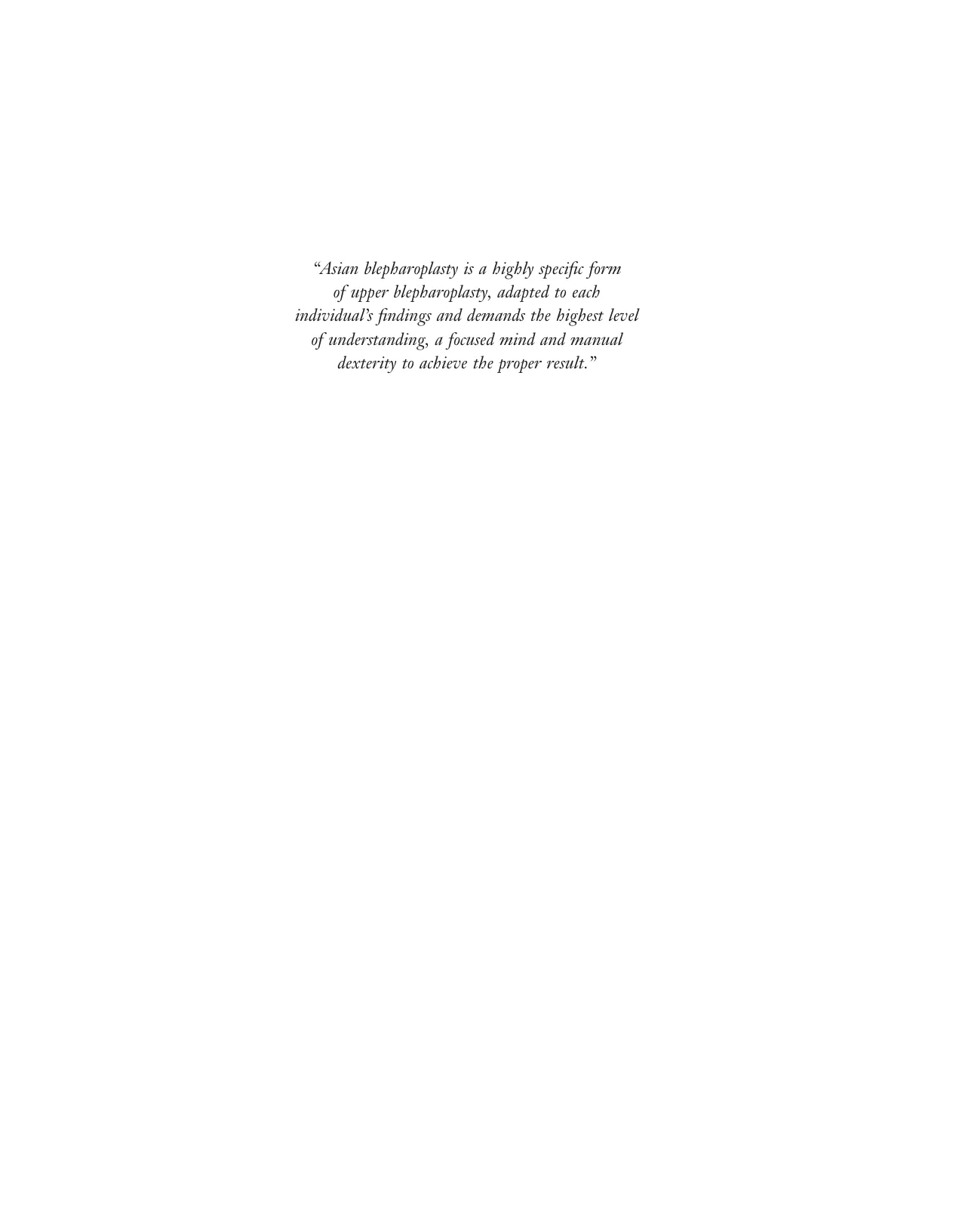
I am continually fascinated by the unpredictability of
the eyelid crease; some of its factors can be represented
by the expression above. This chapter will reveal the
significance of each of these factors, and we will return
to this expression again at the end.
WHAT IS AN UPPER EYELID CREASE?
Let us start gently as an initial attempt to answer the
question posed here. An upper eyelid crease can be
described as an inward folding of the upper lid skin above
the eyelid margin, and is positioned along the superior
tarsal border.
In common usage, the lid crease is less anatomically
defined, and it can be confused to include anything from
a faint wrinkle above the lid margin where tight skin
becomes looser, to a well-formed crease invagination
along the superior tarsal border, to describing what in
actuality is the upper eyelid sulcus. I have personal expe
riences of well-educated patients describing what to them
constitutes an eyelid crease: from anything that occasion
ally appears in the morning, the evening, after a drink
or two, after crying, after reading, after they turn 17, 30
or whatever age, or after a voluntary attempt using a
co-contracting stare-and-frown maneuver (staring acti
vates the levator through the third nerve, frowning
tightens the periorbital orbicularis oculi and frontalis
muscle, mediated through the seventh nerve), to a fold
that forms as a result of where they applied their lid
crease glue, tape or special fiber string. These various
positions are seldom the correct location for an anatomi
cally natural crease.
Most of the above do not appear natural, and therefore
it is just as important for us to try to define what are the
characteristics of a natural crease.
Firstly, a natural crease is present shortly after birth,
is seen in most head positions and is effortless in its
formation. It may be of different shape and height in
different ethnicity, but should share these common
characteristics.
Secondly, a true eyelid crease has a faint crease line on
the skin which can be seen when the upper lid margin is
turned down (or when the eyelid margins oppose
together); its location typically corresponds to the upper
boundary of the superior tarsus. Anatomically it corre
sponds to where the distal fibers of the levator aponeu
rosis terminate into the pretarsal section of orbicularis
oculi1 or skin along the superior tarsal border. Recent
scientific studies using more refined electron microscopy
were able to see attachment of aponeurotic fibers actually
terminating under the skin where the natural crease line
forms, confirming some previous assumptions. When the
eyelids are open, the ‘reinforced’ pretarsal platform of
tarsal plate/orbicularis muscle and attached pretarsal skin
will vector upward as a unit against a passive preseptal
skin–orbicularis: the invagination is the crease, the over
hang is the eyelid fold. The interface for this to occur is
the preaponeurotic fat, which I conceptually describe as
the glide zone.
Thirdly, a true crease is a dynamic crease; it has the
characteristic of fading on downgaze (when the levator
of the lid is relaxed as the inferior rectus contracts to roll
the globe downward), unlike what one sees with an arbi
trarily anchored eyelid crease indentation or one induced
through compression (encircling, buried) sutures.
C
Latt LC F afi pf gz h
s m f
Vax eypo BrPt BrOA h
=
−
+
∫
(
,
,
,
,
,
,
)
( ,
, , ( )
,
,
,
,
, h−)
302
Asian Blepharoplasty and the Eyelid Crease
FIGURE 22-1
CH 2
FIGURE 22-2
(–) Vax
Most individuals who have an upper eyelid crease will
manifest the full crease in a straight-ahead gaze position
(Figure 22-1).
In downgaze, the inferior rectus and superior oblique
muscles contract while the superior rectus and levator
muscles relax (the relaxed levator is at its longest length
in downgaze). Downgaze results in the shallowing or
disappearance of the upper crease (Figure 22-2) in a
person who is born with a crease, though there is still a
faint skin line.
22 Advanced: Summary of Eyelid Crease Factors
303
Head Tilt and the Eyelid Crease
Why is there a difference in apparent depth to the upper
eyelid crease depending on the frontal posture of the face?
The crease is most apparent when the eyelids are open
and the eyes are looking straight upward – where the
levator muscle is presumably maximally contracted (and
at its shortest length). Compare this to when the person
tilts the head’s vertex backward slightly (in a slight chin-
forward position) but still maintains forward horizontal
gaze; as far as the eyeball is concerned, this is similar to
a downgaze and therefore the crease is less manifested
and observable here.
Anatomic
crease
height
Crease
line
FIGURE 22-3
Height of Crease
What about the ‘width’ of the crease? (It should be meas
ured and named as the ‘height’ of the crease, from ciliary
margin to the crease indentation with the lids closed.)
This is the anatomic crease height (Figure 22-3).
Is the crease the observable step-in between the larger
fold of skin above the crease and the exposed pretarsal
skin with the vectored-in lid crease hidden beneath it?
304
Asian Blepharoplasty and the Eyelid Crease
crease; and although some physicians will be clear about
the concepts involved and will subscribe to the anatomic
crease, a significant portion of physicians will also think
like a lay person. The problem occurs not only during
preoperative discussion, but also intraoperatively when
facing the actual challenge of constructing a crease.
The desired crease a patient outlines may be some
thing quite low, and an inexperienced physician may
believe that this is achievable in surgery. A patient may
just as well describe a crease that is abnormally high,
and the practitioner may also think it is achievable
and ‘natural’ when it is not, essentially creating a high-
anchored crease that, when combined with significant fat
excision, will yield a hollow sulcus and a myriad of long-
term issues as described in previous chapters.
The margin of tolerance for error in placement of
crease height is extremely narrow. The knowledge that a
surgically constructed crease needs to mimic nature in
its biodynamics and be positioned along the superior
tarsal border for it to function permanently as a natural
crease requires us to adopt the anatomic definition of a
crease. To create an aesthetically pleasing crease for the
patient, whose only concept of a crease is the ‘apparent’
crease we discussed earlier, the finesse in this type of upper
lid surgery lies in factoring in how much skin we intention
ally allow to be left above the crease, as we construct a
physiological, permanent crease and not just a ‘tied-in’
crease.
The observed apparent crease height as measured from
the eyelash border will be 1–3 mm narrower than if
measured all the way towards the hidden apex of the
crease, which is the tilted crease height (Tch). The eyelid
crease should be defined by the faint crease line we see
with the lids closed (in the absence of acquired ptosis),
and when measured should seem to correspond to the
superior tarsal border’s central measured height; we shall
denote this as the anatomic crease height.
(Note that the tilted crease height is the vertical com
ponent of the sloped pretarsal segment (Figure 22-4).
When there is an overhanging lid fold of x mm, the
apparent crease height is the vertical component of Tch
minus the obstructing (shielding) portion of the lid fold
of x mm, where x may be 1–3 mm.)
Anatomic crease height
Tilted crease height Tch
Apparent
>
>
(
)
crease height
(Remember that anatomic crease height is the true height;
it is always greater than the frontally observed tilted
crease height, and similarly the apparent crease height is
what you see with the hanging fold.)
Should one adopt this location of the anatomic crease
height as the true crease? I firmly believe so. This ambi
guity underlies some of the confusion that occurs between
patients and physicians. Most patients will use their
description of apparent crease for their impression of a
FIGURE 22-4 ■ Tilted crease height (Tch) is further reduced by the overhanging fold; the seen portion is then the apparent crease
height. ‘I’ stands for incline angle of the tarsus, pretarsal skin or levator aponeurosis (Tch − Fold = Apparent crease height).
Tilted crease
height (Tch)
I°
22 Advanced: Summary of Eyelid Crease Factors
305
FIGURE 22-5 ■ A classical representation of Caucasian anatomy, showing a higher point of fusion of the orbital septum on the levator
aponeurosis.
Orbicularis oculi
Preaponeurotic fat
Septum orbitale
Levator aponeurosis
Muller's muscle
..
Inferior tarsus
Inferior oblique
Inferior rectus
In Caucasians, who often have deep-set globes
(eyeballs) or greater prominence of the superior orbital
rim (Figure 22-5), the crease may be quite high (10–
11 mm) and curves posteriorly into a relative half-moon,
semicircular shape. With age-related recession of
(preaponeurotic) fat, a crease may appear flattened and
disappear into a supratarsal sulcus.
306
Asian Blepharoplasty and the Eyelid Crease
the pretarsal platform vectoring upward relative to the
preseptal–periorbital soft tissues (skin, orbicularis, fat) is
such that it favors crease formation.
In Asians with a crease (Figure 22-6), the crease height
is lower than a non-Asian’s. They have good levator func
tion, good terminal aponeurotic fibers that terminate into
the pretarsal section, and the net force-relation between
FIGURE 22-6
22 Advanced: Summary of Eyelid Crease Factors
307
Asians without a crease (‘single eyelid’) (Figure 22-7)
may have only fair to good levator function. Here the force-
relation between preseptal skin/muscle/fat (Zone 2) over
the underlying pretarsal segment plus levator (Zone 1) does
not favor crease invagination. There may be an increased
amount of skin and fat (preaponeurotic, as well as preseptal
fat), or even increased presence of pretarsal orbicularis and
fat to further complicate crease indentation.
FIGURE 22-7
Zone 1
Zone 2
Zone 3
The dominant factor in this category is the relative
absence of distal interdigitations of levator aponeurotic
fibers within the pretarsal orbicularis zone or attachment
towards the skin. The bellowing skin and fat rolling over
the superior tarsal border is secondary. There is no
attachment towards the skin, and therefore no crease.
308
Asian Blepharoplasty and the Eyelid Crease
or under? This latter scenario is far more likely. There
are many clinical examples of elderly patients who had a
true crease previously, then developed ptosis with aging
and yet retained adequate levator excursion greater than
11 mm, but no longer manifest a normally positioned
crease. It is likely that their levator excursion now reflects
the remaining viable levator fibers as well as current con
tractility, and has actually decreased from its former
15–16 mm measurement as the lid became increasingly
ptotic. In other words, this otherwise healthy excursion
of 11 mm represents a relative decline for the patient in
whom excursion may previously have been greater. Other
aging changes that can affect levator excursion will
include the recent research findings that correlate a
decrease in horizontal palpebral width with each decade
of aging. A shorter eyelid margin width can affect the
tensile relationship within the lid closure mechanism and
so it should not be surprising that levator excursion will
drop slightly with aging. (During ptosis surgery, one can
often observe thinning, degenerative and fatty infiltration
of the levator and aponeurosis as well as the canthal
tendons.) In other words, the patient’s levator function in
terms of contractility has truly decreased, though we still
inaccurately use levator excursion, a linear parameter, as
an indicator of three-dimensional health and express this
measurement of 11 mm as indicative of ‘normal levator
function’ (it is a single sampling without comparison to
the same subject’s past, longitudinal changes). Therefore
the use and measurement of what is currently labeled as
‘levator function’ is actually flawed in terms of its true
function as well as setting of the norm.
The above two factors (Latt and LC) are only some of
the early obstacles that determine whether a crease is
clinically manifested. Additionally, the prominence of a
crease is positively influenced by F, afi, pf, gz and h, and
inversely affected by s, m, f, negative Vax, eypo, BrPt,
BrOA, h+, h−.
The anterior soft tissue mass over the supratarsal
region (preseptal segment) consists of skin, orbicularis
and preaponeurotic fat. The normal amount of skin (‘s’)
and muscle (‘m’) acts as a passive weight against which
the posterior lamella (of tarsus, levator and Müller’s
muscle) vectors upward; excess skin tends to shield the
crease from view. Excess orbicularis muscle in the front
decreases the chance of distal fibers of levator aponeuro
sis penetrating towards the subcutaneous plane of the
eyelid crease.
Healthy preaponeurotic fat (healthy fat represented as
‘F’) in the preseptal portion (Zone 2) of the lid acts as a
friction-less glide plane, where the posterior layer vectors
up over the passive weight of the anterior layer.2,3,4 The
posterior lamella has to move only 2–3 mm with respect
to the anterior lamella to give a crease with an overhang
ing eyelid fold.
Shape of Crease
In Asians, the two main categories of shape are the paral
lel crease and the nasally tapered crease (see Chapter 4).
A parallel crease runs parallel and equidistant above the
lid margin. The parallel nature is relative to the eyelid
margin’s curve so it curls around with the natural lid
margin. Near the medial end, whether there is a medial
upper lid fold or not, the crease ends there without con
verging towards the medial commissure.
In the nasally tapered crease, there is often a medial
upper lid fold that overhangs, and the nasal portion of
this crease often converges towards and joins the medial
commissure, appearing as a nasally tapered crease that
tapers to a point. Both these are natural and ideal crease
shapes for Asians.
PREDICTORS OF CREASE
Among the factors that determine the presence of an
upper lid crease, first and foremost is the presence or
absence of the distal ‘attachment’ of the levator aponeu
rotic fibers (‘Latt’); they course towards the subcutaneous
area along the superior tarsal border as well as inferiorly
in the pretarsal orbicularis, terminating as distal inter
digitating fibers within the pretarsal orbicularis. This
determines whether the patient has an eyelid crease on
the skin or not. Furthermore, the ‘tightness’ to which
these aponeurotic fibers (afi) are able to wind around and
insert into these intermuscular septae determines the
relative rigidity (‘firmness’) of the pretarsal skin–muscle,
which moves up together with the tarsal plate as the
levator contracts. The firmer this pretarsal skin–muscle-
tarsal plate is, the better the invagination of crease.
Assuming the presence of these attachments, the con
tractile strength of the levator is indirectly measured
(and poorly assessed) by millimeters of lid margin excur
sion, and incorrectly labeled as indicative of the ‘levator
function’. It is partly indicative of the robustness of the
levator, and correlates somewhat with whether a normal
superoposteriorly directed pull is capable of invaginating
the pretarsal segment (Zone 1) against the preseptal
segment (Zone 2) to give a clinically apparent crease. We
observe that those with poor to borderline levator excur
sion (under 10 mm) are far more likely to have incomplete
manifestation of the crease. Yet those with 11 mm or
greater levator excursion do seem to form a crease more
readily.
Is this a function simply determined by one or two
millimeters? Or is it more likely that a correlation exists
between those with excursion of 11 mm or more are
having a qualitatively stronger levator muscle contractil
ity (‘LC’) compared with those with excursion of 10 mm
22 Advanced: Summary of Eyelid Crease Factors
309
FIGURE 22-8
In a less pristine condition where there is fibrotic fat
(represented as ‘f’ ), whether in untouched lid tissue or
secondary to prior surgery with complete obliteration of
this desirable glide zone (‘gz’), there is more hindrance
to the smooth gliding between these two lamellae of
tissues, and it is less likely that one can attain a 2–3 mm
relative shift of the two layers4 (Figure 22-8).
In some single-lidded individuals (see Figure 22-7),
one may see an abundance of preaponeurotic fat at a
lower than normal position (normal being 5–10 mm
above the superior tarsal border), in association with
absence of crease and a lower point of fusion of the
orbital septum towards the aponeurosis. I believe these
are associative factors rather than necessarily showing
absolute causation of the one leading to the other.
Preaponeurotic (post-septal) fat pads may occasionally
penetrate as far inferiorly as the pretarsal position (mani
festing as Zone 1 preaponeurotic or post-septal fat) and
disrupts any chance of crease formation.
Fat lying in front of the orbital septum within the
pretarsal orbicularis should be labeled pretarsal fat;
technically the fat here may be an accumulation of fat in
front of the orbital septum, or Zone 1 preseptal fat. It
may also disrupt crease formation. In essence almost any
excessive fat in the pretarsal location may affect crease
formation.
In fatty upper eyelids, the combined weight of the
three subcomponents of skin (s), orbicularis muscle (m)
and preaponeurotic fat (F), may be too heavy for the
posterior levator–tarsus complex to lift and it can result
in an absence of crease. Eventually this may even lead to
acquired ptosis of the upper lid at an earlier age than
normal. Of course, an excess of any of these components
or in combination can lead to absent crease. Healthy
preaponeurotic fat (F) can assist as a glide layer, and
facilitates crease formation, while excess of it can impede.
(When F exceeds a certain quantitative value, it can be
seen as a denominator factor, with an inverse function).
A normal patient with large palpebral fissure size (‘pf’)
as measured both vertically and horizontally will tend to
have a large levator muscle complex, both in width as well
as greater vertical excursion. One can infer that a larger
width of levator will span a longer convex arc of the tarsal
plate as it sweeps upward, and that its central midpoint
insertion along the superior tarsal margin will traverse a
greater distance (vertical excursion of the upper lid
margin).
In the negatively affecting cluster of parameters
(denominator), the lower case ‘s’ stands for floppy eyelid
skin, thickened eyelid skin and excess redundancy of
skin. Some individuals even manifest tight eyelid skin,
observable through tissue resistance when their eyelid
skin is being infiltrated with local anesthetic solution
prior to surgery. They show a much delayed resorption
of the injected anesthetic, as well as needing greater
efforts with infolding of a surgically constructed crease
despite extra steps being taken to enhance the crease
formation. Therefore loose skin adds bulk, tight skin adds
resistance.
The lowercase ‘m’ stands for excessive mass of orbicu
laris muscle, and dehiscence of preseptal- or pretarsal-
based orbicularis muscle layers. Fat may also be
intertwined within the orbicularis fibers.
The lowercase ‘f’ represents fat located in the glide
plane which is obstructive rather than facilitative in
nature. The clinical condition is often observed during
surgery for single-lidded individuals – where instead of
observing globular fat, we see plastered-down amorphous
fat strands, or mosaic clusters of ill-defined fat globules
without clear demarcation of where the septum lies in
front of the aponeurosis. It may occur thus in primary
cases; at other times, during revisional blepharoplasty, the
entire glide zone is a solid scarred plaque. The probabil
ity of having a natural crease is obviously an inverse
function to ‘f’ (imagine it as a sort of adverse fat residual),
or proportional to 1/f. Crease construction is easier if
adverse fat (f) is minimal or not present.
310
Asian Blepharoplasty and the Eyelid Crease
FIGURE 22-9
(+) Vax
The levator muscle is at its maximum contraction
on upgaze; here the crease appears the deepest (we can
consider the patient’s visual axis angle [Vax] in this
maximum upgaze position as being its maximally positive
angle away from a straight-ahead view [zero angle])
(Figure 22-9).
22 Advanced: Summary of Eyelid Crease Factors
311
maximally negative angle. With this negative Vax, the
crease’s invagination should naturally shallow or decrease
(Figure 22-10).
The levator is at its most relaxed state when the eyes
look down, with the levator as well as superior rectus in
a relaxed state. Here the visual axis angle (as measured
away from the arbitrary point of zero angle) is at its
FIGURE 22-10
(–) Vax
312
Asian Blepharoplasty and the Eyelid Crease
When a person with a natural crease is looking straight
ahead with a level gaze (set as zero value Vax), the crease
is optimally manifested, neither too deep nor too shallow
(Figure 22-11). The same should be observed in a dynamic
crease that is surgically constructed.
The examiner’s angle of observation (‘ao’) should also
be level and perpendicular to the subject’s erect face (with
the patient’s frontal plane perpendicular to the floor and
his/her forehead’s frontal boundary and chin aligned like
a plumb line); this is the examiner’s zero angle of observa
tion (zero ao), looking straight ahead.
FIGURE 22-11
CH 2
22 Advanced: Summary of Eyelid Crease Factors
313
This is similar to asking the patient to tuck the chin
in towards the sternum while looking ahead, with the
examiner observing from a level position, essentially
placing the patient’s eyes in an upgaze position relative
to its orbit.
If the observer is standing in a relatively higher observ
ing position (Figure 22-12), with the patient looking back
at the observer in this higher position, the crease will
appear deeper due to the patient looking up with a posi
tive Vax as well as a positive angle of observation (+ ao)
from the observer.
FIGURE 22-12
(+) Vax
(+) ao
CH 1
314
Asian Blepharoplasty and the Eyelid Crease
CH 3
(–) Vax
(–) ao
FIGURE 22-13
If the patient sits at a higher position than the exam
iner, the examiner is at a lower position and looking up
(a negative ao) and the patient looking down (a negative
Vax); the crease then looks shallowed (Figure 22-13).
Therefore with either the Vax or ao schema, a positive
value of each is associated with a deepening of the crease,
and a negative value is associated with a shallowing of the
crease. For study purpose, the crease should always be
evaluated with a neutral or zero Vax and ao value.
In elderly patients with involutional ptosis, we often
see what was a normally positioned crease during the
individual’s youth eventually give way to a higher, less
distinctive crease with onset of ptosis. The reasons are
three-fold: there is an overall weakening of levator
aponeurotic muscle (elevator for the eyelids) as well as its
attachment along the superior tarsal border, a weakening
of pretarsal and preseptal orbicularis muscle organization
near the area in front of the superior tarsal zone (less
pulling in of crease line), and a drop in upper lid level
(causing negative Vax on the part of the patient) and a
shallowing of their crease indentation. The weakening of
orbicularis fibers between the superior tarsal border and
its overlying skin results in lessening of attachment of
distal aponeurotic fibers to skin along the original crease
line. The few aponeurotic branches that remain superior
to this zone of dehiscence then constitute the higher
‘migrated crease’ (a misnomer), which is shallower and
not a true anatomic crease since it had not previously
indented at this higher position.
Ironically, many Asians with a single eyelid have a ten
dency to have a noticeable posterior head-tilt. This is
analogous to a slight downward gaze, inclining their
levator muscle more towards a rested state, and further
diminishing the pull on the pretarsal–superior tarsal
border junction. I am uncertain why this is so, but factors
may include the following. In single-lidded patients, their
pretarsal lid fold tends to obstruct the upper 5–10° of their
superior field of vision, and this backward head-tilt may
be a compensatory move for visual improvement, or the
possible co-occurrence of latent ptosis or even presence
of minimal ptosis. Since birth, these individuals may have
adopted a slight head-back posture to clear their visual
axis. They are often quite surprised when this is pointed
out to them using photography from the side or mirrors.
22 Advanced: Summary of Eyelid Crease Factors
315
the superior tarsal border. A crease set too high (h+) will
risk lagophthalmos, pretarsal fullness and secondary
ptosis in addition to other aesthetic ramifications. A
crease that has been arbitrarily set too low (h−) through
a low incision will also adversely affect the outcome,
including failure of crease formation, subsequent fading
and disappearance of crease, obvious incisional scar that
cannot be easily hidden, as well as segmentation of crease
form. An ideal crease height (h) that is in line with natural
anatomy offers the best aesthetic result and least chance
of complications. Therefore here, h is on the top (numer
ator) line; h+ and h− can be adverse factors, and sit as
denominators.
In an occasional patient we see a dehiscence of the
lateral canthus, showing a thinning of the superior and
inferior crus (limb) of the lateral canthal tendon. Often
one sees rounding and a thinned web-like skin over where
the lateral canthal angle once was. The upper eyelid
crease will be less indented laterally as a result of associ
ated weakening of the lateral portion of the levator
muscle. The pretarsal orbicularis, which normally
attaches itself at the lateral orbital rim, is also weakened,
together with the tarsal attachment near the lateral orbital
tubercle – laterally, the crease indent is weak due to weak
ness of the tarsal–levator complex.
At the opposite end, with the medial canthus, a patient
with medial ptosis as a result of dehiscence of the
medial horn of levator and lateral migration of their tarsal
plate typically shows a similar loss of medial crease
indentation.
A dehiscence over the lateral canthal region of the
upper eyelid typically leads to less effective levator-uplift
at the lateral end of the lid. The weakening of levator
coupled with post-surgical hemorrhage from within
orbicularis can lead to an acquired ptosis position. The
crease now appears shallower as well as a bit further from
the ciliary margin.
Having a comprehensive understanding of the physio
logical parameters that form a crease, and its accentuating
and distracting factors, will allow the surgeon to tailor
each individual’s needs and come forth with a set of ideal
surgical solutions with each of these factors in mind. The
Asian eyelid crease procedure thus chosen should have
the capacity to allow the surgeon to make intraoperative
assessment, and to correct those applicable factors fully
in order to optimize crease formation.
The current evolution of the Asian blepharoplasty
technique used by this author has allowed such assess
ments and corrections, and is one of many reasons for
favoring the external incision techniques over the histori
cally cited suture ligation methods.
Returning to the formula that opened this chapter,
which allows us to consider all the factors that play a role
in creating an eyelid crease:
Indeed, even after successful surgical addition of an
upper eyelid crease, patients often shallow their newly
found crease without knowing it when they assume their
previously accustomed slight head-back position. The
well-formed dynamic crease may not fully invaginate
unless the person’s forehead–chin line is vertical and the
face fully frontal. It is to be expected that a dynamic
crease should shallow on downgaze. I routinely advise
my patients ahead of surgery such that they can fully
incorporate this understanding into their postoperative
routine, that the optimal head posture and facial position
to show the crease is with the face pointing straight ahead
(where the forehead–chin line is perpendicular to the
floor). It is not unusual for the patient to take several
months to fully adopt this more normal head position.
The relative eye position (‘eypo’) of the globe within
the orbit is a much more subtle influence towards the
probable outcome of a crease. A slightly more forwardly
positioned eye and thus upper lid, has the levator in a
comparatively more flattened position (excluding patients
with thyroid eye disease where their levator muscles are
often over-stimulated, or fibrotic and shortened as it
recovers). The passive weight of its anterior skin–muscle
layers is lessened as it is distributed over a more horizon
tal plane, resulting in less gravitational weight per surface
area. A forward position tends to somewhat shallow the
crease.
Conversely, a slightly more posteriorly placed (sunken
in) globe and upper lid now has the levator in a more
vertically biased position as well as having a better rela
tionship for the skin–muscle layer to render an apparent
crease.
The eyebrow participates as a natural scaffold-support
for the upper portion of the upper eyelid, as the frontalis
muscle interacts with the upper periorbital portion (Zone
3) of the orbicularis oculi muscle (see Figure 22-7).
When there is brow ptosis (BrPt, or brow droop), a
crease may become shielded from view or totally obliter
ated due to the avalanche of eyebrow soft tissue mass.
The brow may become overactive (BrOA) in involutional
ptosis, as well as in secondary ptosis following high
anchoring of the lid crease stitches onto levator muscle.
In these situations, the lid fold on top of the crease is
partly lifted up by the overacting brow and the true crease
may now be observed, while in some cases it appears less
indented (more shallow) since the passive anterior skin–
orbicularis layer has been lifted up.
Lastly, and of major significance, is the position of
placement of the crease, that is, the crease height (‘h’).
One will notice that height both facilitates (‘h’ in the
numerator) as well as hinders (h+ or h− in the denomina
tor), and in different ways. The optimal height (‘hopt’) that
mimics a physiological crease is usually matched along
the area equivalent to the height of the middle section of
C
Latt LC F afi pf gz h
s m f
Vax eypo BrPt BrOA h
=
−
+
∫
(
,
,
,
,
,
,
)
( ,
, , ( )
,
,
,
,
, h−)
316
Asian Blepharoplasty and the Eyelid Crease
the horizontal axis (each of these two sides will be approx
imately 5 mm each, as it is 7 mm × 1/√2). Therefore the
measured 7 mm crease will appear to the examiner (or
patient’s view through a mirror) to occupy 5 mm in verti
cal height at its apex (tilted crease height), and about
2–4 mm only (apparent crease height) when a 1–3 mm
eyelid fold hanging over it is subtracted out. Therefore,
it is quite normal for a single-eyelid patient to ask for a
3 mm crease as their preferred end result; the practi
tioner must realize that it needs to come from a 7 mm
crease placement.
The question as to whether there are some factors
relating the finding of increased failure rate (fading of
crease) after surgery among patients with borderline
levator excursion (within the 9–11 mm range), or an asso
ciation of minimal to mild ptosis among single-lidded
individuals, is harder to delineate.
This chapter is a synthesis of all the concepts discussed
in this book so far. Some may be too esoteric, but I hope
that for most readers it serves the function of bringing
the concepts together into a coherent scheme such that
it may be helpful in patient care.
REFERENCES
1. Collin JR, Beard C, Wood I. Experimental and clinical data on the
insertion of the levator palpebrae superioris muscle. Am J Ophthal
mol 1978;85:792–801.
2. Chen WPD. Concept of triangular, rectangular and trapezoidal
debulking of eyelid tissues: application in Asian blepharoplasty. Plast
Reconstr Surg 1996;97(1):212–218.
3. Chen WPD. The concept of a glide zone as it relates to upper lid
crease, lid fold, and application in upper blepharoplasty. Plast
Reconstr Surg 2007;119(1):379–386.
4. Chen WPD. Beveled approach for revisional surgery in Asian
blepharoplasty. Plast Reconstr Surg 2007;120(2):545–552.
the formula’s legend now reads: C represents the prob
ability of crease formation and is proportional to the
summation (∫) of various enabling factors including
levator attachment and contractility, preaponeurotic
fat, aponeurotic fibers of the levator aponeurosis, the
palpebral fissure size, intactness of the glide zone
with healthy normal fat, and correct optimal placement
of crease height; it is adversely influenced by the
factors in the denominator, which include texture and
quantity of eyelid skin, muscle, presence of scarred
fat, patient’s negative visual axis angle, eyeball position,
brow ptosis and brow compensatory overaction, and
excessively high or low deviation from the proper crease
height.
ADDITIONAL EXPLANATION OF
CREASE HEIGHT (previously covered in
Chapters 19 and 20)
If we have a patient lying supine and measure the eyelid
crease height with the lids closed, we obtain the true
location of an anatomic crease, which is usually along the
superior tarsal border. Let us assume the anatomic crease
height is 7 mm for an Asian with a natural crease. When
the patient is sitting up and the face is vertically aligned,
with the patient looking ahead, the crease is optimally
manifested and tucked in under its eyelid fold. The supe
rior tarsal platform is tilted posteriorly in an oblique
direction. The measured anatomic crease height of 7 mm
(pretarsal skin) can be thought of as being tilted on the
hypotenuse of a 45° isosceles triangle, while the two
remaining sides of this triangle form the vertical axis and
317
ADVANCED CONCEPT
OF THE EYELID CREASE
AS A STRINGED SERIES OF UNIPOINTS
C H A P T E R 2 3
Concluding this group of chapters, I present my current
thinking with respect to the functional biodynamics of
the eyelid crease.
In 1993 when I was preparing the manuscript for the
original Asian Blepharoplasty atlas, in its last chapter I
made an analogy that the eyelid crease is somewhat like
the Great Wall of China, where from outer space an
astronaut may merely observe a line crawling across the
planet’s surface while in actuality the Wall meanders
along, covering vastly different terrain and with forts
scattered every 80–120 meters. These outposts were built
with different functions in mind, their interiors having
varying sub-sections serving as sleeping quarters, grain
storage, weapon depots, animal shelters, signal and
command stations. Similarly, the eyelid crease changes
subtly as you traverse across the width of the eyelid. It is
attaching to various substrates of tissues as the curvature
of the lid margin, the tarsus and its opposing canthal liga
mentous attachments come into play.
As with a naturally born eyelid crease, there are differ
ent techniques that you can apply within sections of the
single eyelid as you create (or add) a natural crease, cours
ing continuously across the upper eyelid with its outer
appearance showing as a homogeneous line. (We have
discussed the depth of a crease and how to titrate this, the
apparent crease versus the tilted crease height, as well as
attachment to different anatomic terrain across the width
of the crease.) Here we are discussing the linearity of the
crease with respect to the curvature of the eye.
What about the opposing vectors that act across the
length of the eyelid crease? In other words, what about the
forces acting along the crease which allow the anterior
lamella (preseptal layer) to slide down relative to the pos
terior lamella of levator and tarsus when the eyelid opens?
I prefer to conceptualize the eyelid crease as the
external manifestation of a stringed series of unipoints
(unipoint = single point in space). In Figure 23-1 the dif
ferently sized yellow dots represent unipoints that have
FIGURE 23-1 ■ The slightly different sizes of
the yellow dots represent unipoints that have
different magnitudes of vector forces exert
ing on them.
318
Asian Blepharoplasty and the Eyelid Crease
FIGURE 23-2 ■ Each of the yellow spheres represents a highly magnified unipoint (vector interface) where the red vector is in line
with the pull of the levator, and the blue arrow, the gravitating weight of the anterior skin-muscle layer when the facial nerve input
is relaxed. Note that the blue vector’s direction may vary between 45 degrees relative to horizontal, to almost vertical (90 degrees)
if the facial features are comparatively flat, as is often the case with Asian eyes.
vector forces of different magnitudes acting on them.
Each of the yellow dots (Figure 23-2) represents a highly
magnified unipoint (vector interface), where the red vector
is in line with the pull of the levator, and the blue arrow,
the gravitating weight of the anterior skin–muscle layer
when the facial nerve input is relaxed. Note that the blue
vector’s direction may vary between 45° relative to hori
zontal, to almost vertical (90°) if the facial features are
comparatively flat, as is often the case with Asian eyes.
Let us conceptualize that each unipoint is a nano-
scaled virtual point (like rotating nano ball-bearings, or
mathematical definitions of a point in space) that is
bounded by vector forces going in opposite directions
(sloped-up on the backside, and sloped-down on its front
side). This string of unipoints is like a string of nano-
scaled rosary beads that straddles the two lamellae of the
upper lid at a location approximately along the superior
tarsal border. When the levator is activated (‘ON’ for
third nerve) and pulls the upper lid open, there is an
inhibitory nerve signal that happens at the same time
towards the orbicularis muscle (supplied by seventh
nerve), turning it ‘OFF’. The weight of the relaxed
preseptal layer of skin and orbicularis slides down pas
sively, and covers part of the pretarsal skin and becomes
the eyelid fold. The exact interphase between the poste
rior layer going up and the skin–orbicularis layer coming
down occurs under the skin and within the eyelid, and its
external manifestation on the skin side is the eyelid crease.
The phasic change is where these virtual unipoints (or
virtual nano-balls) are located. The slip-slide is facilitated
by healthy fat in the middle space that I have termed the
glide zone (see Chapter 17).
A good analogy is the wise saying that ‘an avalanche
can start with the added weight of a snowflake’. We can
23 Advanced Concept of the Eyelid Crease as a Stringed Series of Unipoints
319
FIGURE 23-3 ■ (A,B) Pre- and postoperative images of a single-lidded individual, after undergoing Asian blepharoplasty by Chen. Yellow
conceptual unipoints are superimposed here to show the lower pretarsal zone vectoring against a relaxed upper preseptal zone.
A
B
see this easily by watching layers of fine colored sand
trapped in liquid between two panes of glass. As we rotate
the glass panel, we can see sand dunes topple, and slip-
slides as the layers on top gain just a bit more sand.
In mimicking a crease through surgery, it is important
to recognize where this occurs. To create it accurately,
the precise placement of the crease along where these
phasic changes (series of unipoints) naturally may occur
(with surgical clearance of impeding tissues) should be
the most important goal. Deviation from it has significant
physiological and functional side effects.
Figure 23-3 shows pre-and postoperative images of a
single-lidded individual, after undergoing Asian blepha
roplasty performed by the author. Yellow conceptual uni
points are pasted here to show the lower pretarsal zone
vectoring against a relaxed upper preseptal zone.
320
Asian Blepharoplasty and the Eyelid Crease
This concept can explain why buried ligatures that
encircle the levator muscle, or a high-anchoring crease
on the aponeurosis, often lead to problems including
high crease height and acquired ptosis (Figure 23-4)
Note the unipoint function is restricted within the confine
of the buried ligature.
FIGURE 23-4 ■ Note the unipoint function is restricted within the confine of the buried ligature.
23 Advanced Concept of the Eyelid Crease as a Stringed Series of Unipoints
321
FIGURE 23-5 ■ Here we see that any chance for forming a normal unipoint pivot is lost when a buried suture traverses through the
orbicularis of the anterior lamella (blue vector), which is supposed to relax, and the posterior levator muscle (green vector), which
is trying to lift the eyelid.
Here we see that any chance for forming a normal
unipoint pivot is lost when a buried suture traverses
through the orbicularis of the anterior lamella, which is
supposed to relax, and the posterior levator muscle, which
is trying to lift the eyelid (Figure 23-5).
A Faden-like fixation effect is created on the lid in at
least three high locations due to placement of the three
buried permanent sutures. The compression ligatures
create an unintentional drag (increased impedance) on
the contractile function of both levator and Müller’s
muscles. (In this case, the Faden effect is both ecto- and
endo- as the fixation location[s] is both anterior to and
beneath the plane of the levator aponeurosis, shown in
Figure 23-5 as a pink layer. The fixation location is often
superior to the tarsal border by several millimeters.)
The magnified inset view of this same drawing (Figure
23-5, Inset) shows the impediment imposed on the uni
point’s two vectors. The high placement on the levator
reduces the effective contractile range of the levator
aponeurosis. The contractile (lifting) force represented
by the green vector arrow parallel to the levator plane is
reduced. Levator excursion is reduced. Load is increased
when more tissues are piled on to the wagon, so to speak.
The inclusion of more orbicularis oculi muscle and
subcutaneous fascia in this buried suture loop similarly
affects the ability of the orbicularis to relax when the
seventh nerve is inhibited and gravity is supposed to allow
the eyelid fold to form over the crease, resulting in a
lessened blue vector. There is a restricting or braking
force holding the anterior lamella up, and less tissue can
come down. The apparent crease height is magnified, and
we see ptosis. The gray orbicularis layer is prevented
from gravitating down at the desired location along the
superior tarsal border, but is hitched up at a higher point
– a high crease may be the result, or a faded crease is the
eventual result when the tissue tension releases with the
suture eventually having worked its way through tissues.
In severe cases, we may see lagophthalmos.
322
Asian Blepharoplasty and the Eyelid Crease
VARIATION IN FUNCTION
OF THE UNIPOINTS
Are there variations in the functions of these unipoints
that are located along the crease line? The answer is most
certainly yes. One can surmise that the maximal direc
tional change in vectors for these unipoints occurs along
the mid-section of the eyelid crease, in other words, the
maximal directional change occurs centrally (along these
nano-dots). It lessens as you approach each canthal
angle (medial and lateral corners of the eyelid opening).
Towards the corners of the eyelid opening, the vector
forces pulling upward are likely to be less (less functional
levator muscle fibers present), and directionally the
levator aponeurotic vector will be flatter and more side
ways oriented. Similarly, the downward weight of the
relaxed anterior skin–orbicularis muscle layer is likely to
be lessened as one approaches each corner too. At the
same time, the skin is relatively thicker towards each
FIGURE 23-6 ■ Nano-dots placed along the crease of a patient who underwent successful Asian blepharoplasty. The smaller sized
dots indicate lesser vector-reversal. Note the slightly smaller yellow dots I superimposed on the medial and lateral sections of the
crease; they represent smaller scaled vectors interaction as well as a less directionally opposite relationship between the green and
blue vectors.
canthus, and firmer with less potential space within
its strata. Additionally, the blue vector may be flatter
or less vertically positioned (smaller incline angle, I) at
each canthal region, so that the phasic change between
the two vectors (green and blue) is not directionally
opposite (close to 180°) as in the central portion of the
crease. It will be less fluid, and occur at a slower pace
(speed), since even though it takes the same time per
blink medially or centrally, the distance covered is less.
So the net force-reversal (or vector-reversal) along the
medial and lateral sections of the crease is likely to be less
abrupt.
Figure 23-6 shows nano-dots placed along the crease
of a patient who underwent successful Asian blepharo
plasty. The smaller-sized dots indicate lesser vector-
reversal. Note the slightly smaller yellow dots I have
positioned on the medial and lateral sections of the
crease; these represent smaller-scaled vectors interaction
as well as a less directionally opposite relationship
between the green and blue vectors.
23 Advanced Concept of the Eyelid Crease as a Stringed Series of Unipoints
323
shallows to a certain degree for a natural crease as the
vector force decreases in its magnitude and directional
shifts. In a surgically created crease using the external
incision approach such as the author’s Asian blepharo
plasty, one would expect the mid-section of the con
structed crease (with maximal phasic changes along the
unipoints) to be easily noticeable and permanence easily
attained, while the medial one-third of the crease is
enhanced by special wound closure techniques and man
agement of the medial upper lid fold, and the lateral-one
third may be allowed to shallow somewhat like a natural
crease does. These conclusions are largely predicated
on the presumption that the patient has healthy levator
function and an adequate-sized eyelid opening, vertically
as well as horizontally, and that a healthy glide zone
(preaponeurotic space and fat) exists.
In complicated cases where there is cicatrix along
the anterior lamella (skin–orbicularis) or glide zone
(preaponeurotic space), where there is rigidity of skin or a
lack of a slip-slide (vector-reversal), then there is lessened
possibility for formation of a crease. Revisional attempts
will be needed in order to re-establish these biodynamics.
(Here again, further revisional attempts using suture tech
niques are almost always unsuccessful for obvious reasons.)
IN CONCLUSION
In the first edition of this book, I suggested some poten
tial directions for surgical research regarding crease for
mation. Unfortunately there has not been any significant
progression in the medical literature using external means
like newer, novel lasers or implantable bio-dissolvable
material for this purpose. Besides, even if there were,
with the vigorous benchmark we have set and an improved
understanding of biodynamics, it is unlikely that any
technique can reach these ideals entirely, in terms of
optimal control and placement of crease height, control
of crease shape, having a natural continuous appearance,
and to achieve permanence, as well as avoid long-term
harm. One should aim to reach all four parameters in one
surgical session, however one should note that due to
variability of each patient’s intrinsic eyelid anatomy and
associated conditions like ptosis, some patients will need
staged procedures since the objectives may exceed what
can be achieved in one procedure, whether it is primary
or revisional blepharoplasty.
With the force-reversal being more forceful centrally
and less so medially and laterally, one should expect the
crease indentation to be greater centrally, and lessen
towards the two ends. Clinically, it helps to explain why
it is more challenging to form the crease well in medial
and lateral areas.
The direction of the blue vector is worth further dis
cussion. You may often see in Caucasians who have deep-
set eyes that their levator muscles pull up and their crease
indents effortlessly. This is seen often with a hanging lid
fold that almost appears to have a reverse cant (slope)
from the superior orbital rim heading backwards; in other
words, the skin above is hung from their eyebrow and
sloping down and backwards (inferoposteriorly). In
Asians who typically may have a flatter-appearing eyelid-
to-orbital rim spatial relationship, that fold of skin is far
more likely to be sloping down and forward (inferoante
riorly), therefore pushing down against the uprising
green levator force, which is acting at an incline angle
near the tarsal plate of 45–50°. For Caucasians, this
downward blue vector (representing skin and orbicularis)
does not impede crease formation as much, owing to a
higher probability that its angle of action is not directly
opposing at 180° the lifting green vector.
One can tailor the crease construction based on topo
graphical variations of the levator along the pretarsal
segment (skin, orbicularis, superior tarsal border), its
spatial location with respect to the orbit, findings regard
ing the hindering elements towards crease formation, size
of the palpebral fissures and specific individualized design
factors for crease height, shape and continuity. The tech
nique used on one eyelid may need to encompass a com
bination of several specific crease-enhancing skills to be
applied over different sections of that crease. The oppo
site eyelid may need something different.
DIFFERENT POINTS ALONG THE
UPPER LID CREASE
In clinical observation, with a naturally born upper lid
crease the crease indentation is often sufficiently promi
nent over the central one-third as well as the medial one-
third (here, it could be that the net balance of a younger
adult’s levator muscle is still strong and the relaxed weight
of the medial canthal fold allows a good crease to occur
medially). The lateral one-third of the crease often
325
ASIAN EYELID SURGERY:
MY THOUGHTS
by Khoo Boo-Chai (manuscript prepared by William PD Chen MD)
C H A P T E R 2 4
Khoo Boo-Chai of Singapore, a pioneer in this field since
the 1960s, kindly furnished all the information in this
chapter, which was abstracted by W.P.D. Chen and
reviewed by Dr Boo-Chai for the second edition of this
book. This was based on his 40 years of hands-on experi
ence with aesthetic (cosmetic) surgery of the upper eyelid.
Boo-Chai1 reported on his experience over five years
with 625 cases of Asian lid crease procedures using the
conjunctival stitch method. He recommended that this
procedure is best used in patients with little upper lid fat
and without a heavy fold that hangs down over the lid
margin. Non-absorbable suture materials are used to
connect the levator aponeurosis to the eyelid skin at a
desired level 5–8 mm from the lid margin. If there is
excessive supraorbital (preaponeurotic) fat, it is first
removed through an additional central skin incision
about one-quarter the width of the crease line designed.
The lid is everted and treated locally with 5% topical
lidocaine solution; 0.5 ml of 1% lidocaine is given sub
conjunctivally. A needle bearing 4-0 nylon suture is
passed through the conjunctiva in a horizontal fashion for
2–3 mm over the superior tarsal border. Each arm is then
repassed through the conjunctiva towards the skin side
overlying it. One arm of the externalized skin stitch is
then passed subcutaneously towards the second arm,
which is often itself passed through a small stab incision
on the skin to facilitate the passage and subsequent
burying of the knot. The two ends are tied, and the knot
is tied down and buried under the skin surface. Usually
three of these pairs of stitches are used.
Boo-Chai evaluated his patients 1 month postopera
tively using the following parameters for a perfect result:
1. The creases on both sides must match in position,
height, length and contour.
2. The position and contour of the upper lid margin
must match, without any notching or peaking.
3. The eyelashes must not be distorted or missing.
4. Blinking must be normal.
5. Both eyelids must close normally during sleep.
6. There must be minimal scarring and no ectropion.
7. The results must be permanent.
According to Boo-Chai, the advantages of this method
include reversibility, minimal swelling and the absence of
an external linear scar.
Boo-Chai2 also discussed the correction of the follow
ing three conditions: discrepancies in the height or shape
of the crease, the absence of crease formation (‘failed
double-eyelid operation’) and a hollowed supratarsal
recess due to excess removal of fat. In the introductory
paragraphs he commented on the two main types of tech
nique: the non-incisional suture techniques versus the
incisional techniques with clearing of skin, fat, and use of
skin–levator–skin fixation.
In the first category, when revising a previous non-
incisional technique to eliminate discrepancies in crease
height and shape, it is important to eliminate the crease
by removing the loops of suture material that connected
the levator to the skin. Boo-Chai prefers to apply the new
crease-forming sutures usually three at the same setting
after the previous loops have been removed.
For those with crease height and contour problems as
a result of previous incisional techniques, he discussed
two options:
1. If the patient wants an excessively low or shielded
crease (caused by residual excess skin) to be cor
rected to a higher level to match the opposite side,
he starts along the existing crease scar and designs
an upper line of incision several millimeters above
this. The excess skin is excised and the crease
reconstructed.
2. In situations where there is no excess skin, he
prefers to use a non-incisional buried suture loop
technique to create the new higher crease without
forming a second skin cut or scar.
He eliminates the previously created crease by going
through the small stab incisions (used for the non-
incisional method) and effectively undermining the adhe
sion between the aponeurosis and the dermis. The
dissolution of this crease is verified intraoperatively by
having the patient look upwards.
He also discussed lowering creases if both sides are
higher than optimal, the emphasis being to include the
existing scar line within the tissues to be excised.
In the second category, correction of poor crease for
mation, the revision involves excision of the previous
fibrous tissue connection between levator and skin, as well
as excision of the previous incisional scar. He then uses six
or seven 4-0 sutures to connect skin to levator to skin.
The third category of revisional blepharoplasty
involves the correction of a deepened sulcus caused by
excess fat removal. Boo-Chai uses fat harvested from a
lower blepharoplasty and the fat is then divided into
numerous 3 × 4 mm pellets. These are then placed behind
the anterior layer of the orbital septum on top of the
levator aponeurosis (with its closely attached posterior
layer/reflection of orbital septum). He prefers to place
more fat over the medial side of the upper lid. He notes
that when observed 6 months later, these fat pellets
seemed to have coalesced to form one piece.3
326
Asian Blepharoplasty and the Eyelid Crease
medially from the superior medial palpebral branch of the
ophthalmic artery and laterally from a branch of the lac
rimal artery. In Asians, the marginal arterial arcade is
covered by the levator aponeurosis, owing to its low inser
tion on the tarsus. This is in contrast to Caucasians, where
the marginal arcade is not covered by the aponeurosis
because its insertion is high up on the upper part of the
tarsus. He stated that his preferred management of bleed
ing in such incidents was to apply ice compresses, and that
the bleeding is self-limiting. He has subsequently called
this form of bleeding the ‘Boo-Chai sign’. It is unusual in
the sense that the bleeding occurs suddenly posterior to
the levator aponeurosis, and spreads widely within an area
not usually touched during incisional methods of Asian
eyelid surgery (Figure 24-1 A, B).
Boo-Chai3 described the occasional presence of a mar
ginal arterial arcade with perforating branches that pierce
the levator aponeurosis near the insertion of the aponeu
rosis on the tarsus. These perforating vessels run perpen
dicularly in a vertical fashion, and lie within the
suborbicularis areolar fatty tissues. They are not common
and are difficult to detect unless specifically sought. When
the lower skin flap is surgically manipulated or cleared,
these perforators may be damaged and bleed, retracting
within the aponeurosis to lie close to the marginal arterial
arcade in the pretarsal (postaponeurotic) space, giving rise
to a hematoma. Boo-Chai observed this in three cases,
with an incidence of 1 in 500 cases. He went on to explain
that the marginal arterial arcade normally lies on the tarsus
3 mm from the lid margin. It receives a contribution
A
B
FIGURE 24-1 ■ (A) Left upper eyelid
of a 25-year-old Chinese woman
undergoing
Asian
blepharoplasty.
A strip of skin and orbicularis oculi
has been removed from the left
upper lid, revealing giant branches of
the marginal arcade running verti
cally
upward.
These
branches
emerge from underneath the anterior
extent of the distal portion of the
aponeurosis on the tarsal plate, and
pierce the levator aponeurosis. They
then course up vertically and lie in the
retromuscular
(suborbicularis)
fat.
Accidental damage to these branches
when dissecting the skin flap will give
rise to bleeding, with the formation of
a hematoma behind and deep to the
levator aponeurosis: the ‘Boo-Chai
sign’.
(B) Right upper eyelid
illustrating a self-limiting hematoma
forming posterior to the levator
aponeurosis.
FIGURE 24-1 ■
24 Asian Eyelid Surgery: My Thoughts
327
the overlying levator aponeurosis and lying within the
suborbicularis oculi fat. This was confirmed in the Japa
nese paper, which showed that the two arterial arcades
are interconnected by vertically running vessels. When
any of these abnormal branches of the marginal arcade
are damaged during surgery, they can retract and end up
lying posterior to the levator aponeurosis, resulting in a
hematoma there.’3–5
THE BOO-CHAI METHOD
The following is a step-by-step description of Boo-Chai’s
conjunctival suture method, as practised by him (in 2006)
among approximately 5–10% of his Asian patients seeking
placement of an upper eyelid crease.
Principle
Using exogenous monofilament nylon slings to connect
the levator palpebrae superioris to the eyelid skin at a
desired level.
Medications and Instruments
Local anesthetic, 1–2% xylocaine with 1 : 100000 dilution
epinephrine; intravenous sedation as needed. Needle
holder, three strands of 4-0 or 5-0 monofilament nylon,
to be applied on a 14–16 mm curved, tapering non-
cutting needle with an eyelet.
The desired level of the eyelid crease is marked with
indelible ink on the skin side. It is important for the levels
on both sides to be exactly the same, otherwise, at the
end of the operation one may notice asymmetry, which
is the most common cause of suboptimal results. The
half-moon-shaped crease is very popular. To achieve this
using three slings, the middle sling should be about
2–3 mm higher than the other two.
The following figures show Khoo Boo-Chai’s tech
nique step by step (Figures 24-2 to 24-7).
Dr Boo-Chai’s Personal Comments
‘In 2001, besides describing its clinical features for the
first time in the British Journal of Plastic Surgery,3 I also
postulated its causation. I surmised that it was due to
damage to an abnormally large branch of the marginal
arterial arcade. That was only an educated guess, because
the detailed anatomy of the vasculature of the upper
eyelid was not then available in an anatomy text.
‘Two years after this publication, in 2003, a group of
Korean plastic surgeons independently confirmed the
clinical features of this bleeding complication in 25 of
their cases.4 They postulated that the bleeding occurred
from damage to a blood vessel lying in the inferolateral
part of the levator palpebrae. As to the exact vessel, they
said that their research was ongoing and that they would
publish a report as soon as they had arrived at a definitive
conclusion.
‘Unbeknown to me and to them, however, a group of
Japanese anatomists and plastic surgeons5 performed a
detailed anatomic study of the vasculature of the upper
eyelid in seven Asian cadavers. They published their find
ings in 2004 in the Journal of Plastic and Reconstructive
Surgery. This study confirmed my findings of the arrange
ment of the blood vessels and that the source of the mys
terious bleeding was from an abnormally large branch of
the marginal arterial arcade. Their study showed (a) that
there are four arterial arcades in the upper lid, one lying
about 3 mm from the margin (marginal arcade), to which
I had called attention; (b) the other, the peripheral arcade,
is situated at the upper border of the tarsal plate (the other
two are the superficial and deep orbital arcades, and they
communicate with the marginal arterial arcade); (c) the
arcades are interconnected by thin vertically oriented
vessels. The small vertical branches running between the
marginal and peripheral arcades, as well as that between
the marginal arcade and the deep orbital arcade, lie in a
plane posterior to the orbicularis oculi muscle. This also
confirms my previous observations during surgery.
‘I stated that very rarely a few (two or three) of these
fine vertically running vessels become larger, penetrating
FIGURE 24-2 ■ Points A and B are on
the lid skin side, 4–5 mm apart. A′ and
B′ are on the conjunctival side 6 mm
superior to points A and B, and are
also 4–5 mm apart. The conjunctival
surface is moist and difficult to mark
with ink. The lid is everted gently with
forceps or with the forefinger, and
0.5 ml of xylocaine is applied locally
through the conjunctiva. This provides
anesthesia as well as turgidity to the
conjunctiva for easier passage of the
needle (right upper lid).
B'
B
A'
A
328
Asian Blepharoplasty and the Eyelid Crease
FIGURE 24-3 ■ Using a curved non-cutting
needle (14–16 mm diameter) and either 4-0
or 5-0 nylon sutures, the needle is passed
from A′ to B′ subconjunctivally just beneath
the surface and exiting through B′ (right
upper lid). (Step 1.)
FIGURE 24-4 ■ (A, B) The needle coming out of
B′ is regripped and passed through B on the
skin side in a trans-lid fashion (right upper lid).
(Step 2.)
B'
B
A'
A
1
2
A
B
24 Asian Eyelid Surgery: My Thoughts
329
FIGURE 24-5 ■ (A, B) The needle itself is removed from the suture at B, and used to rethread the other end of the same suture coming
from A′. It is similarly passed through the lid and exits through A on the skin side (right upper lid). (Step 3.)
B'
B
A'
A
3
A
B
330
Asian Blepharoplasty and the Eyelid Crease
FIGURE 24-6 ■ Using the needle at A, the suture is passed subcutaneously towards B (Step 4). This is repeated for the other two sets
of nylon slings. With light pressure, check to verify that they are all in place. The tying is done only when all three sets of slings
are in place, as it is difficult to evert the lids once the ends are tied. To facilitate the tied knot being buried under the skin surface
at B, one may elect to make a small skin incision there and excise a small amount of subcutaneous tissue, so that the knot can be
buried and remain flat. There is no need to close this small incision. Postoperative dressing is unnecessary. Topical antibiotic oint
ment is applied for the skin wound.
B'
B
A'
A
4
24 Asian Eyelid Surgery: My Thoughts
331
LA
M
C
FIGURE 24-7 ■ Cross-sectional drawing showing the suture sling encompassing the subcutaneous tissues, orbicularis, levator aponeu
rosis and Müller’s muscle. Dr Boo-Chai further commented that although this technique looks simple, it is technically difficult to
execute unless one is experienced. The postoperative period is brief and the patient can go back to work after several days. The
only drawback is that it is not always easy to obtain an equal crease width on both lids. The crease can be revised by removing the
previously placed nylon slings and reapplying them at the desired level to correct any asymmetry. LA, levator aponeurosis; M,
Müller’s muscle; C, conjunctiva. (William PD Chen MD.)
332
Asian Blepharoplasty and the Eyelid Crease
REFERENCES
1. Boo-Chai K. Further experience with cosmetic surgery of the upper
eyelid. Proceedings of the Third International Congress of Plastic
Surgery. Excerpta Medica International Congress Series 1963;No.
66:518–524.
2. Boo-Chai K. Secondary blepharoplasty in Orientals. In: Lehman J
ed., Problems in plastic and reconstructive surgery, Vol. 1. Philadel
phia: JB Lippincott, 1991:520–535.
3. Boo-Chai K. Perioperative bleeding in the pretarsal (post-
aponeurotic) space in oriental blepharoplasty. Br J Plast Surg
2001;54:370.
4. Kim BG, Youn DY, Yoon ES et al. Unexpected bleeding caused by
arterial variation inferolateral to levator palpebrae. Aesth Plast Surg
2003;27:123–125.
5. Kawai K, Imanishi N, Nakajima H et al. Arterial anatomic features
of the upper palpebra. Plast Reconstr Surg 2004;113:479–484.
FURTHER READING
Boo-Chai K. Some aspects of plastic (cosmetic) surgery in Orientals.
Br J Plast Surg 1969;22:60–69.
Boo-Chai K. The surgical anatomy of the oriental upper lid. World
Plast 1996;1:230–236.
Boo-Chai K. Aesthetic surgery for the Oriental. In: Barron JN, Saad
MN, eds. Operative plastic and reconstructive surgery, Vol. 2. Edin
burgh: Churchill Livingstone, 1980: 761–781.
Boo-Chai K. Surgery for the oriental eyelid. In: Lewis JR Jr, ed. The
art of aesthetic plastic surgery. Boston: Little, Brown, 1989:
611–617.
333
TREATMENT OF MEDIAL UPPER LID
FOLD AND THE FOG SURROUNDING
EPICANTHOPLASTY
C H A P T E R 2 5
The treatment of a clinically prominent medial fold of
the upper lid often elicits a spontaneous reaction from
clinicians that an epicanthoplasty is necessary. This idea
has reached an almost epidemic proportion from patients
searching for information on the Internet, and seeing
informational pages on different websites that profess
their expertise in these corrections. Johnson wrote the
first paper on epicanthus, and there have been several
early papers describing the treatment for truly pathologi
cal epicanthus.1–5
I have often been insistent on the notion that the term
epicanthal folds1 refers to a distinct entity seen as a patho
logical condition in association with blepharophimosis
syndrome and other congenital disorders, often with
ptosis, telecanthus and euryblepharon. As such, the papers
(including Mustarde’s) originally written for correction
of these conditions were as much for correction of tele
canthus (through intercanthal wiring), treatment of con
genital ptosis and reduction/elimination of the epicanthus.
The findings of epicanthus tarsalis were generalized to
normal Asians who are single-lidded (with no upper lid
crease) who happen to have a fold similarly located. Here
is where I have a problem. First of all, there are many
normal Asians who present with a mild medial upper lid
fold (consider that there are probably at least 3 billion
Asians on earth); secondly, the folds we see in these
normal healthy Asians are smaller in extent compared to
the ones we occasionally see in the rare congenital blepha
rophimosis syndrome; and thirdly, they are not really
obscuring the caruncle. So we have a dilemma: do we call
someone who is ‘pre-diabetic’ a diabetic? Is someone who
has a medial upper lid fold considered as having an
‘abnormal epicanthus’, or merely a single-lidded person
with a heavier fold of skin medially that may have a
resemblance to that seen in blepharophimosis, but lacks
any blockage of view of the caruncle, and without ptosis
or telecanthus, or euryblepharon of the lower lids?
We see the drastic techniques for the treatment of
congenital epicanthus being modified to treat these more
benign medial upper lid folds (common in normal Asians)
but we have retained the term epicanthoplasty, as a dis
tinct add-on selection choice on the plastic surgery menu.
So now, medial canthal skin manipulations through
complex stick-man figures, flap transpositions and many
variants of these same ideas of Mustarde are being pub
lished, followed and performed, for very simple promi
nent medial upper lid folds (folds that this author feels
can be eliminated as part of the Asian blepharoplasty,
with some simple techniques applicable to the medial end
of the incisions, which we will go over very soon in this
chapter).
I feel this is a wrong direction to go for many reasons.
There is an expansion of unnecessary complexity added
to the medial canthal region where there is not much
pathology. Medial canthal skin near the nasal bridge is
being worked on in normal otherwise-attractive facies.
Often the surgical results in epicanthoplasty (as practiced
now on normal single-lidded Asians) shows an exagger
ated show of the caruncles, and there is a higher than
expected problem of delayed skin healing (and perhaps
complications) through maneuvers over these anatomic
regions as practiced by aesthetic surgeons dealing with
eyelids. It is common to see residual scars – or at least, I
do. That is why in the first edition of this book (Asian
Blepharoplasty – A Surgical Atlas, Butterworth–Heinemann;
1995) I stated that a prominent medial upper lid fold in
a single-lidded individual can be simply excised through
the main body of steps involved in Asian blepharoplasty,
with the crease shape designed to merge medially into
the remnant of the medial fold, and gives a very natural
nasally tapered crease similar to what we see in those
Asian individuals born with a nasally tapered crease shape.
This is very apparent to those of us who work with Asian
patients. Their caruncles are seen easily, or only half-
shielded, they do not have telecanthus, and their palpe
bral dimensions to inter-canthal distance, as well as
proportion to the width of face, falls within the ideal
ratios after simple Asian blepharoplasty.
To this day I feel that the term epicanthal fold has been
massively mis-applied, commercially exploited and epi
canthoplasty unnecessarily recommended. There is con
fusion among those who are shopping for surgeries/
surgeons as well as pressure among surgeons who follow
the trend. I often see less-than-satisfied Korean-American
patients coming in for revision consultation, who did not
know what they had undergone overseas, and who show
me a medical invoice itemizing at least three charges:
double-eyelid crease procedure, ptosis repair and
334
Asian Blepharoplasty and the Eyelid Crease
A
FIGURE 25-1 ■ Reduction of medial upper lid fold with selection of nasally-tapered crease shape.
epicanthoplasty. Perhaps this is the trend, a trend that is
not beneficial to doctors and their patients. (The overuse
of ptosis correction in non-ptotic Asian patients who
simply have a prominent eyelid fold over their upper lid
margin is another issue that has similar repercussions.)
SOLUTIONS FOR REDUCTION OF
MEDIAL UPPER LID FOLD IN RELATION
TO CREASE SHAPE DESIGN
In my practice the majority of single-eyelid patients who
may have a narrowed palpebral fissure medially may
simply have a prominent medial upper lid fold of skin (a
term I favor). In recommending crease shape, I often will
show these patients illustrations of a natural nasally
tapered crease so they have an idea.
(1) If they understand and choose a nasally tapered
crease (NTC), during the procedure the medial end
of the crease design may include a greater degree of
excision of skin tissues between the crease line (lower
line of incision) and the upper line of incision; for
example, while normally there may be only 1–1.5 mm
of skin marked for excision there, I may design and
include 2.5–3.0 mm (Figure 25-1 A). Their medial
upper lid fold mostly consists of skin and a few strands
of pretarsal orbicularis oculi fibers, and can be easily
excised through the lower skin incision line (crease line).
25 Treatment of Medial Upper Lid Fold and the Fog Surrounding Epicanthoplasty
335
B
C
D
FIGURE 25-1, cont’d
Residual skin still present on the end of the lower line
of incision can be carefully undermined and trimmed
(Figure 25-2 B) so the closure fits in nicely under the
natural lid fold that you have just constructed as a nasally
tapered crease. One seldom sees a prominence of orbicu
laris oculi as one does in blepharophimosis syndrome,
nor does one see an elongation of the superficial portion
of the upper limb of the medial canthal tendon. The
reduction of medial upper lid fold is performed within
and is included in the Asian blepharoplasty that I perform.
The simple maneuvers that have worked for me
include:
(a) a slight oblique down-snip (cut) of skin towards the
medial canthus to eliminate dog-ear (Figure 25-1
C), and
(b) down-knotting the most medial interrupted crease-
fixation suture (Figure 25-1 D).
336
Asian Blepharoplasty and the Eyelid Crease
FIGURE 25-2 ■ Reduction of medial upper lid fold with selection of parallel crease shape.
A
(2) For the single-lidded individual with a noticeable
medial upper lid fold who prefers to have a parallel
crease shape, the parallel crease design proceeds as always,
and will include a greater degree of excision of skin
tissues on both sides of the eventual parallel crease form.
There is a more than usual amount of skin included
for excision medially (by 2.0–3.0 mm), and it involves
the skin area that curls around under the overhanging
medial upper lid fold while you should still maintain the
desired parallel crease height at the terminus of that
crease. (The medial end of a parallel crease is slightly
narrower [closer to the lid margin] than what is designed
centrally, while still staying unconnected to the medial
canthal commissure.)
(a) You may find that residual medial upper lid
fold along the lower line of incision will need
more undermining, and that the amount of skin
dog-ear you need to free up and eliminate is greater in
a parallel crease than for the design of a nasally tapered
crease. An oblique down-snip (cut) is necessary (Figure
25-2 A).
25 Treatment of Medial Upper Lid Fold and the Fog Surrounding Epicanthoplasty
337
B
C
D
FIGURE 25-2, cont’d
(b) Since this is a parallel crease design at the medial
end, the crease terminus is millimeters further
away from the medial canthal angle and upper lid
margin. There are therefore more pretarsal orbic
ularis muscle fibers, there is more vascularity
and it requires more effort in careful reduction
and removal of subcutaneous tissue remnants and
muscle fibers until you can reach the medial
horn fibers of the levator aponeurosis, though no
more than what others are doing with flaps in
epicanthoplasty (Figures 25-2 B–C).
(c) The most medial crease fixation suture is passed
from the inferior skin edge to pick up remnants of
the medial canthal aponeurotic tissue (often fibrous
or tendinous), then the upper skin edge and tied
with an up-knot (up-knotting) (Figure 25-2 D).
338
Asian Blepharoplasty and the Eyelid Crease
smooth wound healing between the incised areas and
untouched skin areas.
Finally, I have included in the literature review at the
end of the book 16 papers on epicanthoplasty published
over the last 20 years (see Appendix 4):
• Many of the papers are quite similar, listing solu
tions for a small region of the eyelid.
• Each paper proclaimed success and excellent results.
One should bear in mind, however, that the problem
may not be inherently significant, and that it is fairly
effortless to report excellent results if the condition
is even questionably pathologic.
• The details are more reflective of how the various
authors judged the medial upper lid fold.
REFERENCES
1. Johnson CC. Epicanthus. Am J Ophthalmol 1968;66:939–946.
2. Khoo BC. The Mongolian fold (plica Mongolia). Singapore Med J
1962;3:132–136.
3. Lessa S, Sebastia R. Z-epicanthoplasty. Aesth Plast Surg
1984;8:159–163.
4. del Campo AF. Surgical treatment of the epicanthal fold. Plast
Reconstr Surg 1984;73:566–570.
5. Chen WPD. Asian blepharoplasty. Ophthalmol Plast Reconstr Surg
1987;3:135–140.
Once again, the reduction of medial upper lid fold is
performed within and included as part of the Asian
blepharoplasty that I perform. The extra step that is taken
is minor, subtle in execution and only takes several
minutes. The skin area medial to and beyond the medial
canthal angle is never cut. In my hands, these maneuvers
serve to eliminate the medial upper lid fold without the
need to transect tissues in the intercanthal zone. We will
not see the skin depression, stria, or dimplings often
observed in patients who have undergone epicantho
plasty performed in conjunction with double-eyelid
crease surgery.
Recent papers on Asian eyelid surgery show a pro
liferation of papers reporting on mini-incision tech
niques as well as epicanthoplasty combined with
double-eyelid surgery. The ‘mini-open incisions com
bined with three buried sutures’ methods certainly can
easily combine three separate 3–4 mm incisions across,
making the combined length of the skin cuts 10–12 mm,
so it certainly does not seem ‘minute’, ‘micro’, or even
‘mini’ in any sense; it is more appropriate to call them
partial incisional techniques. There are cases seen that
present as segmental depression in locations along the
upper eyelid where partial tissue debulking was per
formed combined with placements of permanently
buried non-dissolvable sutures, and occasional dysjunc
tion and segmentation of the crease line due to lack of
339
NOVEL TECHNIQUE,
CHALLENGES AND OUTLOOK FOR
THE FUTURE IN AESTHETIC
SURGERY OF THE ASIAN EYELID
C H A P T E R 2 6
COMBINED APPROACH OF
PARTIAL INCISION TECHNIQUE
WITH MEDIAL TUNNEL FIXATION
(INCORPORATING A BURIED
ORBICULARIS–APONEUROSIS SUTURE)
An ideal compromise would be to combine an external
incision approach across the central 50% of the proposed
area for an eyelid crease, combined with buried suture
ligation over the medial end by way of the open central
wound. This avoids any incision through the thicker
medial canthal skin and still achieves a crease as well as
some control over the shape of the crease there, whether
the crease is to be nasally tapered or parallel.
Surgical Steps
A corneal protector is applied. The central incision
involves excision of a 2–3 mm segment of skin–orbicularis,
application of the usual Asian blepharoplasty technique
with a beveled approach through the orbicularis and
septum, 50–70% transverse opening of the orbital
septum, and graded excision of preaponeurotic fat (Figure
26-1), followed by the following.
One may elect to create a medial suborbicularis tunnel
space along the superior tarsal border, using a small
hemostat, needle-tipped cutting cautery and cotton-tip
applicators. The location and height of this medial sub-lid
tunnel space will correlate with the desired shape and
height of the medial end of the crease. One end of a 6-0
PDS or Vicryl suture (with tapering needle) is passed
through the external skin surface along the desired crease
line. It is passed down through levator aponeurosis and
Müller’s muscle above the superior tarsal border, but
without penetrating the conjunctiva (the needle passage
may also be started directly above the levator aponeurosis
fibers along the superior tarsal border). It takes a bite in
a lateral direction for 3 mm, along the crease form
desired. This needle and suture is retrieved through the
FIGURE 26-1 ■ Shows partial incision technique with incision spanning 50-70% of the width of the palpebral fissure.
340
Asian Blepharoplasty and the Eyelid Crease
FIGURE 26-2 ■ 6-0 vicryl suture needle takes a superficial bite of the distal levator aponeurosis along the superior tarsal border in
the medial tunnel.
medial tunnel and looped out through the central wound.
One may then choose as follows:
Option 1: Cut off the second needle of the remaining
arm of the suture that has not yet been passed. This
free end is then looped out through the suborbicu
laris medial tunnel using a strabismus hook. It is
reloaded on a free needle, and used to take a bite of
the orbicularis muscle that lines the roof space
within this medial tunnel. The two ends are tied,
bringing together the layers of the levator aponeu
rosis, the orbicularis and subcutaneous fascia.
Option 2: The second needle is left intact and pulled
through a small stabbed-skin slit along the same
tract where the first arm passed, and retrieved within
the medial tunnel. It is then re-armed on a needle
holder and used to secure a small amount of the
orbicularis along the proposed crease line and then
tied with the other end from the first passage.
Instead of entering the skin through a most-medial
location, a second approach is to come in from a slightly
more lateral position but still over the medial one-third
to one-fourth of the eyelid. After creating the medial
suborbicularis tunnel:
Option 3: The surgeon holds a half-circled 6-0 Vicryl
suture needle that is back-handed, and this is passed
from the central open wound through levator
aponeurosis on the bottom of the tunnel space
(Figure 26-2), and then immediately back towards
fibers of the suborbicularis tissues over the top of
the tunnel space to form a complete 180° hair-pin
loop (Figure 26-3 A). This contains the posterior
lamella tissues of levator aponeurosis and Müller’s
muscle along the superior tarsal border as well as
subcutaneous fascia and orbicularis oculi. A knot is
tied and buried within this medial tunnel space.
Option 4: Still over the medial one-fourth of the eyelid
as in Option 3, a back-loaded needle approaches the
medial horn of the aponeurosis from the central
open wound and takes a 2–3 mm bite of it. The
overlying orbicularis in the tunnel directly over this
needle’s passage is denuded (removed) using cutting
cautery or a radiofrequency knife. The first needle
that had passed through the aponeurosis is then
used to secure some subcutaneous fascia in this
orbicularis-denuded sector of the tunnel space
(Figure 26-3 B). A knot is tied and buried within
this medial tunnel space. This tied knot brings
together the levator aponeurosis to the subcutane
ous fascia and is similar to the crease construction
used in skin–levator–skin closure with the external
incision method.
26 Novel Technique, Challenges and Outlook for the Future in Aesthetic Surgery of the Asian Eyelid
341
FIGURE
26-3A ■ Passage
of
suture
through the overlying orbicularis oculi
fibers directly above the first bite.
FIGURE 26-3B ■ One can excise some
overlying orbicularis oculi such that the
passage of the overlying suture loop is
through subcutaneous fascia rather
than orbicularis, as shown here.
342
Asian Blepharoplasty and the Eyelid Crease
incision and shift (skew) it more laterally, such that the
medial end of the eyelid has an uncut skin zone that
extends about 10 mm, rather than only 6 mm as above.
This has the added advantage of allowing a greater resec
tion of the laterally situated orbicularis and skin, as often
we see a significant amount of hooding there; partial
reduction of the preaponeurotic fat pad can be more
easily approached due to its location. These excess tissues
need to be debulked to avoid subsequent fading of the
lateral extent of the crease.
The essential points to remember for this combined
approach are:
1. The whole length of the crease should be
marked from medial to lateral (even though the
central 50% is the intended incision line). This
helps in coordinating the central crease with the
medial crease to avoid disjunction of the two
segments.
2. The buried medial suture should be applied under
magnification and direct vision. Be mindful of the
superior tarsal arcade as well as Müller’s blood
vessels.
3. There are few, if any, preaponeurotic fat pads
located medially in young adults, which if abundant
can potentially interfere with crease formation.
4. The levator is relatively ineffective over the medial
one-fourth of the tarsus. It is more fibrous in
nature, and combines with the pretarsal and presep
tal orbicularis to form the medial canthal ligamen
tous complex.
5. A lateral shift of this limited skin incision allows
excision of skin fold and access to the laterally situ
ated preaponeurotic fat.
6. The use of buried suture over the medial quadrant
avoids any incision scar over the thicker skin there.
The passage of the needle through tight and vascular
compartments will lead to occasional hemorrhage from
the orbicularis, levator aponeurosis, Müller’s muscle and
the peripheral arcade that runs along the superior tarsal
border.
The location of the medial end of the crease will
depend on where the medial tunnel is fashioned and
where the buried stitches are applied. For a nasally
tapered crease that converges normally, the medial end
of the crease is usually applied at a distance from the lid
margin equal to one-half of the measured central height
of the tarsal plate. When there is a coincidental medial
canthal fold this maneuver will uplift the medial lid fold.
For those patients who desire a rapidly converging nasally
tapered crease (rapid convergence), one may place the
medial end of the crease at one-third of the measured
central height of the tarsus. This is lower than the actual
height of the tarsal plate there, though the needle should
still be aimed towards aponeurotic fibers along the medial
aspect of the superior tarsal border.
Over the external skin incision, which spans 50–60%
of the normal width of the eyelid crease, the wound
is closed using four interrupted 6-0 silk sutures in the
usual fashion for Asian blepharoplasty, taking lower
skin edge–aponeurosis–upper skin. These four external
stitches cover over an area of about 15–18 mm
(Figure 26-4). A 7-0 suture is then placed as a running
skin-skin closure. The lateral one-fourth of the eyelid
skin is uncut and has no buried sutures. The medial sector
is also uncut, but has a buried suture to help form the
medial end of the crease without any risk of residual hyper
trophic scarring.
In this combined approach, the central crease is created
using open technique, and the medial crease is created
using sutures applied through a semi-open wound.
A further option relates to the placement of the exter
nal skin incision. One can maintain a half-eyelid width
FIGURE 26-4 ■ Closure of partial incision wound with 6/0 interrupted sutures as well as 7-0 continuous suture.
26 Novel Technique, Challenges and Outlook for the Future in Aesthetic Surgery of the Asian Eyelid
343
incision, as compared to a full incisional approach with
aponeurotic attachments, which is favored by this author.
THE CHALLENGE OF AESTHETIC
SURGERY FOR THE ASIAN EYELID
The most important point in the variety of techniques
available is a clear understanding that the external inci
sion methods are an attempt to produce surgical connec
tions between the distal fibers of the levator aponeurosis
(or superior tarsal border) with the eyelid skin, or to the
inferior subcutaneous tissues, or to the tarsal platform as
well as skin. The properly configured tarso-aponeurotic
lamella contracts and ‘glides up’ at its junction with the
relaxed and gravitating supratarsal fold to form the
crease-and-fold relationship.
The suture ligation techniques try to produce a tight
tissue compression using permanently buried ligatures
between the distal portion of the levator aponeurosis and
Müller’s muscle (above the upper portion of the tarsus)
and the pretarsal soft tissues, whether they are subcutane
ous tissues or orbicularis oculi, or by eliminating dead
space between pretarsal skin and tarsus. In these buried
sutures methods, there is often a plication effect shorten
ing the distal levator aponeurosis somewhat.
The Challenges
When progressing from preoperative planning to the
actual surgical performance of an Asian blepharoplasty,
the surgeon must shift from a two-dimensional thought
process of crease placement into a three-dimensional
mode, in approaching the layers of the upper eyelids. I
prefer to think of the progression from one end of the lid
incision to the other end as traveling through different
terrains. The challenge in working on the eyelid is to
construct a functional and continuous structure, i.e. the
lid crease even as it passes through areas with varying
topographic features.
Each vertical section of the eyelid is composed of a
different subset of tissues of varying sizes and density. For
example, the vertical height of the tarsus is shorter over
the medial and lateral extents; there is more pretarsal fat
over the central portion of the eyelid; the levator aponeu
rotic attachments on the tarsal plate at the medial and
lateral horns are thin and less vertically oriented; the
medial upper lid fold may interfere with crease forma
tion; the lacrimal gland may interfere with lateral crease
placement; the lateral dermis is thicker and less likely to
invaginate when one attempts to make a crease; and the
presence of well-developed preaponeurotic fat pad can
obliterate an otherwise well positioned crease. In each
section of the upper lid, factors that can interfere with
the optimal formation of a dynamic crease have to be
corrected. The result should be a crease that provides
optimal height, shape, continuity and permanence.
A Look to the Future
The evolution of this popular cosmetic operation has
been quite steady from 1896 to the present. The early
However if there is significant medial eyelid fold or
dermatochalasis there, it will need a full incision.
7. The medial buried suture approach combined with
partial incision can always be converted to a full
incision approach, should the need arise.
The levator muscle and aponeurosis located over the
central half of the upper tarsus has good dynamic excur
sion as compared to its medial and lateral segments. The
vertical excursion of the levator is maximal over the
central area of the upper eyelid, and is the reason why
some partial incision methods with some excision of fat
also seem to work. Although the medial one-fourth of the
eyelid has levator aponeurotic tissues, it has relatively
limited vertical movement. A surgically created crease
here will need to rely on passive fixation of skin to the
underlying aponeurosis, or fixation of the orbicularis to
the levator aponeurosis. Likewise, the lateral one-third of
the levator aponeurosis shows little vertical excursion,
and mostly coalesces with the lateral extent of the pretar
sal and preseptal orbicularis muscle to form the fibrous
lateral canthal raphe. Crease formation here often
depends on skin–orbicularis fixation.
There are additional surgical options involving these
partial* incision methods of crease construction that
require delivery through a centrally located skin incision.
[*Partial refers to limited or incomplete opening of the
horizontal extent of the upper lid skin.]
1. Within the medial subcutaneous tunnel space, one
may apply bipolar cautery with tying forceps plat
forms to cauterize the epi-tarsal tissues at mid-
tarsal height to the superior tarsal border to create
a tightened platform.
2. Excision of subcutaneous soft tissues can be carried
out medially within the tunnel space using an Elle
mann radiofrequency unit (Empire needle tip) for
precise tissue excision, or monopolar cautery
(Colorado needle tip). Bipolar cautery can be used
for added hemostatic control.
3. Excision of subcutaneous soft tissues can also be
carried out laterally within a lateral tunnel space,
again created via a central skin incision. Even in the
lateral quadrant, buried orbicularis–aponeurotic
sutures may be applied, keeping in mind the abun
dance of orbicularis oculi and the vascularity (due
to the occasional presence of lateral septo-
aponeurotic artery, peripheral arcade, communicat
ing branches from the marginal arcade to the
peripheral arcade and subaponeurotic bleeding in
the pretarsal area).
Kim and Lee1 have described the use of Nd-YAG laser
in dealing with the medial and lateral ends of their Asian
blepharoplasty cases.
In the end, it comes down to the comfort level of each
surgeon, in terms of whether the individual feels more
comfortable performing their crease enhancement proce
dure through a limited incisional access (partial, or through
several smaller stab incisions) or through a complete
opening across the superior tarsal border, and whether one
believes that the resultant crease indents better through
linkages of several skin stab incisions, or through partial
344
Asian Blepharoplasty and the Eyelid Crease
tools, such as the PET scan and functional magnetic
resonance imaging (f-MRI), or yet to be invented nano-
devices, that can study and record the dynamics of levator
aponeurosis and Müller’s muscle, tarsus and preaponeu
rotic fat space (glide zone, tarso-ligamentous junction),
and the localization of the author’s nano-ball concept.
There will be continued innovation in new suture
materials and synthetic implants that can be used to facili
tate lid crease formation. (Suborbicularis oculi thermo
plastic cauterization using micro-voltage current on a
needle tip, and radiofrequency cold needles in cautery
mode have already been tried.)
We can expect some of these future trends: minimally
invasive means of excision of tissue redundancy using
endoscopic principles; partial incision over the central
half of the upper lid with preaponeurotic fat debulking
coupled with medial and lateral suborbicularis cautery or
aponeurotic anchoring (described above); as well as rela
tively non-invasive modes/means that promote highly
specific and targeted tissue adhesions, for example, within
the subcutaneous tissue overlying the superior tarsal
border. This can be combined with a small centrally
located or laterally located wound to remove any
preaponeurotic fat.
REFERENCE
1. Kim JW, Lee JO. Asian blepharoplasty using short-pulsed contact
Nd–Yag laser: limited incision resectable laser double fold with
internal medial and lateral functional epicanthoplasty. Aesth Plast
Surg 1998;22(6):433–438.
Japanese medical literature blazed the trail for the subse
quent modifications of both the suture ligation and
the external incision methods as used today. The future
looks bright for the continued refinement of Asian
blepharoplasty.
I anticipate the availability of better suture materials,
perhaps made of polymers that are fully non-reactive and
tissue-compatible. If these new materials can be made to
last 6–12 months before dissolving, it would reduce sub
optimal results and allow more creativity in technique.
Biocompatible polymers could be designed to be
implanted within the eyelid. For example, implanting a
thin film in the pretarsal plane (that promotes a selective
desirable ingrowth) could result in adhesions between the
pretarsal orbicularis muscle and the overlying skin and
produce a firm pretarsal platform without causing notice
able scarring. Similarly, biocompatible polymers or com
posites may be engineered into micro-clips or tacks, such
that they can be inserted along the superior tarsal border
as an expansion or extension of the distal fibers of the
levator aponeurosis. Innovative lasers using elements
other than the argon lasers, YAG (yttrium–aluminum–
garnet) laser, or KTP–YAG (potassium titanyl-phosphate
YAG) laser may become available in the future and could
be used to produce controlled aponeurotic adhesions,
simulating the physiologic adhesions and interdigitations
seen in eyelids that have natural creases.
I see further analysis on the dynamics of the eyelid
crease (as the author has done over the past 15 years). I
see research methods using already available real-time
345
PRE-1952 JAPANESE LITERATURE
ON COSMETIC EYELID SURGERY
(IN JAPANESE)
A P P E N D I X 1
Pre-1952 Japanese Literature on Cosmetic Eyelid Surgery (in Japanese)
Author
K Mikamo
K Uchida
M Maruo
B Hata
Year
1896
1926
1929
1933
Journal
J Chugaishinpo
Jpn J Ophthalmol
30(5):593
Jpn Rev Clin Ophthalmol
24:393–405
Jpn Rev Clin
Ophthalmol
28:491–494
Country
Japan
Japan
Japan
Japan
Crease height
6–8 mm
7–8 mm fan shape
(semilunar)
7 mm
10 mm
Suture method
Yes
Yes
Yes
Incision method
Incise skin
Remove skin
No
No
No
Orbicularis
Sub Q dissection superiorly
and inferiorly to incision
Orbital septum
Preaponeurotic fat
Crease formed by
Three trans-lid
sutures from
conjunctiva to skin
Three trans-lid sutures,
ligate 2 mm tissue
horizontally
Four throws with single 5-0
catgut suture
Three trans-lid sutures
Suture
4-0 silk
Buried catgut
Catgut suture skin/tarsus/
skin
5-0 silk through tarsus
to skin; tied down
with beads
Days left in
2–6
4
8–10
Variations
Depth of crease
related to days
sutures left in
Alternatively, trans-lid
suture from conjunctiva
above STB to anterior
skin surface
Continued
346
Asian Blepharoplasty and the Eyelid Crease
Pre-1952 Japanese Literature on Cosmetic Eyelid Surgery (in Japanese)
Author
K Hayashi
K Hayashi
S Inoue
Y Mitsui
Year
1938
1938
1947
1950
Journal
Jpn Rev Clin Ophthalmol
33:1000–1010
Jpn Rev Clin
Ophthalmol 33:1098–
1110
Jpn Rev Clin
Ophthalmol
27(11):306
Jpn Rev Clin
Ophthalmol 44:19
Country
Japan
Japan
Japan
Japan
Crease height
Medially 5 mm; centrally
6 mm; laterally 7 mm.
Creates medially tapered
crease
Suture method
Yes
Incision method
Yes
Yes
Yes
Remove skin
Orbicularis
Excision of pretarsal
orbicularis
Dissection of
subcutaneous
connective tissue
between crease
incision line and
eyelid margin
Removes pretarsal
orbicularis, and fat
Orbital septum
Preaponeurotic fat
Crease formed by
Central and lateral stitches
above crease; medial
stitch below crease line
and passes through
tarsus
Interrupted skin/tarsus/
skin closure, plus
in-between skin/skin
stitch
Skin/tarsus/skin
Five 5-0 nylon
sutures: Inferior
skin edge to
tarsus (2–3 days)
Skin/skin closure
with 5-0 silk
Suture
4-0 silk
5-0 silk
Days left in
4
2–3
7–8
Variations
Pre-1952 Japanese Literature on Cosmetic Eyelid Surgery (in Japanese)
Author
K Hirose
K Ohashi
T Ikegami
Year
1950
1951
1951
Journal
Jpn Rev Clin Ophthalmol
45:374
Jpn Rev Clin Ophthalmol 46:723
Jpn Rev Clin Ophthalmol
46:706–708
Country
Japan
Japan
Japan
Crease height
Suture method
Cautery method
Incision method
Yes
Yes
Remove skin
Orbicularis
Orbital septum
Preaponeurotic fat
Crease formed by
Electrocoagulation needle; 7 burns along the
crease line and 2 rows of burns below it
Suture
Days left in
Variations
347
MODERN LITERATURE ON ASIAN
EYELID SURGERY (IN ENGLISH)
A P P E N D I X 2
Modern Literature on Asian Eyelid Surgery (in English)
Author
BT Sayoc
DR Millard
LR Fernandez
LR Fernandez
Year
1954
1955
1960
1960
Journal
Am J Ophthalmol
38(4):556–559
Plast Reconstr Surg
16(5):319–336
Plast Reconstr Surg
25:257–264
Plast Reconstr Surg
25:257–264
Country
Philippines
USA/Korea
USA/Hawaii
USA/Hawaii
Conjunctival suturing
Skin incision
Yes
Simple
Radical
Remove skin
3 mm
7–8 mm
8–10 mm
Orbicularis
1–3 mm orbicularis
Trim inf. edge
3–5 mm orbicularis
Orbital septum
Open
Open
3–5 mm
Preaponeurotic fat
Lipectomy
Excised fat
Crease form
Inf. skin/tarsus
Skin to tarsus
Inf. skin/levator
Inf. subcut. tissue/
levator
Suture
Buried 6-0 silk or chromic
catgut
Interrupted silk
Three buried 5-0 nylon
Three 5-0 nylon and
6-0 nylon
Days left in
5
For skin sutures, 3
3
Effectiveness
’Static’ crease
Superficial
Deep, permanent
Comments
Related articles published
in
AJO 1956;41:1040
AJO 1956;42:298
AJO 1961;52:122
AJO 1967;63:155
Included fat excision Clin
Plast Surg 1974;1:157
See also Am J
Ophthalmol
1964;57:646
’Dynamic’
’Dynamic’
Continued
348
Asian Blepharoplasty and the Eyelid Crease
Modern Literature on Asian Eyelid Surgery (in English)
Author
HG Pang
J Uchida
Khoo Boo-Chai
Khoo Boo-Chai
Year
1961
1962
1963
1969
Journal
Arch Ophthalmol
65:783–784
Br J Plast Surg
18:271–276
Plast Reconstr Surg
31:74–78
Br J Plast Surg
22(1):60–69
Country
USA/Hawaii
Japan
Malaysia/Singapore
Singapore
Conjunctival suturing
Yes (3)
Skin incision
Yes
Yes
Three nicks
Remove skin
Sometimes
Orbicularis
Thins pretarsal
orbicularis
Thins pretarsal orbicularis
Orbital septum
Open
Open
Preaponeurotic fat
Trim some fat
Removes some fat
Crease form
Double-armed suture,
from skin to
conjunctiva to skin
Tarsus/inf. skin
Skin–tarsus–skin
Suture
Three 4-0 silk
Three 2-0 chromic
Silk
6-0 silk
Days left in
10
Remove in 3
10
Effectiveness
May disappear
Comments
Discussed pretarsal,
subcutaneous,
submuscular, and
orbital fat
Skin–levator–skin method
discussed in Barron
JN, Saad MN, eds.
Operative plastic and
reconstructive surgery.
Edinburgh: Churchill
Livingstone;
1980:761–781
Mostly a discussion on
trends of plastic
surgery in Asians.
Also discussed skin
incision and excision
of skin, orbicularis
muscle, and fat
Appendix 2 Modern Literature on Asian Eyelid Surgery (in English)
349
Modern Literature on Asian Eyelid Surgery (in English)
Author
S Ohmori
Y Mutou, H Mutou
JH Sheen
CZ Weingarten
Year
1972
1972
1974
1976
Journal
Ch. 19. Transformation of
the Oriental eye into the
Western eye. In
Goldwyn RM (ed.). The
unfavourable results in
plastic surgery:
avoidance and
treatment. Boston: Little,
Brown; 1972: 275–282
Br J Plast Surg
25(3):285–291
Plast Reconstr Surg
54(4):424–431
Trans Am Acad
Ophthalmol
Otolaryngol
82:442–446
Country
Japan
Japan
USA
USA/Thailand
Conjunctival suturing
Yes (2)
Skin incision
Yes
Yes
Yes
Remove skin
Sometimes
Yes
Variable
Orbicularis
Thins pretarsal orbicularis
Remove orbicularis
Trim inferior edge
Orbital septum
Thins submuscular fat
Open
Open
Preaponeurotic fat
Remove variable amounts
of preaponeurotic fat
Excised
Excised
Crease form
Conjunctiva to skin
Levator/inf. orbicularis
STB/inf. subcut. tissue
Suture
3-0 chromic catgut and 6-0
nylon
Buried 6-0 nylon or
synthetic catgut
Buried 7-0 silk
Buried 6-0 chromic
catgut
Days left in
3
Routine closure
Effectiveness
Crease is not obvious
with eyelids closed
Comments
Also described
tarsus-to-orbicularis
and tarsus-to-skin
closure
Surgery performed on
Thai patients
Continued
350
Asian Blepharoplasty and the Eyelid Crease
Modern Literature on Asian Eyelid Surgery (in English)
Author
AM Putterman, MJ Urist
R Rubenzik
Y Hiraga
JS Zubiri
Year
1976
1977
1980
1981
Journal
Arch Ophthalmol
94(11):1941–1954
Ann Ophthalmol
9(9):1189–1192
Clin Plast Surg
7(4):553–567
Clin Plast Surg 8(4):725–737
Country
USA
USA
Japan
Philippines
Conjunctival suturing
Skin incision
Yes
Yes
Yes
Yes
Remove skin
Yes
Orbicularis
Trim inferior edge
Trim orbicularis
2–3 mm
Orbital septum
Open
Open
Excise 2 mm
Preaponeurotic fat
Excised
Excised
Excised
Crease form
STB/inf. subcut. tissue
Skin–levator–skin
Skin–levator–skin
Skin–STB–levator
Suture
Buried 6-0 polyester plus
Pang’s 6-0 silk
6–0 nylon or silk
Nylon
Buried 6-0 nylon,
Plus skin 6-0 nylon
Days left in
5
6
Effectiveness
Comments
Modern Literature on Asian Eyelid Surgery (in English)
Author
LC Hin
KC Chua
JA McCurdy
T Onizuka, M Iwanami
Year
1981
1982
1982
1984
Journal
Ann Plast Surg
7(5):362–374
Aesthet Plast Surg
6(4):221–223
Otolaryngol Head
Neck Surg
90(1):142–145
Aesthet Plast Surg
8(2):97–100
Country
Singapore
Singapore
USA
Japan
Conjunctival suturing
Skin incision
Yes
Yes
Yes
Remove skin
Yes
Variable
Yes
Orbicularis
3–5 mm
Orbital septum
Open
Open
Preaponeurotic fat
Excised
Variable
Crease form
Skin–levator–skin
Levator–inf. subcut.
tissue
Skin–levator–skin
Suture
5-0 silk
Buried 6-0 polyglycolic
sutures; skin closure:
6–0 nylon
6-0 nylon (7 days);
6-0 nylon subcut.
(3 days)
Days left in
5
5
Effectiveness
Comments
See also Ann Plast Surg
1985;14(6):523–534
Discussion of why
people prefer
double eyelids
Appendix 2 Modern Literature on Asian Eyelid Surgery (in English)
351
Modern Literature on Asian Eyelid Surgery (in English)
Author
Y Shirakabe et al.
RS Matsunaga
RY Song
WPD Chen
Year
1985
1985
1985
1987
Journal
Ann Plast Surg
15(3):224–241
Arch Otolaryngol
111(3):149–153
Aesthet Plast Surg
9(3):173–180
Ophthal Plast Reconstr
Surg 3(3):135–140
Country
Japan
USA
China
USA
Conjunctival suturing
Skin incision
Yes
Yes
Three small incisions
Yes
Remove skin
No
Variable
Orbicularis
Undermine pretarsal
connective tissue
Trim pretarsal
orbicularis muscle
and fat
2–3 mm
Orbital septum
Open
Preaponeurotic fat
Variable
Variable
Crease form
Translid suturing with 6
double-armed sutures
tied with beads
Skin–STB–skin
Tarsus–subcut.
tissue
Skin–levator–skin
Suture
6-0 nylon
Buried 6-0 polyglycolic
acid sutures
Buried 6-0 nylon
6-0 and 7-0 nylon or
silk
Days left in
8
5–7
Effectiveness
Comments
Gave account of
Japanese techniques:
1896 Mikamo
1912 Onishi
1926 Uchida
1926 Nakamura
1927 Kitajima
1929 Maruo*
1933 Hata
1938 K Hayashi*
1947 Inoue*
1949 Hirai*
1950 Mazume
1950 Mitsui*
1950 M Hayashi
1950 Takano
1950 Ohashi
1951 Hirose*
1951 Hirai*
1951 Ikegami*
1950 Momo
(* = incision)
First coined the term
Asian
blepharoplasty;
discussed crease
shapes and
technique
Continued