

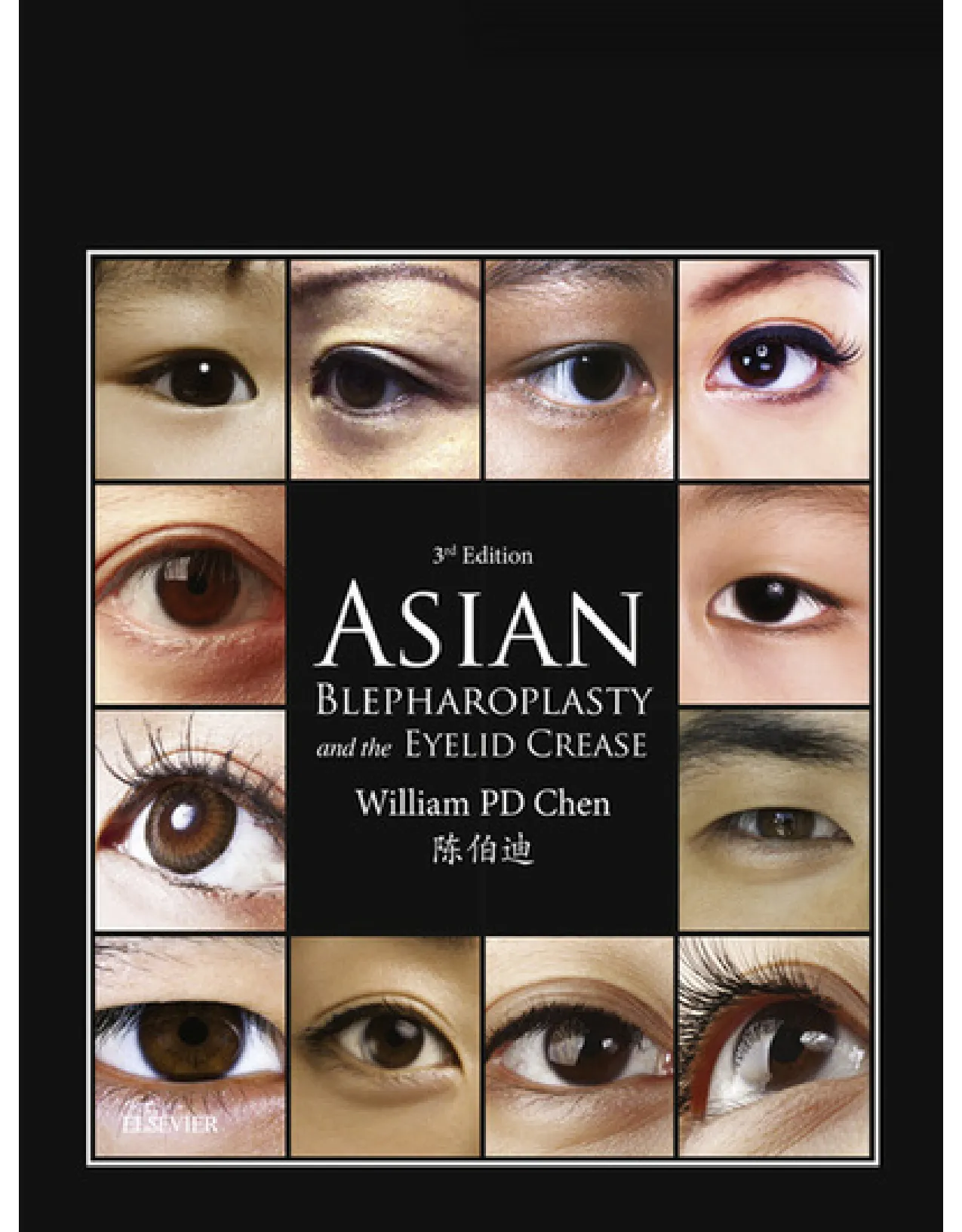
3N Edition ASIAN BLEPHAROPLASTY and the EYELID CREASE William PD Chen Ft 16 th
ASIAN BLEPHAROPLASTY
AND THE EYELID
CREASE
“Asian blepharoplasty is a highly specific form
of upper blepharoplasty, adapted to each
individual’s findings and demands the highest level
of understanding, a focused mind and manual
dexterity to achieve the proper result.”
ASIAN
BLEPHAROPLASTY
AND THE EYELID
CREASE
THIRD EDITION
William Pai-Dei Chen MD FACS
Clinical Professor of Ophthalmology
Department of Ophthalmology
University of California, Los Angeles, School of Medicine
Los Angeles, California, USA
Senior Attending Surgeon
Ophthalmic Plastic Surgery Service
Harbor-UCLA Medical Center
Torrance, California, USA
Private Practice
Irvine and Newport Beach, California, USA
Illustrations by
Paul Kim
Edinburgh London New York Oxford Philadelphia St Louis Sydney Toronto 2016
© 2016 Elsevier Inc. All rights reserved.
No part of this publication may be reproduced or transmitted in any form or by any means, electronic
or mechanical, including photocopying, recording, or any information storage and retrieval system,
without permission in writing from the publisher. Details on how to seek permission, further
information about the Publisher’s permissions policies and our arrangements with organizations such as
the Copyright Clearance Center and the Copyright Licensing Agency, can be found at our website:
This book and the individual contributions contained in it are protected under copyright by the
Publisher (other than as may be noted herein).
First edition 1995
Second edition 2006
Third edition 2016
ISBN 978-0-323-35572-8
British Library Cataloguing in Publication Data
A catalogue record for this book is available from the British Library
Library of Congress Cataloging in Publication Data
A catalog record for this book is available from the Library of Congress
The
publisher’s
policy is to use
paper manufactured
from sustainable forests
Executive Content Strategist: Russell Gabbedy
Content Development Specialist: Carole McMurray
Project Manager: Julie Taylor
Designer: Christian Bilbow
Illustration Manager: Karen Giacomucci
Printed in China
Last digit is the print number: 9 8 7 6 5 4 3 2 1
Notices
Knowledge and best practice in this field are constantly changing. As new research and experience
broaden our understanding, changes in research methods, professional practices, or medical
treatment may become necessary.
Practitioners and researchers must always rely on their own experience and knowledge in
evaluating and using any information, methods, compounds, or experiments described herein. In
using such information or methods they should be mindful of their own safety and the safety of
others, including parties for whom they have a professional responsibility.
With respect to any drug or pharmaceutical products identified, readers are advised to check the
most current information provided (i) on procedures featured or (ii) by the manufacturer of each
product to be administered, to verify the recommended dose or formula, the method and duration of
administration, and contraindications. It is the responsibility of practitioners, relying on their own
experience and knowledge of their patients, to make diagnoses, to determine dosages and the best
treatment for each individual patient, and to take all appropriate safety precautions.
To the fullest extent of the law, neither the Publisher nor the authors, contributors, or editors,
assume any liability for any injury and/or damage to persons or property as a matter of products
liability, negligence or otherwise, or from any use or operation of any methods, products,
instructions, or ideas contained in the material herein.
With a contribution from
(the late) Khoo Boo-Chai MD
Formerly Surgeon Director, Khoo Plastic Surgery
Clinic, Singapore
Visiting Professor, Third Teaching Hospital,
Department of Plastic Surgery
Beijing University, PR China
Visiting Professor, Shanghai Second Medical University
Ninth Hospital, Department of Plastic Surgery
Shanghai, PR China
To my parents, Fred and Katie,
my family (Lydia, Katherine and Andrew),
and my mentor Dr Clinton (Sonny) McCord
viii
VIDEO TABLE OF CONTENTS
All videos are supplied by Dr WPD Chen
1 Primary Asian blepharoplasty for a 29-year-old
female, desired nasally tapered crease, shows treat
ment of medial canthal fold and up-knotting
2 Primary Asian blepharoplasty for a 26-year-old
female, who desired parallel crease. Video shows
up-knotting
3 Revisional Asian blepharoplasty on a woman who
desired parallel crease shape
4 Revisional Asian blepharoplasty for a 19-year-old
female who showed shallowing of crease from resid
ual fat
5 Primary Asian blepharoplasty for a 36-year-old
female. Desired nasally-tapered crease shape, and of
above-average crease height. Shows down-knotting
6 Primary Asian blepharoplasty combined with ptosis
repair for a 65-year-old female
7 Primary Asian blepharoplasty for 62-year-old female
with presence of sulcus and multiple wrinkles, without
true eyelid crease
8 Repeat Asian blepharoplasty for 41-year-old male,
previous Asian blepharoplasty by Chen was 9 years
prior. One can identify the compartments well
9 Primary Asian blepharoplasty for a 48-year-old
female. Has sulcus preoperatively; desires a nasally-
tapered crease shape, with lower-than-average crease
height
10 Revisional Asian blepharoplasty in 28-year-old female
who presented with crease asymmetry. Desired
medium height NTC crease and excised 1 mm scarred
skin. Shows down-knotting
11 An introductory case showing the general concepts
and steps involved in Primary AB on a right upper
eyelid
12 Another case showing the general steps involved in
Primary AB on a right upper eyelid
13 Primary case of a 30-year-old female with OD absent
crease and OS rudimentary crease desiring a higher
than average parallel crease
14 Primary case of a 22-year-old female desiring a paral
lel crease, having previously used adhesive tape over
pretarsal area
15 Primary case of a 22-year-old male with left eye
fissure appearing larger. Patient desires a low-set par
allel crease
16 Primary case of a 31-year-old male with an absent
crease, desiring an average height parallel crease
17 Revisional surgery on a 41-year-old Korean female
who had previously undergone upper blepharoplasty
18 Revisional surgery on a 60-year-old female who had
previously undergone blepharoplasty and a subse
quent revisional attempt whereby abdominal fat had
been injected into the upper lids two years previously
19 Primary case of a 31-year-old female with absent
crease desiring average height NTC
20 Primary case of a 52-year-old male with no crease and
moderate hooding
21 Revisional surgery on a 68-year-old female with per
manent eyeliner. This patient had deepened sulcus
from blepharoplasty and a scarred crease at 8 mm
22 Revisional surgery on a 43-year-old female present
ing for RUL revision
23 Revisional surgery on a 43-year-old female present
ing for LUL revision
24 Revisional surgery on a 35-year-old male presenting
for NTC
25 Primary case of a 28-year-old female with fatty, heavy
lids
26 Primary case of a 31-year-old male with an absent
crease and a truncated upper lid hooding, desiring a
parallel crease of average height
27 Primary case of a 37-year-old female with a shielded
NTC crease OU with more hooding of the left eye,
desiring greater than average NTC
28 Revisional surgery on a 38-year-old female who had
previously undergone revision, desiring the crease to
be reset again
29 Revisional surgery on a 46-year-old female who
had previously undergone three separate crease
procedures
30 Primary case of a 26-year-old male with an absent
crease, desiring a parallel crease
31 Demonstration of effect of high crease placement on
levator muscle function
32 Paper model demonstration of effect of higher-than-
normal attachment on sheets of paper relative to each
other
33 Demonstration of orbital pulley fixation of medial
rectus muscle with recession in strabismus repair for
congenital esotropia in a 3 year-old boy
ix
PREFACE
This new edition of my book on the topic of Asian eyelid
surgery, specifically on the ‘double-eyelid crease’ opera
tion, is a complete update and further expands on what I
feel to be highly relevant functional topics related to the
eyelid crease. Its readership will include house officers/
residents in training, practitioners and seasoned surgeons
in the fields of plastic surgery, ophthalmology (including
oculoplastic surgery), facial plastic/head and neck sur
geons (ears, nose and throat), cosmetic surgeons and der
matologists interested in learning about aesthetic surgery
of the Asian eyelid. (I have also had some patients who
have read the book prior to seeing me.) In essence, it is
a manual for learning upper blepharoplasty at a highest
level of precision.
There are two main schools of techniques, the external
incision methods and the buried sutures methods. (I
prefer to avoid using the misleading terms of ‘non-
incisional’ or ‘no-cut’ method for the buried sutures
method. These are terms often used indiscriminately in
Asia, with patients often unaware of the use of perma
nent, non-dissolvable buried sutures.) Overall the data
are unclear about the incidence for each main category,
but it is probably in the range of 70–90% of cases world
wide using external incisional methods. (My own discus
sion with Dr Khoo Boo-Chai from Singapore 10 years
ago was that in his busy practice he would select only
about 5% of his patients as appropriate candidates for the
buried sutures technique.) This figure is highly variable
region to region, as there are physicians who do either
one or the other method exclusively. Based on statistics
published by the American Society for Aesthetic Plastic
Surgery, there has been a doubling of surgical cosmetic
procedures over the last 15 years in the United States. (If
one adds nonsurgical cosmetic procedures, defined as
Botox, fillers, chemical peels and micro-dermabrasion,
the increase over the 15 years is almost seven-fold.) It is
reasonable to expect this similar explosion in procedures
in Asia, perhaps even more in China and Korea. Even
though there have been more than a dozen papers on
variations of buried suture methods published in the last
five years (in first- and second-level journals), I am unsure
whether this translates into an increase in cases using the
small incision buried sutures methods.
As compared to the original text, Asian Blepharoplasty:
A Surgical Atlas (1995), which described the basic
techniques and concepts, and the second edition, Asian
Blepharoplasty and the Eyelid Crease (2006), which provided
an introduction to the lid crease’s functional dynamics,
this new edition includes a detailed analysis of the biody
namics of the lid crease as well as its functional vulner
ability. Therefore in a sense the book has come full circle,
and could be titled just as well as The Eyelid Crease and
Application in Asian Blepharoplasty. The content here is a
meticulous discussion of the eyelid crease: it is the portal
to the upper lid whenever a physician plans upper eyelid
surgery, whether functional or aesthetic. For an eyelid
surgeon, learning how to enter and exit the upper lid with
functional integrity and ideal wound healing, while con
trolling the appearance of the lid crease, is an essential
skill.
The book’s 26 chapters offer an extensive treatment of
the arrays of techniques available up to present. Seven
new chapters are included on more advanced understand
ing. The advanced concepts start with Chapter 17 on the
concept of the preaponeurotic space and fat as a ‘glide
zone’, followed by Chapter 18 on the beveled approach
in revisional Asian blepharoplasty. (These two chapters
have been carried over from the second edition and are
an essential bridge to advanced understanding.) A greater
emphasis is now placed on correlating the sensitivity and
vulnerability of the lid crease formation to various aspects
of techniques and suture placement. The new advanced
chapters include observation and measurement of crease
height based on head position (Chapter 19), the effect of
tarsal tilt in various clinical conditions (Chapter 20), the
impeding effect of misplacement of buried sutures
and high anchoring (the Faden effect) (Chapter 21), an
advanced summary of eyelid crease factors (Chapter 22),
and a new concept chapter on the eyelid crease as a
stringed series of unipoints (Chapter 23). In addition, I
have added Chapter 25 on my own techniques that may
be used to reduce the medial upper lid fold during Asian
blepharoplasty without the need for more exaggerated
epicanthoplasty cuts – a trend that seems to have reached
a frenzy for the moment in Korea and China. The con
cluding Chapter 26 covers a novel hybrid technique for
those surgeons favoring a less-than-full skin incision,
combining a partial incision with use of a single buried
suture medially. There has been a proliferation of pub
lished papers in the last 10 years, especially from Asia,
the birthplace for this procedure, and I have made great
efforts to be inclusive of these references. However, I
apologize if the need for conciseness and clarity have
prevented me from being all-inclusive, and at times for
taking a subjective, judgmental viewpoint.
The illustration content in this new edition has
doubled, in both photos and drawings, and the use in this
edition of colored three-dimensional cross-sectional
drawings facilitates a quick grasp of the concepts. Detailed
intraoperative findings are included in many of the clini
cal cases from the author’s personal notebook. The
accompanying surgical videos are accessible through
x
Preface
medical English. This new edition is offered in that same
style. The popularity of the second edition was soon fol
lowed by an unforeseen global recession, and it is a joy
for me now to be able to continue my exploration on this
topic.
The book is meant for use by medical professionals in
learning the techniques and concepts of Asian blepharo
plasty, as well as being a comprehensive treatise on this
topic from 1896 to present. It is not meant in any way
for use as an illustrative guide for physicians to show to
their patients, nor for patients searching for medical
guidance or as a substitute for proper medical consulta
tion. The final determinant in surgical outcome always
depends on full understanding as well as an open dialogue
between physician and patient. One should not draw on
these illustrations as examples applicable to any particular
individual nor as a basis to arrive at medical decisions.
As before, I wish to thank Dr Kenichiro Kawai of
Osaka University Graduate School of Medicine for
allowing me to re-use his anatomic drawing and stereo
scopic photograph from his upper eyelid vascular arcade
research paper, as well as sincerely acknowledging with
remembrance the late Dr Khoo Boo-Chai of Singapore,
a pioneer in this field, for his generous teaching when I
first asked him about his techniques and his views on
suture methods versus external incision techniques.
ExpertConsult and have been expanded to 30 surgical
cases. The added videos were recorded using current
generation equipment and provide greater detail.
The current project involved the use of high resolu
tion cameras from Canon, Nikon, Panasonic Lumix with
Leica lens and even an iPhone camera. I use a MacBook
Pro and a Mac Mini computer with a 27” Thunderbolt
display to collate most of the work. For some photo
graphs, only cropping and brightness adjustments were
used. All of the drawings from the previous edition have
been redrawn and colorized; all of the added drawings
are paired with detailed captions.
Favorite features such as ‘Pearls’ and ‘Pitfalls’ have
been carried over from the previous edition, as well as
convenient comprehensive spreadsheet listings of rele
vant literature in the Appendices, which has been brought
fully up to date. There is an additional new Appendix
listing recent papers on epicanthoplasty.
I have been involved in resident-teaching for over 30
years and with teaching the concepts of Asian blepharo
plasty through the American Academy of Ophthalmology
for 25 years, during which time I have published seven
textbooks. My preference, supported by feedback that I
receive from readers, has always been to use a simple
writing style that focuses on conveying ideas rather than
getting entangled in complicated anatomic terms and
xi
ACKNOWLEDGMENTS
I am blessed with a great team from Elsevier in making
this edition possible; their assistance along the way was
highly significant in keeping me on track. As the subject
gets complicated and the demand for detail increases, the
precision I require of myself as an author and what I ask
of the team members rises significantly. The recipients of
this demand included our team leader Rus Gabbedy,
Executive Content Strategist of Elsevier (London),
Carole McMurray (Content Development Specialist,
Edinburgh) and John Leonard (London), who assisted
with manuscript development; and the art team, who
included Karen Giacomucci (Elsevier, Philadelphia) and
her art team members Paul Kim (New Jersey), Vicky
Heim (Atlanta, Georgia) and Jade Myers. The video
effort and online resource development at Inkling is led
by Jonathan Davis, Multimedia Producer of UK/EMEA–
Elsevier Ltd (based in Oxford). Julie Taylor (Publishing
Project Manager, Oxford) and Elaine Leek (copy editor)
helped organize the text and illustrations and correct all
our mistakes. To them, I wish to offer my greatest acco
lades for a job well done, working with me through dif
ferent time zones and work schedules, via emails and
phone calls. I really appreciate their guidance.
I could not have completed the project without the
support of my family, Lydia, Andrew and Katherine, as
well the foundation of knowledge imparted to me during
my formative training under my mentor, Dr Sonny
McCord in Atlanta, Georgia. I am grateful.
1
WHAT IS AN UPPER LID CREASE?
C H A P T E R 1
In common usage, the eyelid crease is often meant to
describe a natural inward creasing of the skin seen in the
upper eyelid, typically dividing it into a lower segment
adjacent to the upper eyelashes, and an upper segment of
skin that runs from the crease to the border of the
eyebrow.
The crease is present in about 50% of the Asian popu
lation, and found to be more prevalent in all non-Asians.
The presence of a crease in an Asian subject is what dif
ferentiates a double eyelid (with a crease unequally divid
ing the lid into two sections, hence ‘double’) from a single
eyelid (or mono-lid).
Double-eyelid crease surgery (procedures) is a form of
surgery to add or supplement an eyelid crease to an indi
vidual who seeks it. This is often for an individual who
does not have a crease, or who finds their crease is insuf
ficient, or in whom the crease is unbalanced between the
two sides.
The configuration of the upper lid crease in Asians
varies greatly. The terminology used to describe these
configurations also varies, depending on the ethnic group
and language concerned. Figure 1-1 illustrates the
Chinese characters for the words ‘double-eyelid fold’.
Figure 1-2 shows the Japanese Kanji writing for ‘single
[one] lid eye’ and ‘double [two] lid eye’. The characters
common to Chinese and Japanese for the operation to
construct a lid crease are illustrated in Figure 1-3.
FIGURE 1-1 ■ Chinese written characters for ‘double eyelid
[skin]’.
FIGURE 1-2 ■ Japanese Kanji characters for ‘single eyelid’ (left)
and ‘double [two] eyelid’ (right).
FIGURE 1-3 ■ Characters common to Chinese and Japanese for
the procedure to construct an eyelid crease, or ‘double-eyelid
procedure’.
2
Asian Blepharoplasty and the Eyelid Crease
FIGURE 1-4 ■ Chen1 has previously described the various forms of Asian eyelids, as illustrated. (A) Single eyelid without crease.
(B) Same size eyelid fissure with crease. (C) Segmented or non-continuous crease. (D) Partial or incomplete crease. (E) Multiple
creases. (F) Asian eyelid with a nasally tapered crease; in a small percentage of cases it shows some lateral flare. (G) Asian eyelid
with a parallel crease. (H) Typical Caucasian semilunar crease.
A
B
C
D
E
F
G
H
As described in previous publications by the author,1–9
the crease may be asymmetric in its presentation, or be
absent in one eye and present in the other. It may be
continuous or segmented (fragmented).
Figure 1-4 shows the various configurations of the
Asian eyelid.
1 What Is an Upper Lid Crease?
3
FIGURE 1-5 ■ Asian lid without crease.
This patient has moderate upper
lid
hooding
causing
downward
rotation of the lashes. Note the
apparent upper lid hooding (fold)
that overshadows the smaller palpe
bral fissure laterally. Strategic place
ment of a crease would make the
palpebral fissure seem larger.
FIGURE 1-6 ■ Asian eyelid with a parallel crease.
FIGURE 1-7 ■ Partial obliteration of crease.
Figure 1-5 shows an eyelid without a crease. There
is a mild degree of upper lid hooding, causing sec
ondary downward rotation of the lashes. Figure 1-6 illus
trates an eyelid with a distinctive crease. This is
the parallel configuration. Figure 1-7 is an eyelid in
which a portion of the crease has been obliterated.
4
Asian Blepharoplasty and the Eyelid Crease
FIGURE 1-8 ■ (A, B) Incomplete or
partial crease.
B
A
FIGURE 1-9 ■ Asian subject with two
well-defined creases that run parallel
to each other but in a nasally tapered
configuration.
An eyelid with an incomplete or partial crease is
shown in Figure 1-8. The crease originates in the
medial canthus and medial upper lid fold (supracanthal
web) and extends halfway across the upper lid. Multiple
creases are illustrated in Figure 1-9, where two
well-defined creases run parallel to each other.
1 What Is an Upper Lid Crease?
5
FIGURE 1-10 ■ Minimal nasally tapered
crease with a mild lateral flare.
Figure 1-10 shows a minimal nasally tapered crease. The
lateral third of the crease may be the same distance from
the eyelash margin as the central third, or it may rise
slightly to form a laterally flared crease, in which the
lateral third of the crease is further from the lash margin
than the central third. A Caucasian upper lid crease is
shown in Figure 1-11, where the central third of the
crease is farthest from the lash margin.
FIGURE 1-11 ■ Caucasian upper lid crease with a
semilunar shape. Note that the widest separation of
the crease from the ciliary border occurs centrally.
6
Asian Blepharoplasty and the Eyelid Crease
FIGURE 1-12 ■ (A) Asian eyelid with a nasally tapered crease. Note the merging of the crease medially into the medial upper lid fold
and the relatively parallel course from the central third of the lid outward. (B) Asian eyelid with a parallel crease.
A
B
In Asians with a continuous eyelid crease, the crease
may be of the nasally tapering type (NTC) (a less desir
able term is ‘inside’ fold) in which the crease converges
toward the medial canthus, coming closer to the lashes
as it reaches the medial canthal angle (Figure 1-12A), or
it may be a parallel crease (PC) (a less desirable term is
‘outside’ fold), in which the crease runs fairly parallel to
the lash margin from the medial canthus to the lateral
canthus (Figure 1-12B).
1 What Is an Upper Lid Crease?
7
FIGURE 1-13 ■ Nasally tapered crease with a variant form showing a lateral flare. The widest separation of the crease from the ciliary
border occurs laterally like that shown in Figure 1-10.
FIGURE 1-14 ■ Nasally tapered crease that runs
level to the eyelash from the central third of the
eyelid outward (see Figure 1-12A).
In eyelids with a nasally tapered crease the crease may
gently flare away from the lid margin as it approaches the
lateral canthal region, forming a laterally flared crease
(LTC) (Figure 1-13). Another configuration is that the
nasally tapering crease may run level to the eyelash
margin from the central third of the eyelid laterally
(Figure 1-14).
8
Asian Blepharoplasty and the Eyelid Crease
FIGURE 1-15 ■ Asian patient after blepharoplasty. Note asymmetry of the two creases and the high placement of the semilunar crease.
Asians rarely have a lid crease that is semilunar in
shape, as is common in Caucasians (see Figure 1-4H). In
a semilunar crease each end of the crease is closer to the
respective lid margin than the central portion of the
crease. Having a semilunar crease is by far the most
frequent complaint heard from Asian patients who have
had blepharoplasty performed in the United States2
(Figure 1-15). This crease is often unnatural, high and
harsh (termed the ‘uhh’ syndrome).
1 What Is an Upper Lid Crease?
9
FIGURE 1-16 ■ When
a
semilunar
crease is placed more than 10–12 mm
in an Asian upper lid, the crease is in
the mid level of the eyelid, halfway to
the brow.
A high crease is one located 8–11 mm from the ciliary
margin. A high crease may result if a surgeon adheres to
an empiric formula for the height of the lid crease, or
uses techniques of supratarsal fixation in which a distance
of 8 mm or more is applied without regard to ethnicity.
Either method results in a crease that looks excessively
high on an Asian patient. To summarize, such a regi
mented approach is counter-effective in Asian blepharo
plasty for the following reasons:
• Asians are usually smaller in build; correspondingly,
the upper tarsus measures only 6.5–8.0 mm in
height on average.
• The distance between the eyebrow and the upper
lid margin is smaller on Asians than on Caucasians.
A crease located 10–12 mm from the lash margin
would look much closer to the mid level of the
upper lid than is natural (Figure 1-16).
10
Asian Blepharoplasty and the Eyelid Crease
FIGURE 1-17 ■ Asian patient who underwent blepharoplasty with excessive removal of preaponeurotic fat. Note the hollow supratarsal
sulcus and the formation of multiple creases.
Not only should ethnicity be a factor in blepharo
plasty, but also each individual’s features. When the
crease is high, it is farther from the lid margin than the
height of the tarsus, the surgically applied crease traverses
thick dermis as it approaches the brow and is likely to be
associated with hypertrophic scarring. The large distance
between the lash margin and the crease also allows little
camouflage by the upper eyelashes, and the crease is
exposed to scrutiny. A crease is harsh when it is overtly
prominent, deep and indurated with dermal reaction.
An unnatural crease describes a shape that is not aes
thetically pleasing on the face of the person. The most
frequent complaint is a semilunar crease. The overall
impression of a crease positioned high and semilunar in
shape is unnatural for Asians. Removal of an excessive
amount of preaponeurotic fat also can cause an unnatural
crease. When all the fat pads are removed from the
preaponeurotic space, the result is a hollowed eye or
‘famined’ look that appears incongruous on the relatively
flat face of an Asian2 (Figure 1-17). For Caucasians, the
same complete excision of preaponeurotic fat pads used
to be a necessary step in the performance of a total cos
metic blepharoplasty.
It is important to recognize that there is a high degree
of variation in the anatomy of the upper eyelids of Asians.
A common misconception is that all Asians are born
without an upper lid crease. In fact, half the Asian popula
tion does have a natural crease. For each person, the
shape and height of the crease and the relation of the
crease to facial configuration should be part of the overall
assessment before a cosmetic surgical procedure is
performed.
The reasons for electing this type of cosmetic enhance
ment may be myriad, and no individuals tend to have the
same reasons. The commonly cited and reasonable indi
cations will include a desire to have the eyelid opening
(fissure) more apparent, since a single eyelid (without
crease) often has a small fold of skin overhanging the
opening, which makes it look covered over and smaller
than it actually is; it is narrower in vertical as well as
perceived horizontal dimensions.
1 What Is an Upper Lid Crease?
11
below the crease, the lashes, the lid margin and the tarsus
(a fibrous plate along the upper lid margin). When the
lids open, the lifting levator muscle is active (by turning
on the oculomotor or third nerve’s upper branch), that
resting section of skin and deeper soft tissues above the
crease (preseptal, above the upper boundary of the tarsus/
fibrous plate) relaxes by inhibition of the facial or seventh
nerve, whose normal function is contraction of the orbic
ularis oculi and facial muscles. There is therefore a facili
tation of the skin that is at the narrow boundary of a
natural crease to fold inward, and it is almost always along
the upper border of the tarsal plate. It is along the inter
face between an active layer of tissues contracting (levator
pulling up on tarsus and small amount of skin adherent
to it) and passively gravitating skin on top (with all its
underlying muscle strands and fat) which is the larger
upper proportion you see in a double eyelid. This is
normal physiology in a natural crease. The crease is just
demonstrating the net force result of a healthy levator
terminating its attachment to the skin’s undersurface.
The reverse happens when the eyelid closes: seventh
nerve ON, third nerve OFF – the orbicularis oculi muscle
that wraps around the eyelid fissure is active and contracts
[ON] shut, while the levator is not lifting due to inhibi
tion [OFF]. (The small skin above, overhanging the
inward crease, is the upper lid fold.)
FIGURE 1-18 ■ (A) A left upper eyelid without a crease; (B) the same upper lid with a parallel crease. The palpebral fissure (eye
opening) is the same in each and yet the visual perception is that this is bigger in (B) than that seen in (A).
A
B
Other reasons may be to enhance the ability to apply
make-up without smudging, to save time, to correct
asymmetry, to create consistency and constancy, or to
fulfill individuals’ often correct impression that having a
crease that simulates a natural crease makes the eye more
attractive. The important word here is ‘natural’. The
single-lidded individual almost always wants the crease to
mimic the dimensions of an Asian double eyelid. Figure
1-18 illustrates two drawings of palpebral fissures of equal
size; visual perception is that the eye opening for the
eyelid with a crease is bigger.
The means by which this can be achieved, over the last
hundred years or more, has been along two surgical
routes: the suture method and the incision method. They
are two totally different approaches, not only in terms of
philosophy but also in the surgical and anatomical bench
marks that each sets (although practitioners may not be
consciously aware of it). This will be explained in greater
details in the ensuing chapters of this book.
The natural infolding of an eyelid crease can be
thought of as the end points of fine muscle fibers from
the opening muscle (levator aponeurosis, a curtain-like
sheet of elevating muscle within the upper lid, like a
garage door motor) of the upper lid attaching onto the
underside of skin; its action contracts the muscle up and
dynamically pulls on the skin to form the upper lid crease.
This levator muscle pulls on the small segment of skin
12
Asian Blepharoplasty and the Eyelid Crease
FIGURE 1-19 ■ Cross-section of the eyebrow, closed eyelids and eye.
Orbicularis oculi
Preaponeurotic fat
Septum orbitale
Levator aponeurosis
Müller's muscle
Inferior tarsus
Inferior oblique
Inferior rectus
Figure 1-19 shows the cross-section of the eyebrow,
closed eyelids and eye. The upper tarsus, which contains
oil glands is usually 10 mm in Caucasian and 6.5–7.5 mm
in Asian women. The inferior tarsus is 3.9–4.0 mm in
both. (Pink layer is levator muscle, with the 10 mm
length of this pink tissue above the upper tarsus being the
aponeurosis segment of the levator.)
1 What Is an Upper Lid Crease?
13
FIGURE 1-20 ■ Diagram showing a simplistic representation of an Asian upper eyelid; here it is shown as in the 50% of subjects who
are without a crease (single eyelid). Zone 1 corresponds to the area in front of the tarsal plate (Pretarsal zone), with its height usually
being 7–8 mm. (Copyright W.P.D. Chen.)
Pretarsal zone
Periorbital zone
Preseptal zone
Figures 1-20 and 1-21 show the basic difference
between a natural Asian eyelid without an eyelid crease
versus the eyelid of a person born naturally with one.
Figure 1-22 shows an upper lid with scarring involving
the anterior skin–orbicularis and the preaponeurotic fat
space.
The issues often raised at academic meetings in recent
times are: ‘If one sees a crease line anywhere on the upper
lid, whether it is lower than the normal insertion point
of the levator aponeurosis on the upper lid skin (which
normally should be precisely along the upper border of
the tarsal plate), or at any point unrelated to the levator,
like one or more wrinkled skin crease lines within the
upper concavity of the upper lid, aren’t these the eyelid
crease also?’ The answer is No! Technically they are just
wrinkles, because they are not caused by contraction of
the levator muscle. Just like a true elbow crease is formed
by the biceps pulling on the forearm bones (radius
and ulnar), and not simply because of any skin wrinkle
left on the arm, or of skin damage or adipose tissue
changes. Nor should we call a sunken sulcus (concavity)
a crease that ‘has migrated upward’. If one adheres to this
biodynamic and anatomically accurate definition of an
eyelid crease, there will be much less confusion among
practitioners as to what a crease is and where to apply a
crease, as well as what form of surgery or procedure
represents a physiologic route. It is indeed curious that
there are just as many medical practitioners who are
unclear about this as there are patients seeking informa
tion on this very point.
The divergence between the suture method and inci
sion method lies at the core of understanding the natural
mechanism of an eyelid crease, and the approaches by
which this can be achieved.
14
Asian Blepharoplasty and the Eyelid Crease
FIGURE 1-21 ■ A representation of an Asian upper lid with crease (‘double eyelid’). The levator muscle endings (pink layer) have
some attachment to the undersurface of the eyelid skin along the upper border of the tarsal plate, where it forms the crease. The
thin arrow indicates the eyelid crease; the darker arrow indicates the overhanging lid fold. (Copyright W.P.D. Chen.)
1 What Is an Upper Lid Crease?
15
REFERENCES
1. Chen WPD. Asian blepharoplasty. Ophthalmol Plast Reconstr Surg
1987;3:135–140.
2. Chen WPD. Review of Aguilar G. Complications of oriental blepha
roplasty. In: Mauriello J, ed. Management and avoidance of compli
cations of eyelid surgery. Vol. 3. Philadelphia: Field & Wood, 1994.
3. Chen WPD. Concept of triangular, rectangular and trapezoidal
debulking of eyelid tissues: application in Asian blepharoplasty. Plast
Reconstr Surg 1996;97:212–218.
4. Chen WPD. Eyelid and eyelid skin diseases. In: Lee D, Higgin
botham E, eds. Clinical guide to comprehensive ophthalmology.
Stuttgart: Thieme, 1999: 137–182.
5. Chen WPD. Upper eyelid blepharoplasty in the Asian patient.
In: Putterman AM, ed. Cosmetic oculoplastic surgery, 3rd edn.
Philadelphia: WB Saunders, 1999: 101–111.
FIGURE 1-22 ■ A representation of one form of scarring involving the skin and the middle space (between the front and back layers)
of the eyelid. There is absence of fat and obliteration of the preaponeurotic space. (Copyright W.P.D. Chen.)
6. Chen WPD. Expert commentary on blepharoplasty and blepharop
tosis surgery in Asians. In: Mauriello J, ed. Unfavorable results of
eyelid and lacrimal surgery. Oxford: Butterworth–Heinemann, 2000:
68–71.
7. Chen WPD. Aesthetic eyelid surgery in Asians: an East–West view.
Hong Kong J Ophthalmol 2000;3:27–31.
8. Chen WPD. Oculoplastic surgery – the essentials. Stuttgart:
Thieme, 2001.
9. Chen WPD, Khan J. Color atlas of cosmetic oculofacial surgery
(with DVD), 2nd edn. Philadelphia: Elsevier Science/Saunders;
2010.
17
C H A P T E R 2
HISTORICAL CONSIDERATIONS
COSMETIC OCULOPLASTIC
SURGERY: EVOLUTION OF
DOUBLE-EYELID COSMETIC SURGERY
IN THE JAPANESE LITERATURE
Publications in the early Japanese medical literature
favored the suture ligation method. The first description
of this method, by Mikamo,1 was published in 1896 (see
Appendix 1, and Appendix 2 under Shirakabe, 1985).
Mikamo performed the procedure on a Japanese woman
who did not have a crease in one of her upper eyelids.
The crease was designed to be 6–8 mm from the ciliary
margin. Three 4-0 braided silk sutures were used, passing
through the full thickness of the lid from the conjunctiva
to the outer layer of skin. The depth of the crease was
adjusted by the number of days the sutures were left in
place, the range being 2–6 days.
As early as 1926, Uchida2 described his suture ligation
method for the double-eyelid operation. He performed
the procedure on 1523 eyelids in 396 male and 444 female
patients. Uchida described the crease configuration as a
fan shape, that is, a somewhat rounded crease. The crease
was designed to be 7–8 mm from the ciliary margin.
Three buried catgut sutures were used on each lid, encom
passing approximately 2 mm of eyelid tissue horizontally.
The sutures were removed 4 days after placement.
The first mention of an external incision method dates
to 1929, when Maruo3 reported on both his suturing
technique and his incision technique. Maruo’s incision
technique required a crease incision across the lid,
designed to be 7 mm from the ciliary margin. The wound
closure technique was a translid passage from the con
junctival side just above the superior tarsal border to the
anterior skin surface. One 5-0 catgut suture was used to
imbricate four throws along the superior tarsal border,
attaching the skin edges to the underlying tarsal plate.
The spacing between each throw of the stitch was about
5–6 mm. Maruo also discussed subcutaneous dissection
5 mm superior and inferior to the incision line.
In 1933, preference for a higher placement of the
crease became evident when Hata4 reported his suture
ligation method. The crease line was placed 10 mm from
the ciliary margin. Hata used three double-armed 5-0
braided silk sutures, passing them from tarsus to skin and
fixing them to the skin surface using small beads. Each
arm of the suture required 1 mm spacing for the bead to
be tied. Stitches were removed after 8–10 days.
In a comprehensive and scholarly article in 1938,
Hayashi5 described the two methods of crease formation.
His suture ligation technique was modeled after
18
Asian Blepharoplasty and the Eyelid Crease
Western readers. As a result, the publication of articles
on this procedure in Western medical journals in the
1950s made the procedure seem new (and Western) in
concept. Between 1896 and 1950, 11 articles relating to
the suture ligation methods and eight articles on external
incision methods were published in the Japanese medical
literature (see Appendix 1 for selected articles).
Much of the later Western literature on this subject
described techniques quite similar to those described in
the early Japanese publications (see Chapters 6 and 7 for
a continuation of historical publications from the 1950s
onwards).
Figure 2-1 illustrates the visual expansion of the eye
size when a natural crease is added.11 From the original
eye size of Width (W1), and Height (H1), the apparent
fissure size is expanded to W2, and H2 when a crease is
there, either through surgery, eyelid tape, glue or attached
thin strings or fibers.
REFERENCES
1. Mikamo K. A technique in the double eyelid operation. J Chu
gaishinpo 1896.
2. Uchida K. The Uchida method for the double-eyelid operation in
1,523 cases. Jpn J Ophthalmol 1926;30:593.
3. Maruo M. Plastic construction of a ‘double-eyelid’. Jpn Rev Clin
Ophthalmol 1929;24:393–406.
4. Hata B. Application of eyelid clamp and beads in ‘double-eyelid’
operation. Jpn Rev Clin Ophthalmol 1933;28:491–494.
5. Hayashi K. The double eyelid operation. Jpn Rev Clin Ophthalmol
1938;33:1000–1010, 1098–1110.
6. Inoue S. The double eyelid operation. Jpn Rev Clin Ophthalmol
1947;27:306.
7. Mitsui Y. Plastic reconstruction of a double eyelid. Jpn Rev Clin
Ophthalmol 1950;44:19.
8. Ohashi K. The double eyelid operation using electrocautery. Jpn
Rev Clin Ophthalmol 1951;46:723.
9. Hirose K. The double eyelid operation. Jpn Rev Clin Ophthalmol
1950;45:374.
10. Ikegami T. Brief discussion on the double eyelid operation. Jpn Rev
Clin Ophthalmol 1951;46:706–707.
11. Chen WPD. Revision and correction of suboptimal results. In
Asian blepharoplasty: a colour atlas. Oxford: Butterworth–
Heinemann; 1995: p. 97, Figure 7-23.
Mikamo’s method but was novel in that it was designed
for a nasally tapered crease. Three sutures were used on
each lid. The central and lateral sutures were applied
superior to the crease line or tarsal plate, whereas the
medial suture was deliberately applied below the crease
line or tarsal plate. Hayashi’s incision method was also
revolutionary, in that he advocated excision of the pre
tarsal orbicularis oculi muscle at the area of the incision.
He also advocated the use of interrupted skin–tarsus–skin
sutures, and in between skin–skin stitches consisting of
4-0 silk for wound closure. The crease was designed so
that medially it was 5 mm from the ciliary margin, cen
trally 6 mm from the margin, and laterally 7 mm from
the margin; in essence it was a nasally tapered crease. The
sutures were removed after 4 days.
Inoue6 in 1947 proposed dissecting the ‘connective
tissues’ in the subcutaneous plane between the incision
line and the ciliary margin. Sutures of 5-0 braided silk
were used for skin–tarsus–skin closure; sutures were
removed after only 2–3 days.
In 1950, Mitsui7 continued the evolution of the double-
eyelid crease procedure when he described the dissection
and removal of pretarsal connective tissue, including pre
tarsal orbicularis muscle and pretarsal fat pads. Wound
closure was carried out in two steps. First, five separate
nylon sutures were used to stitch the inferior skin border
to the anterior surface of the superior tarsal border and
were tied individually. Second, 5-0 braided silk was used to
close the incision site skin to skin. The nylon sutures were
removed after 2–3 days, the silk sutures after 7–8 days.
Ohashi8 described a double-eyelid crease operation
using an electric coagulator. The cautery needle was
applied vertically to the skin surface along the crease line
until the skin blistered; two more rows of cauterization
below the crease line followed. Hirose9 and Ikegami10 in
1951 briefly discussed incision methods but did not offer
any new information.
The foregoing procedures were described only in the
Japanese literature and were not readily available to
FIGURE 2-1 ■ The addition of a crease that is appropriate in nature adds to the apparent width (W2) and height (H2) of the eyelid
fissure.
H1
H2
W1
W2
(W2, H2) > (W1, H1)
H1
W1
19
C H A P T E R 3
COMPARATIVE ANATOMY
OF THE EYELIDS
THE UPPER LID AND CREASE
Studies have shown that about 50% of Asians do not
have an upper eyelid crease; the other 50% have at
least some form of crease. Eyes without a lid crease
are described as having a ‘single eyelid’ (Figure 3-1),
whereas those with two segments of lid between
the eyebrow and the eyelashes have ‘double eyelids’
(Figure 3-2). Most of the plastic surgery literature of the
1950s1,2 was based on the assumption that all Asians are
without an eyelid crease and that all Caucasians have an
upper lid crease.
FIGURE 3-1 ■ Partially shielded crease
over the right upper lid. Absent
crease or ‘single eyelid’ over left
upper lid.
FIGURE 3-2 ■ Double eyelid. Parallel
crease configuration.
20
Asian Blepharoplasty and the Eyelid Crease
FIGURE 3-3 ■ Left palpebral fissure. (A) Without crease. (B) With crease. The crease gives the impression of a larger eye opening.
A
B
Asians born with a crease appear to have a wider palpe
bral fissure and larger eyes and are culturally perceived
to be more alert and friendly than those with a single
eyelid (Figure 3-3). The cultural ideal of feminine beauty
also influences the desire for a double eyelid: having a
double eyelid allows greater latitude in the application of
cosmetics to make the eyes and face more aesthetically
pleasing. It is therefore understandable that some women
without a crease may wish for one, provided the means
are available.
To some Westerners, who presume that all Asians have
eyelids without a crease, such an endeavor equates
to ‘Westernization’ or ‘occidentalization’. In the author’s
opinion, however, it merely represents an attempt to look
like their fellow Asians who do have a crease over their
upper eyelids.3
The growing popularity of Asian blepharoplasty has
been incorrectly interpreted as resulting from the influ
ence of Western culture after World War II and the
Korean war, a manifestation of which was believed to be
a desire on the part of Asians to blend in with Caucasians,
to look Westernized or occidental. Having traveled and
taught extensively in Asia, it is the author’s perception
that the idea of beauty transcends time, geographic
boundaries and ethnicity. For a growing number of
patients undergoing Asian blepharoplasty it has more to
do with an increasing awareness that such procedures are
available than with the cultural influence of the West.
Clinical experience teaches that Asians do not want to
look Caucasian. A frequent postoperative complaint is
that the procedure results in a semilunar crease, a feature
that, although characteristic of Caucasians, is aestheti
cally displeasing in Asians.4
The essential differences in the upper eyelid structure
between Caucasians and Asians have been studied in
cadaver samples by Doxanas5 and Jeong et al.6 The funda
mental difference between the subset of those Asian eyelids
without a crease and a Caucasian eyelid that possesses a
crease appears to be the lower point of fusion of the orbital
septum onto the levator aponeurosis in Asians.
3 Comparative Anatomy of the Eyelids
21
FIGURE 3-4 ■ A typical Caucasian eyelid with a natural upper eyelid crease. Aponeurotic fibers form interdigitations to the pretarsal
orbicularis oculi muscle and a subdermal attachment along the superior tarsal border.
Orbicularis oculi
Preaponeurotic fat
Septum orbitale
Levator aponeurosis
Müller's muscle
Inferior tarsus
Inferior oblique
Inferior rectus
Individuals with a Lid Crease
In Caucasians who have a crease5 (Figure 3-4), the orbital
septum fuses with the levator aponeurosis approximately
5–10 mm above the superior tarsal border. Below this
point, the terminal interdigitations of the levator aponeu
rosis insert towards the subdermal surface of the pretarsal
and preseptal upper lid skin, with maximal concentration
along the superior tarsal border and spreading inferiorly.7
Collin8 has detailed electron microscopic findings of
fusion of the terminal fibers of the levator aponeurosis
with the septae that are between the pretarsal and presep
tal orbicularis muscle fibers; he was unable to show any
direct attachment to the skin in that particular study.
There has been a subsequent study in China using
scanning electron microscopy that described the presence
of aponeurotic fibers penetrating the orbicularis fibers to
fuse with the skin underneath a crease. In this study,
published in 2001, Cheng and Xu9 reported using
scanning electron microscopy and detecting bunched
fibers of levator aponeurosis penetrating through the
orbicularis muscle and fusing with the skin on the area
of the eyelid crease in those patients born with a crease,
versus those without. These differences were not detect
able using conventional light microscopy. The authors
described the arrangement of these bundled fibers as
being different from that of the fibers in the intermuscu
lar septum. The bundles were thinner, unidirectional and
22
Asian Blepharoplasty and the Eyelid Crease
on the anterior surface of the aponeurosis and the under
lying levator aponeurosis, and that these need to be
reflected away before carrying out the skin–aponeurosis
fixation.
Bang et al.13 argued against the conventional theory
that the levator termination inserts into the skin to form
the crease. The authors proposed that this theory is more
accurate, i.e. that the absence of a crease is associated with
a lower position of the septum in a single eyelid without
a crease, hence there is more inferiorly migrated fat and
the eyelid is thicker than one that has a crease, which is
thinner and has a tighter pretarsal skin zone. The crease
in this theory corresponds to the lowermost edge of the
orbital fat, or the lower level of the orbital septum, which
is usually 2–3 mm above the highest point of insertion of
the levator aponeurosis. In an excellent brief discussion
following the above-mentioned paper by Bang et al.,
Khoo Boo-Chai14 stated that below the lowermost edge
of the orbital fat there are fine filamentous condensations
of the connective tissue that connect the aponeurosis to
the connective tissue septa between the fibers of the
orbicularis oculi muscles. The crease lies in the pretarsal
skin at the superior insertion of these fibers and serves as
a useful external landmark of the common boundary
between the lowermost edge of the orbital fat and the
filamentous connective tissue condensation fibers. When
the eye opens, the pull of the levator is transmitted via
these fibers to the pretarsal skin–muscle complex. The
pretarsal skin below the crease moves as a single unit
upward and backward, like the visor of a helmet. The
orbital fat moves back into the orbit, and the superior
palpebral fold is formed by the lid skin scrolling down at
the upper lid crease. Khoo Boo-Chai further observed
that the pretarsal skin is soft and very thin, with little
space between the dermis and the subcutaneous areolar
plane to the orbicularis beneath. The skin above the
upper lid crease is comparatively thick, with some subcu
taneous fat, with the crease lying at the junction of this
region. He favored the continued use of the term levator
expansion (extension).
THE FAT PADS OF THE UPPER LIDS
In terms of fat distribution and compartments, Uchida15
described the presence of four areas of fat pads in Asian
upper eyelids. He described the subcutaneous fat, the
pretarsal fat, the ‘central’ (submuscular or preseptal) fat
pads and the ‘orbital’ fat pad, which is now better known
as the preaponeurotic fat pad (see Figure 3-7).
Miyake et al.16 described upper eyelid MRI findings in
those with a crease versus those without. He observed
that the ‘orbital fat’ normally returns into the orbit as the
upper eyelid opens in someone with a crease, but that
when the fat does not return then crease formation is
prevented. He correctly observed that the crease folds in
at the junction between the thin skin without subcutane
ous fat (pretarsal area) and the thick skin with subcutane
ous fat (preseptal area).
The preaponeurotic fat pad is limited in its inferior
most position by the junction (or reflection) of the septum
with the levator aponeurosis and does not tend to
aligned as threads. They noted that where these fibers
pass through the orbicularis to attach to the subcutaneous
fibers they were linear in shape, closely aligned and
clearly visualized, whereas the fibers of the intermuscular
septum were thicker and aligned in a disorderly fashion.
Of interest is the observation that in those eyelids with a
crease the orbicularis bundles lying transversely were
arrayed sparsely and loosely in a single layer, in contrast
to those in a single eyelid (i.e. without a crease), which
had muscle tissues that were dense and muscle bundles
arrayed in a stratified manner. Their overall conclusion
is that a fiber link between levator aponeurosis and the
upper eyelid skin results in the formation of the palpebral
sulcus (crease) in the double eyelid. They draw the infer
ence that the purpose of most double-eyelid procedures
should be to establish a stable attachment between levator
aponeurosis and the eyelid skin. They further stated that
the obstructing effect of the orbicularis in single-lidded
individuals could explain why the surgical outcome is
unpredictable with the suturing method. Using the inci
sion method, the excision of a suitable amount of orbicu
laris muscle changes the dynamics of the upper eyelid and
assures a good aesthetic outcome. The authors recom
mended the incision method with supratarsal fixation in
order to establish a stable attachment between the levator
aponeurosis and the eyelid skin.
A similar study published in the same year by Mori
kawa et al.10 described the scanning electron microscopic
findings in single- versus double-eyelid samples taken
from Japanese cadaver specimens. They were able to
trace the collagen fibers that branched off from the
levator aponeurosis, running through the orbicularis
oculi muscle layer and inserting at the subcutaneous layer
just within the crease space indentation. These fibers do
not contact the skin directly, but become continuous with
the collagen fibers in the subcutaneous tissues.
Hwang et al.11 attempted to show that the orbital
septum consists of an outer (whitish, superficial) layer and
an inner layer which, upon meeting the levator aponeu
rosis inferiorly, then reflects superiorly and continues
with the sheath of the levator muscle, which they termed
the levator sheath. Several line drawings in their article
attempted to illustrate this concept, and a light micro
scopic slide showed what purported to be the inner layer
of the orbital septum and the sheath of the levator, but
the higher-magnification slide did not have a portion
showing that the one continues into the other, which was
their premise. There had been a previous description of
the anterior lining of the levator (levator sheath) descend
ing and then reflecting up the back surface of the orbital
septum to reach the superior orbital rim,12 but there had
been no concrete study illustrating that the reverse is
true, i.e. that the orbital septum has two layers – a pos
terior layer that actually reflects back on to the levator
surface to form its sheath. Hwang et al. further postulate
that the reason for some crease procedures failing to form
a crease is owing to the presence of remnants of the inner
layer of the orbital septum on the aponeurotic surface,
which may have been attached to skin surgically instead
of the desired skin–aponeurosis attachment. They also
stated that there may be remnants of fat behind this
fourth layer, the inner layer of the orbital septum lying
3 Comparative Anatomy of the Eyelids
23
FIGURE 3-5 ■ Caucasian upper lid
creases that are high and semilunar
in shape.
interfere with the terminal insertions of the aponeurotic
fibers. When the levator contracts and pulls the tarsal
plate up, the lid forms a crease just above the superior
tarsal border, with the skin superior to the crease forming
the fold (Figure 3-5). The rigidity of the upper tarsus and
the firm adherence of the skin over the pretarsal region
FIGURE 3-6 ■ Elderly Caucasian patient
before
blepharoplasty.
Redundant
preaponeurotic fat pads and dermato
chalasis of the upper lid form a hood
over the previously distinctive crease.
effectively results in a pretarsal platform vectoring
superoposteriorly underneath the overhanging fold of
skin and the preaponeurotic tissue platform. In elderly
people there is frequently a lack of preaponeurotic fat
pads and the presence of dermatochalasis causing hooding
over the previously distinctive lid crease (Figure 3-6).
24
Asian Blepharoplasty and the Eyelid Crease
FIGURE 3-7 ■ Asian upper eyelid without a crease. Note the absence of terminal interdigitations of the levator aponeurosis and the
relatively lower point of fusion of the orbital septum.
In those Asians who do not have a lid crease
(Figure 3-7) the anatomic studies of Doxanas5 and
Anderson7 appeared to confirm that they have a lower
point of attachment of the orbital septum to the levator
aponeurosis,
frequently
as
low
as
the
superior
tarsal border. The author has seen patients whose
orbital septum fuses with the upper tarsus below the
superior tarsal border, halfway down its anterior surface
3 Comparative Anatomy of the Eyelids
25
FIGURE 3-8 ■ Right upper lid with a
very low point of fusion of the orbital
septum along the anterior surface of
the upper tarsus. Preaponeurotic fat
can be seen posterior to the surgi
cally opened septum.
(Figure 3-8). This lower point of fusion permits the pres
ence of the preaponeurotic fat pad at a lower point on
the aponeurosis, giving the eyelid a fuller appearance.
The lower preaponeurotic fat pad may in turn prevent
the attachment of the terminal interdigitations of the
levator aponeurosis along the superior tarsal border to
the pretarsal orbicularis oculi muscle fibers.
26
Asian Blepharoplasty and the Eyelid Crease
FIGURE 3-10 ■ Elderly Asian patient
before blepharoplasty. The patient
has redundant dermatochalasis rather
than an active prolapse of preaponeu
rotic fat pads.
FIGURE 3-9 ■ Single eyelid with upper
eyelid hooding.
Subcutaneous fat, sub-brow (submuscular, suborbicu
laris, or preseptal) fat and pretarsal fat infiltration as
described by Uchida15 may be seen. The presence of pre
tarsal fat pads may also disrupt the terminal interdigita
tions of the aponeurosis, if we are to presume that Collin’s8
and Cheng’s9 scanning electron microscopy findings are
accurate and applicable to those eyelids in Asians that are
without a crease. The clinical picture is a puffy ‘single
eyelid’ without a crease (Figure 3-9). With age, the inter
val change in Caucasians tends to be an increase in
prominence of the preaponeurotic and nasal fat pads as
they migrate forward and inferiorly. The elderly Asian
eyelid tends to simply manifest more skin redundancy, as
the lid has always been comparatively full owing to the
lower position of the preaponeurotic fat and fascial tissues
(Figure 3-10). Some degree of gravitational inward set
tling of the orbital fat in the upper half of the orbital space
does occur with aging, therefore the volume of fat seen
clinically over the upper eyelid may be variable.
3 Comparative Anatomy of the Eyelids
27
An intriguing hypothetical explanation for the findings
of Collin8 and Cheng9 that in upper eyelids with a crease
there are interdigitations of the terminal aponeurotic
fibers to the septae8 between the pretarsal and preseptal
orbicularis muscle, or insertion9 into the subcutaneous
area of the crease, giving a firm pretarsal platform to ‘tele
scope’ back to form a crease, is that in those Asians who
have only a rudimentary crease the crease is not obvious,
owing to the presence of pretarsal fat pads in the pretarsal
orbicularis muscles (Figure 3-11). The presence of these
fine fat pads may dilute the effect of the aponeurotic fiber
interdigitations among the pretarsal orbicularis fibers,
creating a ‘puffier’ and less rigid pretarsal platform, and in
conjunction with the inferior migration of the orbital
septum and preaponeurotic fat pads it produces a full lid
with no opportunity for the clear demarcation of a crease.
A corollary to this would be that in those Asians who do
have a crease, there is a paucity of pretarsal fat pads: the
terminal fibers from the aponeurosis interdigitate with the
pretarsal orbicularis muscle as before, and they still have a
rigid pretarsal platform to form a crease, even though their
preaponeurotic fat pads are at a lower level than in Cauca
sians. Caucasians who previously had a crease may lose it
with age, as the pretarsal platform loses its rigidity owing
to dehiscence of the orbicularis fibers and infiltration of
fat into that area (see Figure 3-6). In both Asians and Cau
casians who do not have an eyelid crease it is possible to
create one by placing sutures that attach the skin to the
FIGURE 3-11 ■ The presence of pretar
sal fat pads within pretarsal orbicula
ris oculi muscle in an Asian upper lid
without a crease.
28
Asian Blepharoplasty and the Eyelid Crease
levator aponeurosis. Excessive pretarsal dissection and
debulking can lead to multiple crease formation (Figure
3-12).
Yoo17 formed a crease simply by trimming the pretarsal
fat and placing ‘basting’ sutures. This procedure elimi
nated the dead space formed by the removal of pretarsal
tissues but did not involve direct stitch attachment of the
levator aponeurosis or tarsal plate. The net result was
a rigid pretarsal platform that allowed crease formation.
It is highly unlikely, however, that there would be no
aponeurotic adhesions to the pretarsal tissues after such
a maneuver.
FIGURE 3-12 ■ Multiple creases and a hollow supratarsal sulcus secondary to excessive dissection in the pretarsal and supratarsal
space. The patient has undergone a lid crease procedure through an anterior approach.
3 Comparative Anatomy of the Eyelids
29
FIGURE 3-13 ■ Mild epicanthus supraciliaris.
FIGURE 3-14 ■ Two-year-old Eurasian child with epicanthus palpebralis.
THE MEDIAL CANTHUS
According to Johnson,18 the epicanthal folds can be
divided into at least four clinical types: epicanthus supra
ciliaris, epicanthus palpebralis, epicanthus tarsalis and
epicanthus inversus. Epicanthus supraciliaris is very
uncommon. It originates from the brow and curves down
towards the lacrimal sac (Figure 3-13). Epicanthus palpe
bralis rises above the upper tarsus and extends to the
inferior orbital rim (Figure 3-14). Epicanthus tarsalis
30
Asian Blepharoplasty and the Eyelid Crease
FIGURE 3-16 ■ Child with epicanthus
inversus, part of the congenital
blepharophimosis syndrome.
rises from the upper lid crease and merges into the skin
near the medial canthus (Figure 3-15). Johnson called
this configuration ‘Mongolian eye’. In epicanthus inver
sus, the fold rises from the lower lid and extends to the
upper lid over the medial canthus (Figure 3-16).
Of the four types of epicanthus described by
Johnson,18 Asians usually have epicanthus tarsalis. This
configuration is a subtle fold of skin that arises from the
medial canthal angle and courses laterally, forming a
small medial upper lid fold, toward a point between the
FIGURE 3-15 ■ Six-year-old Eurasian
child with epicanthus tarsalis.
3 Comparative Anatomy of the Eyelids
31
FIGURE 3-17 ■ (A) A 3-year-old child
and (B) a 1-year-old child with slight
epicanthal fold (a more appropriate
term would be medial upper lid fold).
The presence of this fold in Asian chil
dren frequently makes them appear
to have esotropia (inward turning of
the visual axis).
A
B
medial third and the medial half of the upper lid. Some
Asian children may appear to have an epicanthus palpe
bralis (Figure 3-17), in which the fold covers the upper
and lower lids equally across the medial canthus. With
growth and development, however, the fold appears to
be transformed into a medial upper lid fold. This may
32
Asian Blepharoplasty and the Eyelid Crease
FIGURE 3-18 ■ Same child as in Figure
3-14, now 6 years of age and showing
much less of the epicanthus palpe
bralis, with development of the nasal
bridge.
FIGURE 3-19 ■ Medial upper lid fold in
an Asian teenager born with a nasally
tapered crease.
be the result of the development of the nasal bridge
(Figure 3-18). The medial upper lid fold is often seen in
patients who already have a natural crease (Figure 3-19).
Most articles19–21 in the literature seem to focus on the
correction of epicanthus palpebralis and epicanthus
inversus. The latter is a condition more often associated
with the syndrome of congenital blepharophimosis. Most
repairs are a variation of the double-Z- or Y–V-plasty.
These techniques are ill-suited to the correction of the
medial upper lid fold in Asians.
The author3,4,22,23 tends to be conservative in the cor
rection of these medial upper lid folds, for the following
reasons. First, it is hard to call something pathologic
when it occurs naturally in a large percentage of the
population, considering that there are at least 2–3 billion
Asians living on this planet. Second, Asians tend to have
thicker skin near the nasal bridge, which is more reactive
and prone to hypertrophic scarring in the thick medial
canthal skin area. Third, a medially tapered crease that
merges with the origin of a mild medial upper lid fold
or ‘epicanthus tarsalis’ provides an aesthetically natural
crease, as seen in those Asians who do have a lid crease.
Fourth, the fold often evolves and regresses as the indi
vidual reaches adulthood. These small medial canthal
folds are almost always a very small version of a true
epicanthus and do not warrant being labeled with a term
implying pathologic status.
UPPER TARSUS
Asians tend to be smaller built than Caucasians. Their
upper tarsus is often only 6.5–8.5 mm in vertical dimen
sion compared to Caucasians,22,23 in whom the average is
10 mm, ranging from 9.5 mm upwards.
3 Comparative Anatomy of the Eyelids
33
FIGURE 3-20 ■ There are four arterial arcades in the upper eyelid: the marginal arcade (MA), the peripheral arcade (PA), the superficial
orbital arcade (SOA), and the deep orbital arcade (DOA). Each provides small vertical branches running on both sides of the orbicu
laris oculi muscle or on both sides of the tarsal plate. From these small vertical branches, fine vessels branch off to the skin, muscle
and tarsal plate. FB, frontal branch of the superficial temporal artery; ZOA, zygomatico-orbital artery; LPA, lateral palpebral artery;
STA, supratrochlear artery. (Reproduced with permission from Kawai et al.,24 Journal of Plastic and Reconstructive Surgery. Lippincott,
Williams and Wilkins.)
SOA
DOA
PA
MA
Orbicularis oculi muscle
Tarsal plate
FB
ZOA
PA
MA
LPA
DOA
SOA
STA
VASCULAR SUPPLY OF THE
UPPER EYELID
Kawai et al.24 studied the upper eyelids in seven fresh
cadaver specimens, presumably of Asian origin. The spec
imens were systemically injected with a lead oxide–gelatin
mixture and stereoscopic radiographic records of the anas
tomotic vessels were made, followed by macroscopic dis
section. The authors found four main arterial arcades in
the upper eyelids, i.e. marginal, peripheral, superficial
orbital and deep orbital. They described vertical branches
arising from each arcade, with the vertical branches of the
superficial orbital arcade lying anterior to the preseptal
orbicularis, the vertical branches of the deep orbital arcade
behind the same orbicularis, and the vertical branches
between the marginal and peripheral arcades lying both
in front of and beneath the upper tarsal plate (Figure
3-20).
The marginal and peripheral arcades were formed by
the anastomosis of the medial and lateral palpebral arteries,
which were branches of the ophthalmic and lacrimal arter
ies. The marginal arcade was situated between the orbicu
laris and the tarsal plate; it lay just anterior to the lower
margin of the tarsal plate and gave off small vertical branches
that ascended tortuously on both sides of the orbicularis
and on both sides of the tarsal plate. These branches
provided fine vessels to the skin, muscle and tarsal plate.
In addition to these small vertical branches, the marginal
arcades provided fine vessels to the free edge of the upper
lid. The peripheral arcade coursed in Müller’s muscle
along the superior tarsal border, and gave off vertical
branches that descended on both sides of the tarsal plate.
34
Asian Blepharoplasty and the Eyelid Crease
over the orbicularis oculi traversed obliquely (connecting
the marginal to the superficial arcade) rather than verti
cally, as with those that ran under the orbicularis, which
connected the deep orbital arcade to the marginal arcade.
They did not observe any single dominant intramuscular
vessel within the orbicularis and deduced that its blood
supply must therefore come from the fine vessels from
the vertical branches. The authors implied that their
method of study provided an undistorted assessment of
the arterial structure of the upper eyelid.
Kim et al.25 reported an incidence of 25 among 230
eyelids where an artery thought to be a variation of
the lacrimal artery was found superficial to the orbital
septum and inferolateral to the levator muscle, at a
location 4–5 mm medial from the lateral canthus. After
piercing the levator it was observed to connect with
the lateral palpebral artery behind the levator muscle.
This lateral vessel was nicked in 14 cases and the bleeding
arterial end retracted posteriorly behind the levator
to form a hematoma in the postaponeurotic space.
This resulted in swelling, secondary ptosis and difficulty
in designing the lateral portion of the eyelid crease.
The descending branches (of the peripheral arcade)
running over the tarsal plate anastomosed with the ascend
ing branches arising from the marginal arcade, whereas
the descending branches running beneath the tarsus sepa
rated into fine vessels and formed a vascular plexus with
the ascending branches arising from the marginal arcade
(Figure 3-21).
The superficial and deep orbital arcades were formed
by the anastomosis of the branches of the zygomatico-
orbital artery, the transverse facial artery, or the frontal
branch of the superficial temporal artery laterally and the
branches of the supratrochlear artery, the ophthalmic
artery, or the medial palpebral artery medially. Of these,
the supratrochlear artery contributed significantly to
both the superficial and deep orbital arcades, the
latter running along the superior orbital rim. The terms
‘superficial’ and ‘deep’ refer to the level of the preseptal
orbicularis oculi layer. Both the superficial and deep
orbital arcades gave off vertical branches that descended
anterior and posterior to the orbicularis and anasto
mosed with ascending branches from the marginal arcade.
The authors further noted that the vertical vessels running
FIGURE 3-21 ■ Stereoscopic radiographic angiograms of the right upper lid. To superimpose the images of the left and right black
dots, the reader should cross (converge) their eyes and then elevate their gaze towards the center of the paired images, which for
someone with binocular vision will then yield a stereoscopic view. It should be observed that each arcade branches off the small
vertical vessels (large and small arrows), and that the vertical vessels from the marginal arcade (MA) anastomoses with the vertical
vessels from the other three arcades. Large single arrow, vertical vessel connecting the marginal arcade and the deep orbital arcade
(DOA); large double arrows, vertical vessel connecting the marginal arcade and the superficial orbital arcade (SOA); small arrows,
vertical vessels connecting the marginal arcade and the peripheral arcade (PA). (Reproduced with permission from Kawai et al.,24 Journal
of Plastic and Reconstructive Surgery. Lippincott, Williams and Wilkins.)
3 Comparative Anatomy of the Eyelids
35
Clamping the bleeding vessel was noted to be more effec
tive than electrocautery when this occurred. Besides pos
tulating that this might be a variant of a branch of the
lacrimal artery, the authors also mentioned the possibility
of a communicating artery between the peripheral arcade
and the marginal arcade, or that this might be a medially
displaced lacrimal artery. They mentioned that a cadaver
dissection with latex injected into the ophthalmic artery
was in progress.
In a somewhat similar study, Hwang et al.26 described
the occasional presence of a laterally located artery,
named the lateral septoaponeurotic artery (LaSA), as a
branch of the superior lateral palpebral artery after it
divides into the peripheral arcade, the other being the
marginal arcade. It was detected in 50 eyelids (11%) out
of 460 operated on. (The superior lateral palpebral artery
is a branch of the lacrimal artery, itself a branch of the
ophthalmic artery.) The LaSA arises from the superior
portion of the peripheral arcade, which runs along the
superior tarsal border. From the peripheral arcade it
pierces the levator aponeurosis and orbital septum at the
upper level of the tarsal plate and is seen coursing on the
surface of the orbital septum, about 5 mm inside the
FIGURE 3-22 ■ Lateral septoaponeu
rotic artery found on the left upper
eyelid during blepharoplasty: the
skin-orbicularis layer had been peeled
off after incision on the upper and
lower skin edges using Ellmann’s
radiofrequency unit. (Image courtesy
of W.P.D. Chen.)
lateral canthus (Figure 3-22). It anastomoses with a
branch of the supraorbital artery at the superior aspect
of the orbit. The authors commented that care should
be taken in incising the lateral aspect of the septum,
as severance of the LaSA may cause severe bleeding,
hematoma formation and temporary ptosis, as well as
retraction of the bleeding point and the formation of a
hematoma in the postaponeurotic plane. This may occur
during use of the incisional method as well as the stitch
method, which involves the placement of stitches over
the lateral portion of the upper eyelid. This makes the
surgeon’s task of following the designed crease difficult.
The authors recommended specific visual examination of
the lateral aspect of the orbital septum prior to any hori
zontal transection.
Hematoma may also occur separately following injury
to the perforating branch of the marginal arcade. There
is occasionally a larger branch of the arcade running
perpendicularly upward and piercing the aponeurosis
near its insertion on the anterior surface of the upper
tarsus, where it may be injured when the lower incisional
skin edge is manipulated during surgery. This, however,
is separate from the LaSA.
36
Asian Blepharoplasty and the Eyelid Crease
FIGURE 3-23 ■ Rule
of
horizontal
thirds and vertical fifths. The sym
metric thirds are the distances from
the hairline to the brow, from the
brow to the base of the nose, and
from the base of the nose to the chin.
The fifths are the distances from the
auditory canal to the lateral canthus,
from the lateral canthus to the medial
canthus, from the medial canthus to
the opposite medial canthus, from
the medial canthus to the lateral
canthus and from the lateral canthus
to the external auditory canal.
1⁄3
1⁄5
1⁄5
1⁄5
1⁄5
1⁄5
1⁄3
1⁄3
FACIAL ANATOMY
Onizuka and Iwanami27 noted that the Japanese charac
teristically have a flat face, a mesocephalic head shape and
eyes that are not as deeply recessed in the orbit as in
Caucasians. They noted that the lateral canthus is often
10° superior to the medial canthus. To produce an aes
thetically pleasing result, they made an upper lid crease
and removed any upper lid hooding to make the palpe
bral fissure appear wider and more open. Although the
present author does not believe the contention that most
Asians have a lateral canthus 10° above the medial
canthus, some of Onizuka and Iwanami’s observations
may be correct.
CONCEPT OF FACIAL SYMMETRY
Song,28 in 1988, wrote that since ancient times Chinese
portrait artists have followed the rule of ‘horizontal
thirds, vertical fifths’ when portraying the ideal face
(Figure 3-23). An ideal palpebral fissure should be equal
3 Comparative Anatomy of the Eyelids
37
to one-fifth the width of the face. Interestingly, most
patients with a single eyelid who seek to have a lid crease
tend to have a palpebral fissure that is narrower than the
ideal (Figure 3-24); their eyes appear inharmonious with
the rest of the face.
In attempting to match eyelid and crease configuration
to the facial configuration, Song also mentioned six facial
shapes and the eight factors that influence a crease. The
six possible facial shapes seen in the average Asian popu
lation include square, rectangular, round, oval, triangular
(with the base down) and diamond.
The eight factors that influence a crease are really
the three variants of crease height (broad, average
and narrow) and the five combinations of crease
shape:
1. Parallel crease, favored by Song for a person with
a stable temperament and a mild personality, a
cheerful and happy person.
2. Medially narrow (<7–8 mm) and laterally broad
crease (7–10 mm). Used for a person with a rectan
gular, stern face, a serious person.
3. Laterally narrow and medially broad crease. The
eyelid appears hooded down laterally, producing a
sad face and a triangular eye configuration. Not
recommended.
4. Broader medially and laterally than centrally. Con
sidered inharmonious and not recommended.
5. Broader centrally than medially and laterally. Con
sidered inharmonious and not recommended.
A broad crease (>10 mm) was advocated by Song
for people with a rectangular face and a strong
character, such as performers and actors. An average
crease (7–8 mm) is chosen most often. A narrow crease
(4–5 mm) is recommended for people who want a crease
but are self-conscious about others knowing about the
operation. A nasally tapered crease and a parallel crease
based on tarsal height give the face the most aesthetic
harmony.
The conclusion is similar to what the present author
stated a year earlier in 1987,22 i.e. that a nasally tapered
crease and a parallel crease based on tarsal height meas
urement give the best aesthetic appearance to the face.13
FIGURE 3-24 ■ Asian patient before
Asian blepharoplasty. Note that the
width of the palpebral fissure appears
to be less than the intercanthal dis
tance or the distance from the lateral
canthus to the ear canal.
38
Asian Blepharoplasty and the Eyelid Crease
11. Hwang K, Kim DJ, Chung RS, Lee SI, Hiraga Y. An anatomical
study of the junction of the orbital septum and the levator aponeu
rosis in Orientals. Br J Plast Surg 1998;51:594–598.
12. Fink WH. An anatomic study of the check mechanism of the verti
cal muscles of the eyes. Am J Ophthalmol 1957;44:800–811.
13. Bang YH, Chu HH, Park SH, Kim JH, Cho JW, Kim YS. The
fallacy of the levator expansion theory. Plast Reconstr Surg
1999;103:1788–1791.
14. Boo-Chai K. [Discussion following paper by Bang13.] Plast Recon
str Surg 1999;103:1792–1793.
15. Uchida J. A surgical procedure for blepharoptosis vera and for
pseudo-blepharoptosis orientalis. Br J Plast Surg 1962;15:
271–276.
16. Miyake I, Tange I, Hirage Y. MRI findings of the upper eyelid and
their relationship with single and double-eyelid formation. Aesth
Plast Surg 1994;88:183–187.
17. Yoo HB. The double eyelid operation without supratarsal fixation.
Plast Reconstruct Surg 1991;88:12–17.
18. Johnson CC. Epicanthus. Am J Ophthalmol 1968;66:939–946.
19. Khoo BC. The Mongolian fold (plica Mongolia). Singapore Med
J 1962;3:132–136.
20. Lessa S, Sebastia R. Z-epicanthoplasty. Aesth Plast Surg
1984;8:159–163.
21. del Campo AF. Surgical treatment of the epicanthal fold. Plast
Reconstr Surg 1984;73:566–570.
22. Chen WPD. Asian blepharoplasty. Ophthalmol Plast Reconstr
Surg 1987;3:135–140.
23. Chen WPD. A comparison of Caucasian and Asian blepharoplasty.
Ophthalm Pract 1991;9:216–222.
24. Kawai K, Imanishi N, Nakajima H, et al. Arterial anatomic features
of the upper palpebra. Plast Reconstr Surg 2004;113:479–484.
25. Kim BG, Youn DY, Yoon ES, et al. Unexpected bleeding caused by
arterial variation inferolateral to levator palpebrae. Aesth Plast Surg
2003;27:123–125.
26. Hwang K, Kim BG, Kim YJ, Chung IH. Lateral septoaponeurotic
artery: source of bleeding in blepharoplasty performed in Asians.
Ann Plast Surg 2003;50:16–159.
27. Onizuka T, Iwanami M. Blepharoplasty in Japan. Aesth Plast Surg
1984;8:97–100.
28. Song RY. Further comment on double eyelid operation. [In
Chinese] Chin J Plast Surg Burn 1988;4:6–9.
SUMMARY
It is apparent that there is more than a superficial differ
ence between Asian and Caucasian eyelids. In addition to
the readily apparent differences in the size of the lids,
crease shape and crease height, there are also anatomic
differences in the tarsus and fat pads, differences in the
structure of the medial canthus, differences in facial con
figuration and differences in the concept of beauty. All
these factors should be taken into account in the per
formance of Asian blepharoplasty.
REFERENCES
1. Millard DR Jr. Oriental peregrinations. Plast Reconstr Surg
1955;16:319–336.
2. Millard DR Jr. The oriental eyelid and its surgical revision. Am J
Ophthalmol 1964;57:646–649.
3. Chen WPD. Upper blepharoplasty in the Asian patient. In: Putter
man AM, ed. Cosmetic oculoplastic surgery, 3rd edn. Philadelphia:
WB Saunders; 2000: ch. 11.
4. Chen WPD. Review of Aguilar G. Complications of oriental
blepharoplasty. In: Mauriello J, ed. Management and avoidance of
complications of eyelid surgery. Vol. 3. Philadelphia: Field &
Wood; 1994.
5. Doxanas MT, Anderson RL. Oriental eyelids: anatomic studies.
Arch Ophthalmol 1984;102:1232–1235.
6. Jeong S, Lemke B, Dortzbach R. Comparison of Asian and Cau
casian upper eyelid. Arch Ophthalmol 1999;117:907–912.
7. Anderson RL, Beard C. The levator aponeurosis. Arch Ophthalmol
1977;95:1437–1441.
8. Collin JR, Beard C, Wood I. Experimental and clinical data on the
insertion of the levator palpebrae superioris muscle. Am J Ophthal
mol 1978;85:792–801.
9. Cheng J, Xu Feng-Zhi. Anatomic microstructure of the upper
eyelid in the oriental double eyelid. Plast Reconstr Surg 2001;
107:1665–1668.
10. Morikawa K, Yamamoto H, Uchinuma E, Yamashina S. Scanning
electron microscopic study on double and single eyelids in Orien
tals. Aesth Plast Surg 2001;25:20–24.
39
C H A P T E R 4
THE EYELID CREASE:
GOALS AND BENCHMARKS
In the opening chapter of this book I discussed, in
common terms but in an exacting way, what an eyelid
crease is, both from a layman’s point of view as well as
from a scientific neuromuscular standpoint. The two are
complementary. Figure 4-1 shows a typical Asian eyelid
without a crease, usually denoted as a ‘single eyelid’ or
‘mono-lid’ (but remember that the incidence of absent
crease among Asians is 50%, meaning the other 50% are
born with an eyelid crease).
FIGURE 4-1 ■ A normal Asian eyelid without an upper lid crease.
40
Asian Blepharoplasty and the Eyelid Crease
exposed to scrutiny by the individual and their peers. It
also brings in an associated functional problem that will
be covered later (in Chapter 21). I consider a crease as
harsh when it is overtly prominent, deep and indurated
with dermal reaction.
By ‘unnatural’, I mean that the crease assumes a shape
that is aesthetically not attractive on the face of the indi
vidual. The main offender is a semilunar crease. The
overall impression created by a crease positioned high
and with a semilunar shape is of an unnatural look for an
Asian individual. Another cause for an unnatural crease
is if an excessive amount of the preaponeurotic fat pad is
removed. When a major portion of the fat pad is removed
in the preaponeurotic space the result is a hollowed-eye
(‘famined’) look which appears incongruous in the rela
tively flat facies of an Asian person.1 By contrast, removal
of preaponeurotic fat may be a necessary step in age-
related cosmetic blepharoplasty for those Caucasian
patients who in their youth had a deep-set supratarsal
sulcus. (Of course, not all Caucasians have a deep-set
sulcus and for those who had full eyelids, some fat pres
ervation is desirable.)
A crease that is located at a height of 8–10 mm from
the ciliary margin is considered ‘too high’ for Asians.
This may be a result of surgeons adhering to an empirical
formula for the height of the lid crease; or following
techniques of supratarsal fixation where a distance of
9–10 mm or more is applied without regard to ethnicity,
since for Caucasians the upper tarsus usually measures
10 mm in its vertical dimension. In either case, the crease
looks excessively high when it is applied on an Asian
patient for the following reasons. First, Asians are usually
smaller in build and their upper tarsus measures only
6.5–8 mm in height on the average. Second, the distance
between the eyebrow and the upper lid margin is propor
tionately less in Asians. Therefore, if one were to apply
a crease at 10–12 mm from the lash margin, it would look
much closer to the mid section of the upper lid.
When the crease is farther from the lid margin than
the height of the tarsus for that patient, the surgically
applied crease traverses through thicker dermis as we get
closer to the brow and is more likely to be associated with
hypertrophic scarring. Being farther away results in less
camouflage by the upper eyelashes and the crease is more