

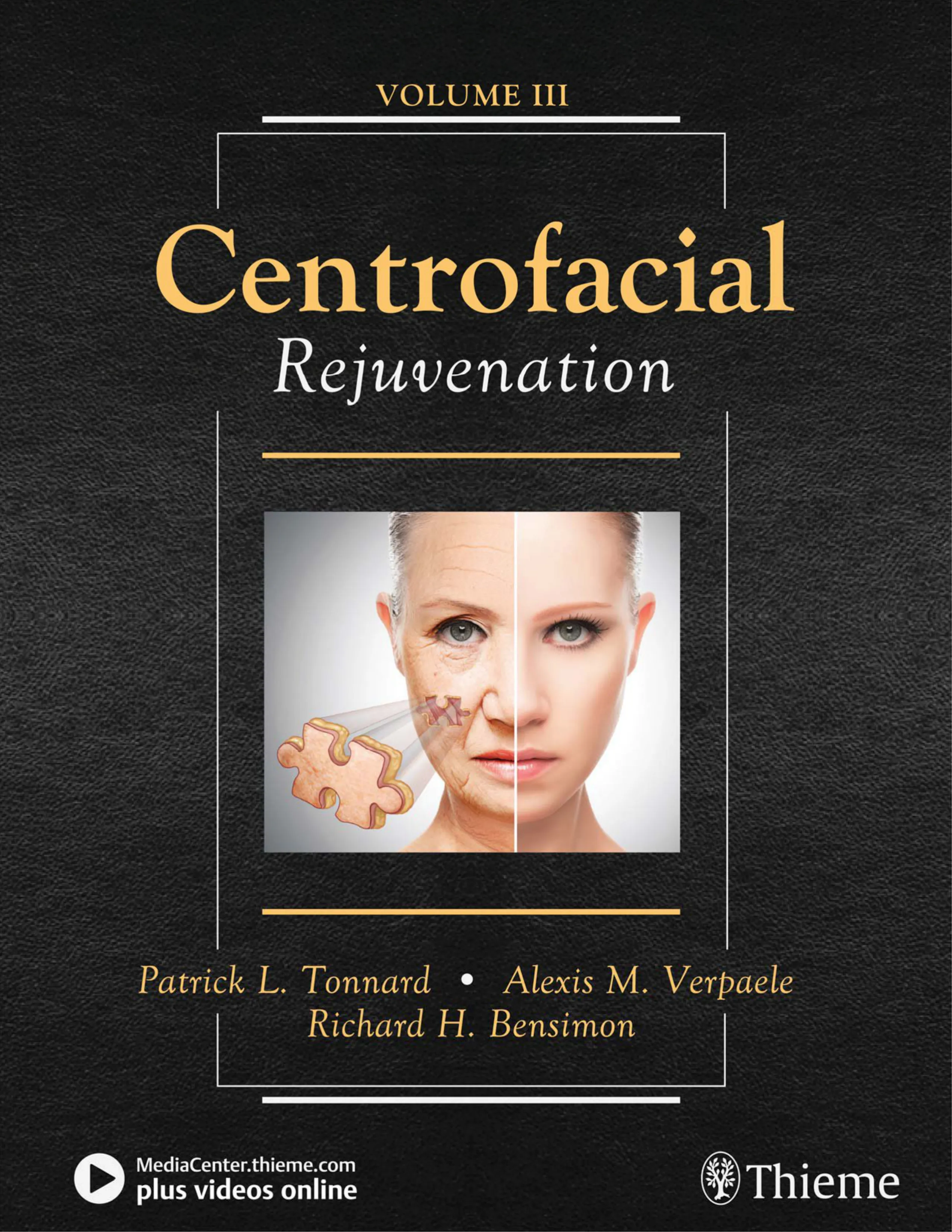
• Centrofacial Rejuvenation: Putting It All Together
Surgical Plan
The surgical treatment involved the following:
• Simple MACS lift
• Short-scar temporal lift
• Liposuction of the inferior part of the jowls
• Augmentation blepharoplasty
– Upper eyelid
Microfat grafting of medial infrabrow area (1 cc bilaterally)
– Lower eyelid
Transconjunctival fat removal of the medial and middle fat
compartment
Augmentation of malar area (4 cc of microfat on the right side
and 4.8 cc on the left side)
•
from the abdomen
– Malar: right, 4 cc; left, 4.8 cc
– SNIF, 21-gauge needle:
White roll, upper lip: 1 cc
White roll, lower lip: 1 cc
Philtral columns: 0.8 cc
– SNIF, 23-gauge needle:
Small nasolabial rhytids: 2 cc
The results are shown at 1 year and at 4 years postoperatively (Fig. 7-21). This
surgical rejuvenation procedure was a typical refreshing procedure, a conser-
vative correction of the surgical laxity in the peripheral part of the face and a
discrete correction of the volum e loss in the central part of the face. A natural
replenishm ent of the volum e of the face toward its appearance at the age of
24 years is evident. A stable correction of the orbitomalar groove was achieved
by the com bination of transconjunctival fat rem oval and m alar augm entation
procedure. At the age of 56 years (4 years postoperatively), the tired expression
seen in the preoperative pictures is still corrected.
• Fat Grafting: From Volume Restoration to Regenerative Cell Therapy
Fig. 7-21 A, At 24 years. B, Preoperatively at age 52 years. C, At 1 year postoperatively. D, At
4 years postoperatively.
B
A
D
C
• Centrofacial Rejuvenation: Putting It All Together
Fig. 7-21, cont’d Left, Preoperatively at age 52 years. Center, At 1 year postoperatively.
Right, At 4 years postoperatively.
F
E
G
H
I
J
• Fat Grafting: From Volume Restoration to Regenerative Cell Therapy
The patient in Fig. 7-22 presented for facial rejuvenation at 51 years of age. When
her appearance was compared with pictures taken at younger ages, the follow-
ing features were observed:
• Laxity of peripheral facial tissues, w ith temporal hooding, m ild jowling,
and submental skin laxity with obvious platysmal bands
• -
mainly manifests as deep nasolabial folds, a barcode at the upper lip, and
deep rhytids along the nasolabial and marionette lines.
A
B
C
Fig. 7-22 A, At age 19 years. B, At age 32 years. C, Preoperatively at age 51 years.
• Centrofacial Rejuvenation: Putting It All Together
Nonsurgical Plan
The nonsurgical treatment plan consisted of the following:
• A Retin-A skin care program
• Botulinum toxin in glabellar and frontal areas
Surgical Plan
The surgical treatment involved the following:
• Simple MACS lift
• Short-scar temporal lifting by galeapexy
• Upper and lower augmentation blepharoplasty
– Upper eyelid
Conservative skin resection
Microfat grafting of upper eyelid sulcus (right, 1.5 cc; left, 1.5 cc)
– Lower eyelid
Augmentation of malar area (4.5 cc of microfat per side)
Skin-pinch blepharoplasty
• SNIF, 23-gauge needle:
– Glabella: 0.5 cc
– Upper lip rhytids: 1.3 cc
– Nasolabial area: 2.2 cc
– Medial cheek rhytids: 1.8 cc
• SNIF, 21-gauge needle:
– White roll upper and lower lip: 2.2 cc
– Philtral columns: 0.4 cc
• Erbium-YAG laser resurfacing of upper lip and chin with a 5 mm
The 1.5- and 6-year postoperative results are shown (Fig. 7-23). The peripheral
laxity has been corrected by the surgical lifting procedures; correction of the
neck laxity are also evident.
The restoration of centrofacial volum es is apparent in the m alar volum e, cor-
-
ening of the facial features, and an obvious change in the emotional expression
from sadness toward an energetic vivacity. Although after 6 years a degree of
relapse of neck laxity has occurred, the centrofacial volumetric rejuvenation has
remained stable.
• Fat Grafting: From Volume Restoration to Regenerative Cell Therapy
Fig. 7-23 A, At 19 years. B, Preoperatively at age 51 years. C, At 18 m onths postoperatively.
D, At 6 years postoperatively.
B
A
D
C
• Centrofacial Rejuvenation: Putting It All Together
Fig. 7-23, cont’d E, At 32 years. F, Preoperatively at age 51 years. G, At 18 months postoperatively.
H, At 6 years postoperatively.
F
E
H
G
• Fat Grafting: From Volume Restoration to Regenerative Cell Therapy
The woman in Fig. 7-24 presented at 62 years of age for facial rejuvenation. She
had a combination of laxity in the peripheral part of the face, including the neck,
and the midface. Skin quality changes are seen in the frontal and perioral area.
-
tysma only.
Fig. 7-24 A, At age 28 years. B, Preoperatively at age 62 years.
B
A
• Centrofacial Rejuvenation: Putting It All Together
Nonsurgical Plan
The nonsurgical treatment plan consisted of a Retin-A skin care program.
Surgical Plan
The surgical treatment involved the following:
• Simple MACS lift with retroauricular incision
• Open neck procedure with platysmarrhaphy and partial transec-
tion of the platysma at the level of the hyoid
• Short-scar temporal lift
• Upper augmentation blepharoplasty (skin only, plus fat grafting)
• Lower transconjunctival blepharoplasty and skin-pinch excision
• Microfat grafting, 33 cc, harvested from the abdomen
– Infrabrow: 1 cc/side
– Malar: 8 cc/side
– Marionette grooves: 2 cc/side
– SNIF, 21-gauge needle:
White roll, upper and lower: 2.0 cc
Philtrum: 1.0 cc
– SNIF, 23-gauge needle:
Barcode (upper lip): 2 cc
Frontal wrinkles: 2.3 cc
Glabella: 0.7 cc
Cheek rhytids: 4 cc
• Erbium-YAG laser, frontal and perioral
• Fat Grafting: From Volume Restoration to Regenerative Cell Therapy
The 1-year postoperative result demonstrates the correction of the neck laxity
restored by correction of the jowling. This result could not have been obtained
without opening the neck and perform ing the anterior platysm arrhaphy. Cen-
-
viously. A nice reconstruction of the facial architecture is evident on the oblique
view, especially in the m alar area. Compared w ith the photograph of her ap-
pearance at 28 years, the rejuvenation is very natural, w ith replenishm ent of
the anterior cheek area is m arked. Correction of the frontal and glabellar lines
should be maintained with botulinum toxin, which she refused.
Fig. 7-25 Left, Preoperatively at age 62 years. Right, At 1 year postoperatively.
• Centrofacial Rejuvenation: Putting It All Together
Fig. 7-25, cont’d Left, Preoperatively at age 62 years. Right, At 1 year postoperatively.
• Fat Grafting: From Volume Restoration to Regenerative Cell Therapy
The patient in Fig. 7-26 presented at 65 years of age for facial rejuvenation and
body contouring. The following features were observed:
• Peripheral descent of facial features: temporal brow, cheeks, oral commis-
sures, jowls
• Neck laxity with dynamic platysmal bands and important skin laxity
Fig. 7-26 A, At 19 years. B, Preoperatively at 65 years.
B
A
Surgical Plan
The surgical treatment involved the following:
• Simple MACS lift with a posterior cervicoplasty
• Short-scar temporal lifting by galeapexy
• Upper augmentation blepharoplasty
– Upper eyelid
Conservative skin resection
Microfat grafting of upper eyelid sulcus (right, 2 cc; left, 2 cc)
– Lower eyelid
Augmentation of the malar area and tear trough with 8 cc of
microfat per side
• Microfat grafting of the nasolabial folds (2 cc bilaterally) with sub-
cision, and of the marionette grooves (2 cc bilaterally)
microfat per side.
• Centrofacial Rejuvenation: Putting It All Together
Fig. 7-27 Left, Preoperatively at age 65 years. Center, At 1 year postoperatively. Right, At 2 years
postoperatively.
The 1- and 2-year postoperative results are shown (Fig. 7-27). She has correction
of the peripheral laxity by the surgical lifting procedures, including correction of
the temporal hooding, lifting of the oral commissures, restoration of the jawline
were still present, amenable to treatment with botulinum toxin.
The midfacial aging is slightly undercorrected after 1 year. On the 2-year postop-
of the malar area is visible.
• Fat Grafting: From Volume Restoration to Regenerative Cell Therapy
The woman in Fig. 7-28 presented at 65 years of age for facial rejuvenation. She
had a m oderate sagging in the cervicom ental region with obvious jowling and
-
ent in the upper eyelids, the malar area, the nasolabial area, and the marionette
groove. Skin quality changes were mainly visible on the upper lip. The upper lip
-
m ilion border and the philtral colum ns. She had discrete bulging of the lower
eyelid fat pads as well as a marked eyelid-cheek junction.
Fig. 7-28 A, At age 28 years. B, Preoperatively at age 65 years.
B
A
• Centrofacial Rejuvenation: Putting It All Together
Nonsurgical Plan
The nonsurgical treatment plan consisted of a Retin-A skin care program
(started before laser treatment and restarted 6 weeks after).
Surgical Plan
The surgical treatment consisted of the following:
• Simple MACS lift
• Short-scar temporal lift
• Augmentation blepharoplasty
– Upper eyelid
Conservative skin resection
Microfat grafting of infrabrow area (1 cc bilaterally)
– Lower eyelid
Fat redraping lower blepharoplasty
Augmentation of malar area (5.8 cc of microfat per side)
• Microfat grafting, 22 cc, harvested from the abdomen
– Infrabrow: 1 cc bilaterally
– Malar: 5.8 cc/side
– Nasolabial groove: 1.7 cc/side
– Marionette grooves: 1.0 cc/side
– SNIF, 21-gauge needle:
White roll, upper lip: 1 cc
White roll, lower lip: 1 cc
Philtral columns: 0.8 cc
– SNIF, 23-gauge needle:
Barcode (upper lip): 0.8 cc
Sublabial groove: 0.8 cc
• Liplift (approximately 3 mm)
• Perioral Erbium-YAG laser resurfacing
• Fat Grafting: From Volume Restoration to Regenerative Cell Therapy
The postoperative results are shown at 2 and 6 years (Fig. 7-29). Aside from
the surgical correction of the sagginess in the peripheral part of the face (neck,
jowling, and temporal hooding), most of the facial rejuvenation comes from the
centrofacial work w ith volum e correction and perioral resurfacing. A natural
rejuvenation of the periorbital region is a notable result of the replenishm ent
of the upper eyelids, the volum ization of the m alar area w ith blending of the
eyelid-cheek junction, and shortening of the vertical height of the lower eyelid.
-
tral columns. The emotional expression of the face has transformed from a tired
look into a m ore vigorous, dynam ic look, even at the age of 72 years (6 years
postoperatively).
Fig. 7-29 Left, At 28 years. Right, At 2 years postoperatively.
• Centrofacial Rejuvenation: Putting It All Together
Fig. 7-29, cont’d Left, Preoperatively at age 65 years. Center, At 2 years postoperatively. Right, At
6 years postoperatively.
• Fat Grafting: From Volume Restoration to Regenerative Cell Therapy
The wom an in Fig. 7-30 requested facial rejuvenation at 65 years of age. Com -
pared w ith her appearance at age 24 years, she presented w ith the follow ing
the centrofacial area; full-face deterioration of the skin quality with the appear-
ance of wrinkles in the frontal area, crow’s-feet, and perioral area; and craqueléd
skin in the anterior neck.
Fig. 7-30 A, At age 24 years. B, Preoperatively at age 65 years.
B
A
• Centrofacial Rejuvenation: Putting It All Together
Surgical Plan
The surgical treatment involved the following:
• Simple MACS lift with a full preauricular and retroauricular inci-
sion for adequate neck skin redraping
• Submental liposuction
• Submental incision and subplatysmal fat resection with anterior
platysmaplasty
• Short-scar temporal lift
• Upper augmentation blepharoplasty with skin-only resection
• Lower skin-pinch blepharoplasty
• Microfat grafting, 62 cc, harvested from the abdomen
– Infrabrow: 2 cc/side
– Malar: 9.5 cc/side
– Marionette grooves: 2 cc/side
– SNIF, 21-gauge needle:
Upper white roll: 1.3 cc
Lower white roll: 1.0 cc
Philtrum: 0.4 cc
– SNIF, 23-gauge needle:
Frontal wrinkles: 3.4 cc
Glabella: 0.8 cc
Perioral wrinkles: 3.8 cc
– Nanofat neck: 17 cc
• Perioral Erbium-YAG laser resurfacing
• Fat Grafting: From Volume Restoration to Regenerative Cell Therapy
The result is shown 14 m onths postoperatively (Fig. 7-31). The surgical lifting
areas and the temporal hooding.
The central part of the face exhibits restoration of the facial volume in the perior-
bital, malar, and perioral area and dramatic improvement of the neck skin quality
by the nanofat injection. Despite already quite full upper eyelids preoperatively,
the crisp appearance of her upper eyelids and infrabrow region.
The eyelid-cheek junction has been blended by the malar augmentation, and the
projection of her cheekbone area has increased, resulting in a m ore triangular
shape of the face. In the perioral area, the vertical rhytids have been reduced and
Fig. 7-31 Left, At age 24 years. Right, At 14 months postoperatively.
• Centrofacial Rejuvenation: Putting It All Together
Fig. 7-31, cont’d Left, Preoperatively at age 65 years. Right, At 14 months postoperatively.
• Fat Grafting: From Volume Restoration to Regenerative Cell Therapy
The woman in Fig. 7-32 presented at 68 years of age for facial rejuvenation but
was not willing to undergo a surgical facelift procedure because of fear of a long
scar and prolonged recovery. She had three typical aspects of facial aging: sagging
the periorbital and perioral area, and skin quality changes on the whole surface
of the face (rhytids and pigmentation disorders), especially in the perioral area
and the cheeks. She underwent neurosurgery in the past for a bleeding aneu-
rysm, which was why she refused to undergo major facelift surgery. This made
only to minimal invasive techniques under local anesthesia.
Fig. 7-32 A, At age 25 years. B, Preoperatively at age 68 years.
B
A
• Centrofacial Rejuvenation: Putting It All Together
Nonsurgical Plan
The nonsurgical treatment plan consisted of a Retin-A skin care program
(started before laser treatment and restarted 6 weeks after).
Surgical Plan
The surgical treatment was done under local anesthesia and involved the
following:
• Upper augmentation blepharoplasty: excision of skin only and fat
grafting
• Microfat grafting, 40 cc, harvested from the abdomen
– Infrabrow: 3.0 cc bilaterally
– Malar: 5 cc/side
– Marionette groove: 2 cc
– Upper lip vermilion: 2 cc
– Lower lip vermilion: 2 cc
– Trepanation surface deformity in right temporal area (not visible
on photos): 9 cc
– SNIF, 21-gauge needle:
White roll, upper lip: 1.8 cc
White roll, lower lip: 0.8 cc
Philtrum: 0.5 cc
– SNIF, 23-gauge needle:
Barcode (upper lip): 5.25 cc
• Nanofat in the anterior neck area: 12 cc
• Microliposuction of the lower part of the jowls with a 2 mm Mer-
cedes cannula (2 cc/side)
• Liplift (approximately 4 mm)
• Full-facial Erbium-YAG laser resurfacing
• Fat Grafting: From Volume Restoration to Regenerative Cell Therapy
Fig. 7-33 A, At 25 years. B and D, At 1 year postoperatively. C, Preoperatively at age 68 years.
The results are presented 1 year postoperatively (Fig. 7-33). This case illustrates
the power of centrofacial rejuvenation, because no lifting procedure was per-
by altering the periorbital and perioral region. Compared with the picture taken
at age 25 years, replenishm ent of the upper eyelids, correction of the m alar
volum es with blending of the eyelid-cheek junction, shortening of the vertical
A
C
D
B
• Centrofacial Rejuvenation: Putting It All Together
E
F
G
H
Fig. 7-33, cont’d Left, Preoperatively at age 68 years. Right, At 1 year postoperatively.
height of the upper lip, and general improvement of the quality of the skin with
norm alization of the pigm entation and reduction of w rinkling are evident. A
comparison of the preoperative and postoperative appearance of the lips reveals
the change in vertical length and shape of the upper lip. The white roll borders
volum e augm entation by fat grafting, liplift, and skin Erbium -YAG laser resur-
lower part of the jowl.
• Fat Grafting: From Volume Restoration to Regenerative Cell Therapy
The 52-year-old man in Fig. 7-34 presented with premature aging; his main con-
cerns were about eyelid, facial, and neck laxity and frontal rhytids. In addition
to the moderate sagging in the peripheral parts of the face and neck, there is an
Fig. 7-34 A, At age 24 years. B, Preoperatively at age 52 years.
B
A
• Centrofacial Rejuvenation: Putting It All Together
Nonsurgical Plan
The nonsurgical plan consisted of botulinum toxin in the glabella and
frontal area.
Surgical Plan
The surgical treatment involved the following:
• Simple MACS lift
• Submental liposculpture
• Subplatysmal fat resection and anterior platysmarrhaphy
• Augmentation blepharoplasty
– Upper eyelid: conservative skin resection
– Lower eyelid: microfat grafting (malar and tear trough) and skin-
pinch resection
• Microfat grafting, 220 cc, harvested from the abdomen
– Tear trough and orbitomalar groove: 3 cc bilaterally
– Malar: 4 cc bilaterally
– Nasolabial grooves: 4 cc bilaterally
– Marionette grooves: 2 cc bilaterally
The 1.5-year postoperative result is shown (Fig. 7-35). There is a marked change
in the em otional expression of the m an’s face, from sad and fatigued to cheer-
ful, healthy, and energetic. Correction of the peripheral sagging is evident, with
correct positioning of the sideburn and the natural hairless area in front of the
tragus despite the retrotragal incision. Most striking is the change in the inten-
sity of the gaze resulting from the augmentation blepharoplasty. This produced
a natural replenishment of the upper eyelid and a softening of the orbitomalar
groove with blending of the eyelid-cheek junction.
• Fat Grafting: From Volume Restoration to Regenerative Cell Therapy
Fig. 7-35 A, At 24 years. B, Preoperatively at age 52 years. C, At 1.5 year postoperatively.
B
C
A
• Centrofacial Rejuvenation: Putting It All Together
D
E
F
G
Fig. 7-35, cont’d Left, Preoperatively at age 52 years. Right, At 1.5 year postoperatively.
• Fat Grafting: From Volume Restoration to Regenerative Cell Therapy
The m an in Fig. 7-36 presented at the age of 71 years for facial rejuvenation.
-
groove. Skin quality changes were visible as spotty pigmentation and keratotic
changes on the whole facial skin surface. The upper lip also had a lengthening
marked eyelid-cheek junction.
Fig. 7-36 A, At age 32. B, Preoperatively at age 71.
B
A
Nonsurgical Plan
The nonsurgical treatment plan consisted of a Retin-A skin care program.
• Centrofacial Rejuvenation: Putting It All Together
Surgical Plan
The surgical treatment involved the following:
• Simple MACS lift
• Short-scar temporal lift
• Submental incision and subplatysmal fat resection with anterior
platysmaplasty
• Augmentation blepharoplasty
– Upper eyelid
Resection of skin only
– Lower eyelid
Augmentation of malar area (14 cc of microfat per side)
•
from the abdomen
– Malar: 14 cc/side
– Nasolabial groove: 3 cc/side, plus 0.5 cc/side after subcision
– Marionette grooves: 2.5 cc/side
• Liplift (approximately 3 mm)
The postoperative results are shown at 2 years at the age of 73 years (Fig. 7-37).
Aside from the surgical correction of the sagginess in the peripheral part of the
face (neck, jowling, and temporal hooding), most of the facial rejuvenation comes
-
ural rejuvenation of the periorbital region has resulted from the volum ization
of the malar area with subsequent triangularization of the facial shape and the
reduction of shadowing in the face, especially in the nasolabial folds. A discrete
reduction of the vertical height of the upper lip is evident. The change in emo-
tional expression of the face from a tired look into a more vigorous dynamic look,
even at the age of 74 years (2 years postoperatively), is remarkable.
• Fat Grafting: From Volume Restoration to Regenerative Cell Therapy
Fig. 7-37 Left, Preoperatively at age 71 years. Right, At 2 years postoperatively.
• Centrofacial Rejuvenation: Putting It All Together
The 58-year-old m an in Fig. 7-38 presented 10 years after a facelift that was
obvious in the periorbital area. The oblique view revealed a triangular aspect to
his face, with a wide base below and a more narrow aspect above.
Surgical Plan
The surgical treatment involved the following:
• Secondary simple MACS lift
• Submental liposculpture
• Short-scar temporal lift
• Microfat grafting, 30 cc, harvested from the abdomen
– Infrabrow: 1.2 cc bilaterally
– Malar: 10 cc bilaterally
– Nasolabial grooves: 2 cc, plus 0.5 cc after subcision
– Marionette grooves: 2 cc bilaterally
The 1- and 5-year postoperative results are shown (Fig. 7-38). Stable volumetric
enhancement of the periorbital area has been achieved, and the skin quality has
improved (less m elasm a). The facial proportions have been transform ed from
an upright triangle into the inversed triangle of youth, a result of the enhanced
-
auricular scar with conservation of the position of the sideburn.
Fig. 7-38 Left, Preoperatively at age 58 years. Center, At 1 year postoperatively. Right, At 5 years
postoperatively.
Continued
• Fat Grafting: From Volume Restoration to Regenerative Cell Therapy
REFERENCE
1. Yarbus AL. Eye Movements and Vision. New York: Plenum Press, 1967.
Fig. 7-38, cont’d Left, Preoperatively at age 58 years. Center, At 1 year postoperatively. Right, At 5
years postoperatively.
The Place of
Toxins and Fillers in
the Modern Plastic
Surgery Practice
Part II
Beauty is only skin deep,
but ugly goes clean to the bone.
DOROTHY PARKER
(Trang không có chữ)
Chapter
5
The MACS-Lift
Short-Scar Face Lift
Technical and Strategic
Considerations
Mark Laurence Jewell • Mark Laurence Jewell
Chapter
8
Midface Volume
Rejuvenation
With Fillers
F. Javier Beut • Glenn W. Jelks • Christopher C. Surek
Jerome Lamb
. . . thou know ’st the mask of night is on my face.
W ILLIAM SHAKESPEARE
Chapter Videos
8-1 Layered Midface Anatomy (From Surek CC, Beut J, Stephens R, et al. Pertinent
anatomy and analysis for midface volumizing procedures. Plast Reconstr Surg
135(5):818e-829e, 2015.)
8-2 Midface Injection Analysis (From Surek CC, Beut J, Stephens R, et al. Pertinent
anatomy and analysis for midface volumizing procedures. Plast Reconstr Surg
135(5):818e-829e, 2015.)
8-3 Beut Type 1 (From Surek C, Beut J, Stephens R, et al. Volumizing viaducts of the
midface. Aesthet Surg J 35:121-134, 2015.)
8-4 Beut Type 2 (From Surek C, Beut J, Stephens R, et al. Volumizing viaducts of the
midface. Aesthet Surg J 35:121-134, 2015.)
8-5 Beut Type 3 (From Surek C, Beut J, Stephens R, et al. Volumizing viaducts of the
midface. Aesthet Surg J 35:121-134, 2015.)
8-6 Beut Type 4 (From Surek C, Beut J, Stephens R, et al. Volumizing viaducts of the
midface. Aesthet Surg J 35:121-134, 2015.)
8-7 Dynamic Midface Rejuvenation Summary (From Surek CC, Beut J, Stephens R,
et al. Pertinent anatomy and analysis for midface volumizing procedures. Plast Reconstr
Surg 135(5):818e-829e, 2015.)
Additional procedural video and author discussion of technique can be
found on Media Center.
• Midface Volume Rejuvenation With Fillers
This chapter examines the layered anatomic array of the midface and translates
these anatom ic principles into consistent m idface injection analysis and tech-
nique. The cadaver dissections, video dem onstrations, and three-dim ensional
patient photography provide injectors with a comprehensive armamentarium to
deliver accurate and reproducible results in m idface volume rejuvenation with
INDICATIONS AND CONTRAINDICATIONS
Primary indications for midface volumizing procedures include generalized facial
-
plete history and physical examination, documentation of drug allergies, and a
listing of the patient’s current m edications. Prophylactic m edications such as
antiherpetics, medications to decrease bruising, and a short course of postpro-
cedure antibiotics need to be discussed during the consultation.
Relative contraindications to volumizing procedures include but are not limited
to vascular malformations, trigeminal neuralgia, prior silicone injections, and a
documented history of allergic reaction. Patients with autoimmune skin disease
or history of excessive scarring should be examined carefully.
requires a stereotaxic anatomic knowledge of the vasculature, ligamentous sup-
port, soft tissue spaces, and fat compartments of the face.
The aged appearance of the midface is the result of multifactorial processes. Val
Lambros1 painted a great analogy of the aging midface when he wrote, “the mid-
face behaves like a tectonic plate sliding down the face to form the nasolabial
fold, leaving in its wake the nasojugal groove and the exposed and dim inished
malar eminence.” Beginning at the skeletal level, Kahn and Shaw 2 demonstrated
rotational changes of the orbit along with posterior retrusion and enlargement
of the piriform aperture. Over areas of bony support and the m asseter, the re-
taining ligaments of the face as described by Furnas3 prevent large-scale ptosis
• The Place of Toxins and Fillers in the Modern Plastic Surgery Practice
of the midface, chin, and lateral face. With age, these retaining ligaments become
more vertically oriented. Lambros1 postulated that midface changes were largely
volume related and not ptosis dependent. This has become accepted as the new
paradigm guiding midface rejuvenation. Furthermore, the fat of the face has been
-
ments are intimate with the mimetic muscles and neurovascular structures and
4
fat compartments are separated by vascularized septae that are ligamentous in
nature over the bony and masseteric supporting structures. Based upon adipo-
cyte size, fat within the deep compartm ents is postulated to atrophy with age,
5
The deep compartments of the midface contain three relevant anatomic spaces.
Anatomic spaces are areolar in nature and are devoid of crossing neurovascular
structures and ligaments.6-7
Volumization procedures should focus on reestablishing support in the “pillars
-
animation will assist the injector in aesthetic visualization of a patient’s face in
youth.
Prezygomatic Space
First described by Bryan Mendelson,6 the prezygomatic space lies between the
orbital retaining ligam ent and the zygom atic retaining ligam ents (Fig. 8-1 and
Video 8-1). The space extends laterally in a cephalic direction to the lateral or-
bital thickening. The space is enveloped by a capsule analogous to the SMAS
space resides the preperiosteal fat resting on the maxilla (Fig. 8-3). This is a dis-
The SOOF is a loosely oriented fat pad residing between the prezygomatic space
capsule and the posterior surface of the orbicularis muscle (Fig. 8-4).
• Midface Volume Rejuvenation With Fillers
Fig. 8-1 The depth of anatomic layers in the upper midface. The infraorbital fat compartment lies
-
cles. The prezygomatic space is highlighted in blue. (Copyright Levant Efe, courtesy Jerome Lamb.)
Fig. 8-2
to the prezygomatic space capsule. The prezygomatic space capsule was percutaneously stained
with m ethylene blue before dissection. (From Surek CC, Beut J, Stephens R, et al. Pertinent anat-
omy and analysis for midface volumizing procedures. Plast Reconstr Surg 135:818e-829e, 2015.)
• The Place of Toxins and Fillers in the Modern Plastic Surgery Practice
Fig. 8-3 Cadaver dissections. A, The preperiosteal fat pad has been percutaneously dyed with
The palpebral branch of the infraorbital artery is marked with arrows. B, The preperiosteal fat pad
(PPF) (NLF) is la-
beled for reference. (From Surek CC, Beut J, Stephens R, et al. Pertinent anatomy and analysis for
midface volumizing procedures. Plast Reconstr Surg 135:818e-829e, 2015.)
B
A
Fig. 8-4 Bird’s-eye view of a cadaver dissection. The layers of the upper midface are labeled. (SOOF,
Suborbicularis oculi fat.) (From Surek CC, Beut J, Stephens R, et al. Pertinent anatomy and analysis
for m idface volum izing procedures. Plast Reconstr Surg 135:818e-829e, 2015.)
• Midface Volume Rejuvenation With Fillers
to the orbicularis oculi m uscle. We believe this is the corollary to the “m alar”
fat pad whose traditional description refers to the fat pad above the orbicularis
m uscle. Therefore distinction m ust be m ade regarding this published m alar or
from its caudal counterparts by the cutaneous insertion of the zygom atico-
cutaneous ligam ents (see Fig. 8-1). This compartm ent is believed to have poor
lymphatic drainage, and injection into this region can be problematic, potentially
resulting in an iatrogenic malar mound. Clinically we have seen retained lymph-
edema in this compartment after injection (Figs. 8-5 and 8-6; see also Fig. 8-1).
Fig. 8-5 Iatrogenic m alar m ound resulting from
compartm ent. Cutaneous insertions of the orbito-
m alar ligam ent superiorly and the zygom atico-
cutaneous ligam ents inferiorly are evident. (Copy-
right Levant Efe, courtesy Jerom e Lam b.)
Fig. 8-6 Infraorbital fat compartm ent. (From Surek CC, Beut J, Stephens R, et al. Pertinent anat-
omy and analysis for midface volumizing procedures. Plast Reconstr Surg 135:818e-829e, 2015.)
• The Place of Toxins and Fillers in the Modern Plastic Surgery Practice
Regarding the prezygomatic space and its role in midface injections, an impor-
tant point to consider is the dynamic gliding nature of the midface during facial
-
tures glide over it. In regards to the intim ate vasculature, authors describe the
-
graphic landmark for the infraorbital neurovascular bundle. An ascending branch
percentage of angular arteries that have detoured from their norm al course;
injections (see Fig. 8-3).
Prema xillary and Deep Piriform Spaces, Deep Medial Cheek
Compartment
The deep medial cheek compartment overlies the maxilla and lies lateral to the
deep piriform space (Figs. 8-7 and 8-8). The m ore lateral component of deep
Deep lateral injections in this region can be troublesome, because material can
descend caudally into the buccal space (Fig. 8-9). We recommend more medially
oriented injections in the deep piriform space or the m edial component of the
deep to the SMAS (see Fig. 8-8). The lateral boundary of the premaxillary space
contains the angular vein.
Fig. 8-7 The deep medial cheek fat compartment. (From Surek CC, Beut J, Stephens R, et al. Perti-
nent anatomy and analysis for midface volumizing procedures. Plast Reconstr Surg 135:818e-829e,
2015.)
• Midface Volume Rejuvenation With Fillers
Fig. 8-9 A,
cheek fat and medial suborbicularis oculi fat (SOOF), stained with methylene blue. The zygomaticus
major (ZM), levator anguli oris (LAO), levator labii superioris (LLS), and levator labii superioris alaeque
nasi (LLSAN)
the lateral SOOF overlying the preperiosteal fat compartment. The orbicularis has been removed to
B,
retracted. The preperiosteal fat is stained with methylene blue, and remnants of lateral SOOF are
noted resting on the capsule of the preperiosteal fat. The loose areolar consistency of the deep me-
dial cheek fat is noted lateral to the levator anguli oris (cut). A cavernous communication into the
buccal recess denotes a deep injection adverse-event zone. (IO, Infraorbital nerve.) (From Surek CC,
Beut J, Stephens R, et al. Pertinent anatomy and analysis for midface volumizing procedures. Plast
Reconstr Surg 135:818e-829e, 2015.)
B
A
Fig. 8-8 The deep piriform space and important adjacent structures. (Copyright James Vargo.)
Prezygomatic
space
Deep piriform
space
Premaxillary
space
Levator labii
superioris
Depressor
septi nasi
Deep medial
cheek fat
Angular artery
Zygomaticus
minor
Zygomaticus
major
Orbicularis oris
Orbicularis oris
• The Place of Toxins and Fillers in the Modern Plastic Surgery Practice
Both the deep piriform and the premaxillary spaces are devoid of crossing neu-
rovascular and ligam ent structures. The cephalic extension of both spaces lies
deep to the orbicularis oculi muscle and is superiorly bounded by the tear trough
ligament.8 The angular artery is tethered to undersurface of the levator muscu-
lature and does not reside in the space (Fig. 8-10). Medial m axillary recession
with age likely enlarges the deep piriform space, making it a plausible location
space. In addition, volumizing of this space provides support for the draping lip
Fig. 8-10 The deep piriform space is cradled m edially by the piriform aperture and depressor
nasale. The angular artery courses between the space and the deep m edial cheek fat compart-
the roof of the space. Postorbicularis oris fat (POOF) is stained green.
• Midface Volume Rejuvenation With Fillers
Fig. 8-11 Fat compartments of the face. A, Nasolabial. B, (From Surek CC, Beut
J, Stephens R, et al. Pertinent anatomy and analysis for m idface volum izing procedures. Plast Re-
constr Surg 135:818e-829e, 2015.)
B
A
septae divide these compartments. These septae can be readily felt with the end
-
cial fat compartments glide upward with facial expression. Volume placed within
these compartm ents can restore turgor to the skin. Consideration is also given
that can alter cheek convexity.
• The Place of Toxins and Fillers in the Modern Plastic Surgery Practice
PREOPERATIVE PLANNING AND PREPARATION
Many surgeons rely on their “aesthetic eyeball” to analyze and plan volumizing
approach using an anatomically inspired topographic analysis (Video 8-2). This
m ethod divides the cheek into “target areas” and “injection caution zones” us-
analysis videos accompanying this chapter for a m ore detailed description of
this planning approach.
Fig. 8-12 Midface analysis. A, Step 1. The malar equator (turquoise line) bisects the midface and
anatomically correlates with the zygomatico-cutaneous ligaments. B, Step 2. The medial vertical
reference mark (blue line) anatom ically corresponds with the palpebral branch of the infraorbital
artery and the lip elevators. C, Step 3. The lateral vertical reference m ark (green line) anatomically
corresponds with the zygomaticus muscle origin and retaining ligament. This marking divides the
“immobile” lateral face from the “mobile” anterior face.
B
A
C
• Midface Volume Rejuvenation With Fillers
D
E
F
Fig. 8-12, cont’d D, Step 4. The upper malar partition (red dotted line) from the medial canthus to
the predicted origin of the zygom aticus major m uscle. E, Step 5. The lower m alar partition (lower
red dotted line) from the corner of the m outh to the predicted origin of the zygom aticus m ajor.
F, Sum m ary of an anatom ically correlated m idface analysis. (IOF, Infraorbital fat compartm ent;
LAO, levator anguli oris; LLS, levator labii superioris; LMP, lower malar partition; LVR, lateral ver-
tical reference; ME, m alar equator; MVR, m edial vertical reference; OML, orbitom alar ligam ent;
OO, orbicularis oris; SIRM, safety inferior orbital rim margin; UMP, upper m alar partition.) (Copy-
right Levant Efe, courtesy Jerome Lamb.)
OML
OO
IOF
LAO
OO
LLS
ME
UMP
SIRM
LMP
MVR
LVR
Zygomaticus
major
Zygomaticus
minor
Buccal
fat pad
Maxillary ligaments
Deep
medial
cheek fat
compartment
Angular
vein
• The Place of Toxins and Fillers in the Modern Plastic Surgery Practice
Malar Equator
the tragus. This line is the malar equator and anatomically corresponds with the
zygomatico-cutaneous retaining ligaments and upper labial buccal sulcus. These
anatomic landmarks distinctly separate the upper midface from the lower mid-
face (see Fig. 8-12, A).
Medial Vertical Reference
A vertical line is draw n from the m edial corneal lim bus to the corner of the
mouth to form the medial vertical reference. Anatomically this line corresponds
with the ascending branch of the infraorbital artery in the upper m idface and
the lip elevators in the lower midface (see Fig. 8-12, B).
Lateral Vertical Reference
The lateral vertical reference is a vertical line drawn from the lateral orbital rim
to the angle of the mandible. Anatomically this line corresponds with the origin
of the zygomaticus major muscle at the anterior masseteric border and divides
the lateral “immobile” face from the anterior “mobile face” (see Fig. 8-12, C).
Upper Malar Partition
The upper malar partition is an oblique line drawn from the medial canthus to
the predicted origin of the zygomaticus major muscle (see Fig. 8-12, D).
Lower Malar Partition
The lower malar partition is an oblique line drawn from the corner of the mouth
to the predicted origin of the zygomaticus major (see Fig. 8-12, E).
Injection Target Areas
1. Medial malar target area: Outlines the anatomic location of the deep me-
dial cheek fat compartment and deep piriform space
2. Middle m alar target area: Outlines the anatom ic location of the m edial
3. Lateral m alar target area: Outlines the anatom ic location of the lateral
SOOF and the gliding prezygomatic space