

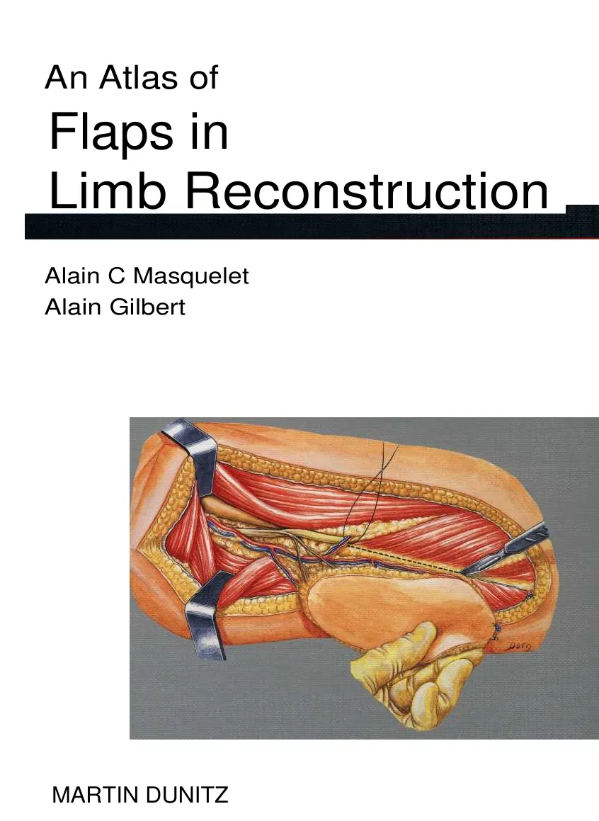

An Atlas of
Flaps in
Limb Reconstruction
Alain C Masquelet
Alain Gilbert
MARTIN DUNITZ
An Atlas of
~
Flaps in
Limb Reconstruction
Alain C Masquelet, MD
Professor of Orthopaedic and Reconstructive Surgery, University of Paris
Consultant Orthopaedic Surgeon, Hbpital Avicenne, Bobigny
Alain Gilbert, MD
Professor of Orthopaedic Surgery, University of Paris
Consultant Orthopaedic Surgeon, Institut de la Main, Paris
With illustrations by Leon Dorn
Alain C Masquelet and Alain Gllbert 1995
Artwork and design © Martin Dunitz Ltd 1995 (except where otherwise indicated)
MIA published in the United Kingdom in 1995
by Martin Dunitz Ltd, The Livery House, 7-9 Pratt Street, London NWI OAE
Reprinted 1997
All rights reserved. No part of this publication may be reproduced, stored in a retrieval
system, or transmitted, in any form or by any means, without the prior permission of
the publisher.
A CIP record for this book is available from the British Library.
ISBN 1 85317 172 7
A 35-mtn colour slide set collection based on 200 images in this hook is also available.
Composition by Scribe Design, Gillingham, Kent, UK
Colour origination by Imago Publishing Ltd
Manufactured by Imago Publishing Ltd
Printed and bound in Singapore
Foreword, by Raoul Tubiana
Historical note
viii
Preface
ix
Acknowledgements
x
1 Historical review
1
2 Vascular anatomy
9
Contents
Vascularization of the skin of the limbs and
l
ifi
ti
f
ki fls 10
casscaon osn a
Vastus lateralis flap
96
Tensor fasciae latae myocutaneous flap
101
Gracilis flap
104
Biceps femoris flap (long head)
107
Lateral and distal thigh flap
111
Osteoperiosteal flap (from the femur)
115
Leg
118
Fasciocutaneous saphenous artery flap
118
Gastrocnemius flaps
122
Soleus flap
130
Flexor flaps
138
Extensor flaps
143
Peroneus flap
146
Lateral supramalleolar flap
148
Neurocutaneous flaps
158
Vascularized fibula transfer
162
Foot
169
Dorsalis pedis flap
169
Extensor digitorum brevis flap
177
The pelvic girdle
252
The knee
255
The middle third of the leg
255
The distal third of the leg
257
Foot and ankle
258
Bibliography
261
Index
267
Medial plantar flap
183
Vascular anatomy of muscles: classification
18
Medialis pedis flap
189
The venous drainage in the reverse arterial
Abductor hallucis flap
193
flow flap
24
Second toe transfer
196
Upper limb: tree of flaps and blood supply
28
Lower limb: tree of flaps and blood supply
36
6 Transfers from the thorax,
3 Preoperative planning
45
abdomen and pelvis
203
Shoulder girdle
204
4 Transfers from the upper limb
51
Latissimus dorsi flap
206
Arm and forearm
52
Serratus anterior flap
211
Posterior arm flap
52
Scapular and parascapular flaps
215
Lateral arm flap
55
Abdomen and pelvis
219
Brachioradialis flap
62
Rectus abdominis flap
220
Forearm flaps
66
Groin flap
223
Posterior interosseous flap
72
Vascularized iliac crest transfer
226
Distal ulnar artery flap
78
Osteomyoperiosteal flap
231
Hand and digits
83
Gluteus maximus flap
233
Kite flap
83
Island flaps from the digits
88
7 Indications for pedicled island
Intermetacarpal and dorsocommissural
flaps
241
flaps
93
Pedicled flaps of the upper extremity
244
5 Transfers from the lower limb
95
Tissue losses from the shoulder
244
Tissue losses from the arm and hand
246
Thigh
96
Pedicled flaps of the lower extremity
251
(Trang không có chữ)
Foreword
Several factors explain the rapid advance in the
domain of flaps over the last 20 years: this evolution
was driven by the development of reconstructioe
surgery and the increasing demands of surgeons to
ensure the optimal quality of care, to have a reliable
technique and to reduce the number of stages and the
overall length of treatment.
A major step forward was taken when Bakamjian
(1965) and McGregor and Jackson (1972) defined
flaps based on a known axial blood supply: these are
increasingly popular, since they allow a more reliable
single stage procedure than the older flaps which
relied on random blood supply and were subject to
the limitations of length-to-breadth ratios. In fact, axial
pattern flaps had been used before by John Wood,
who described the first groin flap in 1863; and by
Dunham in 1882, Monks in 1898 and Esser in 1915,
who used island flaps which could be taken from
a
distance when based on a vascular pedicle.
Ombedanne reported in 1906 a pectoralis minor flap
for breast reconstruction. Tansini (1896) and Este in
1912
used latissimus dorsi
myocutaneous flaps.
However, all these techniques were forgotten and
only recently rediscovered.
Likewise, studies of skin blood supply are not new.
We owe a particular debt to pioneers in this field such
as Manchot (1989) and Salmon (1936) whose exten-
sive works have remained tmrecognized for so long.
The introduction of microsurgical techniques has
been a turning point in the field of reconstructive
surgery and has led plastic, orthopaedic and hand
surgeons to a deeper study of the vascular and nervous
topography, with emphasis on its relationship to skin,
muscle and skeleton. This knowledge had obvious
repercussions on the design of many new flaps: myocu-
taneous, fasciocutaneous and osteocutaneous flaps,
reversed arterial flow flaps, free skin flaps and free
composite flaps. Reliable microvascular surgery now
allows any composite tissue with an identifiable vascu-
lar pedicle to be transferred to a new recipient site.
With all the new procedures of skin cover and
tissue transfer, surgeons now have an arsenal of
techniques to cover all needs for reconstruction in the
limbs. To choose the right flap for each defect is often
the main problem facing them.
It is for me a real pleasure to have been invited by
Alain Masquelet and Alain Gilbert to write a Foreword
for their book because I had the privilege to follow,
in the course of more than 15 years, their anatomical
research and their clinical work. Not only were they
the first to describe some of the new flaps and tissue
transfers published in this book, but they also
organized instructional courses on flaps, undertaken
by hundreds of young surgeons, in the microsurgery
laboratory. Their teaching, reflected in this book,
gives a comprehensive classification of skin flaps and
of the vascular supply of the muscles in the upper and
lower limbs. They emphasize the role of secondary
arteries, the skin arteries and the dominant pedicles
to the muscles, so important in flaps surgery. Their
original description of `trees of flaps' in the upper and
lower limbs, where flaps are considered as leaves at
the extremities of small arterial branches along the
course of the main trunk, is particularly appealing.
The beautiful illustrations by Leon Dorn allow one
to follow the surgical technique for each flap,
described step by step.
The most important chapter of the book, and
probably the most difficult topic, concerns the
indications. The vascular anatomy suggests that most
tissue losses at the level of the extremities may be
covered with a cutaneous flap or a neighbouring
muscular flap. Free flaps can be extremely useful in
rather infrequent circumstances where other types of
one stage flaps are not available. The authors' prefer-
ence is to use as far as possible a one stage pedicled
island flap, which is more reliable than any other
flaps. These can be fasciocutaneous or muscular.
Fasciocutaneous flaps are preferentially used to resur-
face mobile skin areas and find their best indications
in the upper extremity, where few muscles can be
sacrificed without functional detriment. Muscle flaps
are used for the coverage of exposed bone, the filling
of cavities and the treatment of bone infection. They
are preferentially indicated in the lower extremity
where numerous muscle flaps can be elevated
without impairment.
Raoul Tubiana
Historical note
'Flaps Workshop' at the Amphitheatre d'Anatomie des Hopitaux de Paris: a painting
by Leroy Garioud (7 x 0.8 m, oil on canvas), photograph by courtesy of Gilles Puech.
Leon Dorn (on the left) and his pupil (on the right) are drawing the main steps of a
surgical procedure as demonstrated by the dissection: all the techniques in this book
have been illustrated by this method. The illustration behind the demonstrator
represents the dissection room at the end of the nineteenth century: note the old
fountain for washing hands.
The Amphitheatre was founded in 1830 and was
initially devoted to the teaching of anatomy and to
anatomical studies that were performed by the physi-
cians of the Hopitaux de 1'Assistance Publique de
Paris. The institution is now called the Ecole de
Chirurgie des Hopitaux de Paris: old building struc-
tures have been removed and the recently built
complex comprises a dissection room for anatomical
researches and the training of residents; an operating
theatre
devoted to experimental surgery; and a
microsurgery laboratory, where the first teaching
in France of microsurgical techniques was organized
by Alain Gilbert in 1977. Every year numerous
courses on theory and practice in all surgical special-
ties are conducted; in particular, there is a course on
flaps.
Preface
Over the last 20 years, the surgery of flaps has under-
gone a great deal of development. Initiated by plastic
surgeons, it was at first considered with suspicion by
surgeons of other specialties, but nowadays one can
assert that all surgeons need to utilize tissue trans-
fers. Many recent advances in surgery are due to the
possibility
of
mobilizing
vascularized tissue to
restore function or to repair a large defect. 'rhe field
of application of flaps has defined a new and non-
academic specialty, reconstructive surgery, i.e. the
surgery of loss of substance.
The rapid development of surgical repair proce-
dures has led to a true revolution in the approach to
the repair of compound defects. The repair of soft
tissue has become a prerequisite for the reconstruc-
tion of underlying structures such as bone, joint
tendons and nerves. In some specific fields such as
surgery of the hand, the routine utilization of flaps
has permitted the treatment of multiple lesions using
one-stage repair.
Orthopaedic surgery has gained much from the use
of coverage techniques: the prognosis for open
fractures has been completely transformed. The treat-
ment of pressure sores, the management of deglov-
ing lesions of the foot, and the excision of tumours
are now routinely associated with flap techniques.
The reliability of the procedures allows ambitious
reconstructive surgery of the limbs using the combi-
nation of prosthetic implants and bone allografts.
The surgery of flaps and tissue transfers has empha-
sized the importance of anatomy and the need for a
revision of the instruction of residents.
The aim of this book is limited. Obviously it is not
a treatise on reconstructive surgery, which would
involve the consideration of indications in specific
pathologies. We have collected in this book all the
basic procedures for flap surgery that we have used
clinically for more than 15 years. All the techniques
presented have been repeated and promoted through
personal dissections and the educational courses that
we organize twice a year in Paris. As we are both
basically orthopaedic surgeons, we have restricted the
field of applications to the limbs. Limbs are commonly
involved in trauma injuries. Great differences have
appeared in recent years between the upper and the
lower extremities. The physiology, pathology and
principles of surgical treatment are specific to each
limb. Even the use of flaps is quite different.
However, one should keep in mind that flaps and
other tissue transfers are no more than techniques. In
order to be applied effectively, techniques should be
integrated in a general plan according to the pathology
Alain Alasquelet
Alain Gilbert
Acknowledgements
The painting by Leroy Garioud on page viii is repro-
duced by kind permission of the artist and photo-
grapher.
Figure 3 on page 3 is reproduced by kind permission
of the Royal College of Surgeons of England, London.
Figure 4 on page 4 is reproduced by kind permis-
sion of Springer-Verlag, Berlin.
Pictures E a-d on pages 15-17 are adapted from
BGH Lamberty and GC Cormack, The arterial anatomy
of upper-limb skin flaps, in A Gilbert, AC Masquelet,
VR Hentz (eds), Pedicle Flaps of the Upper Limb
(Martin Dunitz, London, and Little, Brown, Boston,
1992;
first
published by Expansion Scientifique
Frangaise, Paris, 1990), with kind permission of the
authors.
Pictures A-D on pages 26-7 are adapted from JTK
Wee, Venous flow in the distally pedicled radial
forearm flap, in A Gilbert, AC Masquelet, VR Hentz
(eds),
Pedicle Flaps of the Upper Limb
(Martin
Dunitz, London, and Little, Brown, Boston, 1992; first
published by Expansion Scientifique Francuise, Paris,
1990), with kind permission of the author.
Historical
review
Historical review
Introduction
The tissue defect has always been a challenge for the
reconstructive surgeon- The first systematic proce-
dure using a flap for repairing a mutilated nose in an
adult female is attributed to Susruta, who lived in the
sixth or seventh century BC (see Figure 1). More
recently (four centuries ago!) the Italian surgeon
Gaspare Tagliacozzi described a delayed flap taken
from the arm and also used for reconstruction of the
nose (Figure 2).
We have come to forget all the surgical attempts
performed at the end of the nineteenth century and
in the early twentieth century, which were not always
reported in the literature; but in many ways, the
extraordinary advances in reconstructive surgery, and
particularly the surgery of flaps, are a rediscovery. The
concepts and the technical and anatomical knowledge
that form the basis of flap surgery were formulated
before World War 11, often as offshoots of anaesthe-
sia.
We may be astonished by the discrepancy
between the sum knowledge available in the early
twentieth century and the applications that have been
performed in the past 20 years.
We believe that three concomitant axes of devel-
opment can be considered from a historical point of
view:
I
The empirical development of surgical concepts.
2
Anatomical knowledge.
i
The technical achievements of vascular anasto-
moscs and the use of the binocular microscope.
Figure 1
Plastic surgery on the nose using a pedicled flap from the
patient's forehead. This procedure of rhinoplasty is probably
at least 2000 years old in India (from The Gentleman's
Magazine of Calcutta, October 1794).
Figure 2
I llustrations from De curtorum chirurgia per
insitionem (1597). This procedure of
rhinoplasty uses a bipedicled flap elevated
from the anterior aspect of the arm. The flap
was probably raised in two stages, with some
delay between them.
The pioneers
The empirical development of surgical
concepts
It would be erroneous to believe that the now famil-
iar concepts of reconstructive surgery, such as flap
pedicle, vascular axis and cutaneous territory, have
only been developed over the past two decades. Axial
pattern cutaneous
flaps and myocutaneous and
muscle flaps were used in clinical practice a long time
ago, and they have been rediscovered only recently.
'to our knowledge, the first true axial pattern
cutaneous flap was performed in 1862 by John Wood,
a surgeon at the Medical Department of King's
College in London (Figure 3). He was well-known
worldwide for his dissection work elucidating the
anatomy of hernias and his main interest lay in the
surgery of repair.
In 1863, Wood reported the case of an 8-year-old
girl with a severe burns deformity of the hand:
The wrist joint was completely reversed and the
hand drawn backwards upon the forearm: the
metacarpophalangeal and finger joints curved
backwards... I ultimately determined to attempt an
operation of remote transplantation from another
part of the body, upon the Tahacotian principle.
In the choice of the donor site, Wood was guided by
the need to incorporate an artery into the design of
the flap. Therefore, because he had extensive experi-
ence of the anatomy of the groin, he chose the super-
ficial epigastric vessels.
The surgical
procedure
was
performed in
November 1862 under chloroform anaesthesia. The
hand was released from the forearm, the wrist was
straightened and the peninsular flap was sutured on
the defect. The flap survived entirely, and Wood
decided `to cut it by small instalments at distant inter-
vals'. Two months later the last bridge of the pedicle
was divided. Wood called the flap the `groin flap'.
There is no doubt that Wood's groin flap signifi-
cantly predates that described by Shaw and Payne
(1946),
also based on the superficial epigastric
vessels. It also corresponds to the axial pattern flap,
which was introduced by McGregor and Jackson in
1972.
Tansini and the first latissimus dorsi flap
(1892)
Three decades after the first groin flap, the Italian
surgeon, Ignio Tansini, Professor of Surgery at the
University of Pavia, perfected the method of breast
Figure 3
The pioneers
John Wood, who described the first groin flap in 1863
(reproduced by kind permission of the Royal College of
Surgeons of England).
amputation for cancer by radical removal of the
tumour with the overlying skin. On an early attempt,
Tansini covered the resulting defect by a dorsal skin
flap whose pedicle was based in the armpit. However,
the flap did not survive completely, and Tansini then
studied the vascular anatomy of the region with the
anatomist Sala. He wrote:
I observed that large arterial branches from the
subscapular artery extended as far as the pedicle of
my flap. In order to reach its surface the arterial
branch passed between teres major and teres minor
and across latissimus dorsi. This observation
convinced me that if my flap was to remain viable,
I had to include the latissimus dorsi muscle.
Thus Tansini established the fundamental notion of
pedicled muscle flap and myocutaneous flap with
corresponding overlying skin territory.
Unfortunately,
Tansini's
procedure
of `radical
mastectomy' was swiftly supplanted by Halsted's
technique. The latissimus dorsi flap was abandoned
and forgotten for many years before being rediscov-
ered in 1976 by Ofivari and by Quillan et al.
Historical review
Figure 4
Ill ustrations from Die Hautarterien des menschlichen Korpers (1889). Cutaneous arteries supply the medial and the posterior
aspect of the leg.
Esser and the concept of the island flap
At the beginning of this century, and even later, the
method used by all surgeons in elevating skin flaps
followed the dogma of the never-to-be-exceeded
length/breadth radio of 1:1.
The Dutch surgeon, Esser, who was consultant to
the Austro-Hungarian and German armies during
World War I, felt that the notion of including a band
of skin as large as possible in the pedicle of a flap was
wrong. He developed the concept of `biological' or
`artery' flaps, which, in fact, followed logically from
the concept of Wood:
But Esser saw further than Wood, as his method was
to elevate flaps whose pedicle did not contain skin.
He also wrote: `the idea behind this new method,
then, is that the skin itself is in no way necessary to
keep the flap of tissue alive' and the actual term
`island flap' was first used by Esser to refer to a special
type of flap where the skin had been completely
removed around the pedicle.
The biological flap is the name given to a skin flap
which may contain tissues other than muscle and
fat, etc, all of which belong to the territory of
certain arteries and their branches and are drained
by their associated venae comitantes; their pedicles
contain lymph channels, blood vessels and nerves.
The name `island flap' is only practical but it should
not be forgotten that they are only of value if they
are well cleansed and nourished as the principles
of biological flaps require ... Artery flaps have great
possibilities in facial operations. On other parts of
the body, many different arteries can be used,
notably the inferior epigastric, which gives suitable
flaps for treating wounds on the lower limbs, on
the abdomen and on the genitals, and also the inter-
costal arteries.
Indeed, it is impossible to say if Esser had read the
work of Wood, but it is certain that Esser presented
the concept of pedicled flaps a long time before
modern authors (Littler, 1953, and Moberg, 1955).
Anatomical knowledge
Until the end of the last century, there was little inter-
est in the blood supply to the skin. The anatomists
were interested in gross anatomy. The main pedicles
of the muscles were quite well identified but nobody
could see the purpose of studying the vascular
network of the skin. The earliest study of value is that
Figure 5
Dr Michel Salmon (1903-19731.
Figure 6
Ill ustration from Les Arteres
de la peau (1936). The
location and the course of the
superficial nerves contributing
to the skin supply are well
i dentified.
The pioneers
of Carl Manchot from Hamburg. In his book Die
Hautarterien des menschlicben Korpers, published
in Leipzig in 1889, he gives a detailed description of
the deep cutaneous arteries and their emergence from
the underlying muscles (Figure 4). The last part of the
study is devoted to the cutaneous territory of the
main arteries. However, Manchot does not accord
importance to the small but continuous vessels which
arise from the main arteries and directly supply the
skin.
Dieulafe and his pupil, Bellocq, were interested in
the vascular network of the skin, but their descrip-
tions concern mainly the anastomotic systems in the
dermis and subcutaneous tissue and do not have
great importance from our current surgical point of
view.
In fact, the definitive work on the cutaneous arter-
ies was clone by the French anatomist and surgeon
Michel Salmon (Figure 5) in 1936 under the title Les
Arteres de la peau (Figure 6). The work of Salmon
was
complete,
innovative and is always valid.
Nonetheless, it has been neglected for 50 years, as
surgeons and Salmon himself did not appreciate the
huge potential in surgical applications of his detailed
study. Today, Salmon's book is a mine of information
and several authors have thought of new flaps as a
result of reading it. In Chapter 2, on vascular
anatomy, we apply some of Salmon's ideas to the
surgical systemization of flaps and blood supply to
the tissues.
The technical achievement of vascular
anastomoses and the use of binocular
microscopes
Recent advances in reconstructive surgery would not
have been possible without the ability to suture
vessels and to perform microsurgical anastomoses
under the microscope.
The earliest and most fundamental work was
probably done by Alexis Carrel in 1902. Carrel was a
French surgeon who emigrated to the USA. One of his
most famous experimental studies concerned vascular
end-to-end anastomosis, which has since been a
fundamental technique of vascular surgery.
He
worked with Charles Guthrie to perform organ trans-
plants in animals.
The other basic advance was the introduction of
the operative microscope by the Swedish surgeon,
Carl Nylen. He used it experimentally to produce
labyrinthine fistulae in rabbits and operated on a case
of chronic otitis for the first time in 1921.
Historical review
Applications
This historical review of the pioneers demonstrates,
in our opinion, an essential notion. At the beginning
of the 1920s all the concepts, anatomical knowledge
and technical abilities were sufficiently established to
make possible flap surgery and even the free vascular
flap as we know it now. However, it has been neces-
sary to wait more than 30 years, and 50 years in some
fields, to see the true development of surgical proce-
dures which were described a long time ago. How
can we explain this delay in application?
We believe that the principal obstacle to develop-
ment was the dogma of the never-to-be-exceeded
length/breadth ratio of 1:1, which did not constrain
Wood in his time. Retrospectively, we can observe an
intermediate period of rediscovery at the same time
as the first applications of vascular surgery in the field
of limb replantation. In 1946, Shaw and Payne redis-
covered the hypogastric flap based on the superficial
epigastric artery. In 1950, Littler conceived the island
flap, probably without knowing the initial work of
Esser.
But,
undoubtedly,
microsurgery, and especially
microvascular surgery, has been the catalyst for the
explosive development of modern reconstructive
surgery. At almost the same time, at the end of the
1950s, experimental work on the replantation of
amputated extremities was being performed in Japan,
in the LISA and in the USSR. All researches and clini-
cal applications were dramatically stimulated by the
publication of the work of Jacobson and Suarez on the
anastomosis of 1 mm blood vessels in 1960. Buncke,
who was called `the father of microsurgery', reported
the successful replantation of amputated rabbit ears,
and other successful clinical replantations
were
subsequently reported in the 1960s. In 1962, Malt
achieved the world's first arm replantation in a 12-
year-old boy (without a microscope). In 1965 Tamai
performed the first microsurgical replantation of a
completely amputated thumb, and Cobbett in 1969
reported the first hallux to thumb transfer.
Microvascular surgery also made possible free tissue
transfers in experimental research. The first free skin
transplant, an abdominal skin flap based on the super-
ficial epigastric vessels, was performed by Krizek et
al in 1965. In 1968 Tamai et al achieved the first trans-
plantation of rectus femoris in a dog with microneu-
rovascular anastomoses. The first clinical applications
of free tissue transfers were carried out at almost the
same time. Harii was probably the first author, in
1972, to perform a skin flap transfer in a patient. In
1973, Daniel and Taylor reported a free groin flap
transfer which had been defined by McGregor and
Jackson in 1972. Free muscle transplantations were
also being clinically performed at this time by Chinese
and Japanese authors. Simultaneously, several authors
explored the possibilities of coverage of pedicled
flaps: Bakamjian had described the deltopectoral skin
flap in 1965, and Orticochea in 1972 and Ger in 1971
published a series of clinical applications of muscle
flaps.
The early 1970s saw the fusion of the concept of
the flap, the technical possibility of using the opera-
tive microscope for microvascular surgery and the
anatomical knowledge necessary for the procedures.
It became increasingly clear that all tissues could be
transferred with their blood supply. Numerous studies
were carried out to demonstrate the possibility of the
transplantation of vascularized bone graft. The efforts
of Taylor in this field were crucial; in 1974 he
performed vascularized fibular grafts to the tibia. In
1972 McGregor and Jackson had considered two
types of skin flap, `blood supply axial pattern' and `at
random'. This concept, which was presented as an
advance, was, in fact, less elaborate than that which
could be deduced from Salmon's studies.
In the later 1970s, events accelerated. Olivari redis-
covered the latissimus dorsi flap in 1976. In the same
year, Baudet proposed the term `musculocutaneous
flap' and pointed out the usefulness of the latissimus
dorsi musculocutaneous flap based on the thora-
codorsal vessels. In 1977, McCraw refined and gener-
alized the concept of the musculocutaneous flap, and
in 1979 Mathes and Nahai established a systematic and
always valid classification of muscle vascularization.
Three major advances took place in the early 1980s.
First,
a Chinese
military surgeon,
Yang
Kuofan,
demonstrated the possibility of elevating a skin flap
based on a main artery which is not the vascular axis
of the flap (the radial forearm flap). 'thus a new type
of vascularization of the skin was defined and charac-
terized by the septal arteries. Second, in 1981, Ponten
from England demonstrated empirically the survival of
a
pedicled
flap
taken
from the leg with a
length/breadth ratio of 4:1 when the skin is elevated
with the subcutaneous tissue and the fascia en bloc.
Ponten, at that time, did not explain the basis of this
technique, but clearly emphasized the role of the
fascia. Third, in 1982, Chinese authors took a new
step in proposing the distally based pedicled island
flap, which seemed extravagant as it defied Harvey's
law for the venous return. This type of flap raised
several controversies but its reliability has revolution-
ized the repair of soft-tissue defects at the extremities.
All these new discoveries stimulated in the 1980s
the interest in anatomy in order to find new applica-
tions and undescribed flaps. Today, clinical research
is orientated towards compound transfers and prefab-
ricated microsurgical tissue units.
The possibilities of anatomy are still being pursued
vigorously in many directions:
First,
the reverse-flow phenomenon and the
presence of intervening joints, which are variable
vascular anatomical crossroads, permit at every level
of each limb the elevation of island flaps with a
reversed pedicle or peninsular flap with a distal skin
hinge. Such flaps are possible at the elbow, the wrist,
the metacarpal phalangeal joints and the interpha-
langeal joints of the fingers. Analogous applications of
flaps can be found at the knee and the ankle.
Next,
secondary vascular axes, such as the
interosseous arteries of the forearm, intermetacarpal
arteries of the hand or ramus perforans of the
peroncal artery at the ankle, may be utilized to design
large flaps. It is worth pointing out in this regard that
the entire posterior cutaneous surface of the forearm
can be elevated on the single posterior interosseous
artery fed only by the distal anastomosis of a diame-
ter usually smaller than 0.5 mm. Large cutaneous
territories can also be elevated on arterial axes of
minor importance because of the concept of territo-
rial extension of flaps by the suprafascial network.
The advantage of arteries that have a relatively small
inflow is that this feature prevents the high pressures
that are observed, for example, during the elevation
of the Chinese flap when the high flow of the princi-
pal artery is rerouted to a cutaneous paddle of small
Applications
dimensions. When a reversed pedicle is utilized,
venous congestion appears to be less significant in -
those flaps with a weak supply, but this empirical
observation needs to be verified.
Finally, pedicle flaps may be developed that are
drained or fed by a superficial vein or a superficial
sensitive nerve. These techniques, whose applications
are still relatively untested, are the basis of continuing
controversies concerning the physiology of flaps.
Twenty years after the first free revascularized skin
flap, we believe that the solutions provided by recon-
structive surgery are limited only by the imagination
of the surgeon.
We would like to conclude with two quotations
which seem contradictory but are in fact comple-
mentary. The first one concerns the pioneers:
We are dwarfs perched on the shoulders of these
giants.
Karl Marx
The second concerns the future:
Faisons comme si rien n'etait fait et que tout reste
a faire.
Magendie
(Trang không có chữ)
2
Vascular
anatomy
Vascular anatomy
Vascularization of the skin of
the limbs and classification of
skin flaps
I ntroduction
Many modern studies in this area have led to the redis-
covery and refinement of old descriptions of the
vascular anatomy of the skin. The treatises of Manchot
and, chiefly, Salmon concerning the vascularization of
the skin remained unacknowledged for a long time.
The anatomical vascular studies of Diculafe and
Bellocq gave descriptions of subcutaneous tissue and
dermis, and the works of Dubreuil-Chambardel, TeStut
and Paturet supplied equally important information
regarding variations of the vascular axes.
For a long time the vascularization of the skin has
appeared too confusingly complex to permit an easy
understanding of the vascular anatomy of flaps.
Confusion arising from the lack of precision of
anatomical description at the beginning of the
century has meant that these ideas have been applied
only slowly to a systematic analysis of the vascular
anatomy of flaps.
Until the 1970s, only two types of cutaneous flap
were recognized: the 'random flap', which obeyed
strict rules regarding dimensions depending upon its
site of elevation, and the `bipedicle flap', whose clcva-
tion and migration were equally troublesome for the
surgeon and patient. In 1973, McGregor and Morgan,
in demonstrating the vascular basis of the groin flap,
established the term `axial flap'. The axial flap is
superimposed over the territory of a definite or
named artery, in this case the superficial circumflex
iliac artery. Another important step occurred in 1981,
when Ponten demonstrated that large cutaneous flaps
could be mobilized by inclusion of the underlying
fascia. Here the plane of dissection is just above the
muscle mass itself. In the same year, the Chinese
author Yang Kuofan, with the description of the
Chinese forearm flap, defined a new mode of vascu-
larization, termed `septocutaneous'. Several publica-
tions, particularly from France, reported experience
with fasciocutaneous flaps.
Since that time there has been a profusion of terms
- fasciocutaneous, cutaneo-aponeurotic - which are
still applied indiscriminately to the blood supply or
the component,tissues of the flaps. Today, we need a
clarification of semantic distinction between the
vascularization of the skin attributable to the descrip-
tive anatomy, and the blood supply to the flaps
derived from studies of surgical anatomy and the
component tissues of a flap derived from the
technique of elevation.
Skin vascularization
In terms of the descriptive vascular cutaneous
anatomy, Salmon distinguished between direct and
indirect arteries. This distinction is always valid.
Direct arteries are destined directly for the skin.
They arise from the deep tissues and pass through the
fascia. They can be classified into two groups accord-
ing to their size, their length and their direction,
namely arteries with a long course and interstitial
arteries.
A
1
muscle body
2 fascia
B
4
5
1
muscle body
2 deep artery
3 fascia
4 superficial sensitive nerve
5 neurocutaneous artery
Vascularization of the skin of the limbs
3
2
A
Long-course arteries perforate
the deep fascia obliquely and then
follow a pathway in the depth of
the subcutaneous tissue. These
arteries are of limited number at
the level of the girdle and the
extremities and are of significant
size, between 1 and 2 mm, at their
origin from a main vessel.
B
Emphasis should be placed on
the role of the neurocutaneous
arteries that accompany the super-
ficial sensory nerves. These arter-
ies of small diameter constitute a
network and a true vascular axis
that supplies the nerves and deliv-
ers many short and small arteries
for
skin
vascularization.
Along
their course, they have numerous
anastomoses with the deep vascu-
lar axis of the limbs. The neuro-
cutaneous
arteries
should
be
included in the group of long-
course arteries, as they are the
basis of the neurocutaneous flaps
assimilated to the axial pattern
flap.
Vascular anatomy
C Interstitial
arteries
are
branches from a main axial artery.
They course in a space situated
generally between two muscles
and run perpendicular to the
principal artery. In some localities,
the space between the bellies of
two muscles takes the form of a
true fibrous septum inserted on
bone. After they have perforated
the fascia the interstitial arteries
run in a tortuous manner, with
linking
anastomoses
in
the
immediate
suprafascial
plane.
These anastomoses constitute an
extremely important axial plexi-
form network when the fascia is
included in the elevation, as
advocated empirically by Ponten
in 1981. These anastomosee curls
or ringlets give birth to branches
that comprise a second plexiform
network in the plane of the subcu-
taneous tissue where the terminal
arteries of the skin originate.
D
Indirect vascularization to the
skin is more simple. It is provided
by the arteries of muscular origin
that traverse the fascia and are
distributed to the skin.
C
1
septum
2 fascia
3 suprafascial vascular network
D
1
direct skin artery
2 musculocutaneous artery
3 arterial branch supplying the muscle
4 arterial branch supplying the skin
5 fascia
Vascular anatomy of the flaps
There exists, then, a correlation between the type of
skin vascularization and the type of blood supply to
the different flaps.
The long-course arteries are the basis of those flaps
with axial vascularization, of which the prime
example is the groin flap, described as the first
axial pattern flap.
The neurocutaneous arteries are the basis of small
neurocutaneous flaps that can be elevated without
prejudicing functional sensitivity. These flaps are
limited in number in the lower limb.
The interstitial arteries constitute the foundation of
a flap with loose connective tissue (so-called
`meso'), i.e. the space containing the arteries and
the main vascular axis. It is necessary to include the
meso during the elevation of the flap. The Chinese
forearm flap is a good example of a flap based on
meso. In some locations the meso is replaced by a
septum, but the principle of elevation remains the
same. The posterior interosseous flap from the
forearm is typically a septocutaneous flap because
the posterior interosseous artery is included in a
fibrous tunnel that is inserted on the ulna.
Finally, indirect vascularization of the
muscular
origin is the basis for the musculocutaneous flap, a
fact which has been known for a long time.
Skin territories and flaps
It is remarkable that the same cutaneous territory can
give rise to the description of three flaps with differ-
ent modes of vascularization. In effect, at the level of
the limbs, certain privileged sites are nourished by all
three types of cutaneous vascularization described
above,
with, in addition,
multiple
anastomoses
between these three systems. Likewise, the cutaneous
surface of the medial side of the leg is vascularized by
the neurocutaneous artery of the greater saphenous
nerve (axial vascularization), by the interstitial arteri-
oles from the posterior tibial artery (interstitial vascu-
larization) and by muscular branches from the medial
head of the gastrocnemius muscle (indirect vascular-
ization). Thus, for the same cutaneous territory, it is
possible to elevate an axial-based flap on the medial
saphenous artery (i.e. a neurocutaneous flap), an
interstitial-based flap on the posterior tibia] artery or
a musculocutaneous flap with the gastrocnemius.
Another example of such a cutaneous territory is
the anterolateral aspect of the proximal third of the
forearm. The blood supply to this area is provided by
a long-course artery issuing from the proximal radial
artery, or the anterior recurrent radial artery, by inter-
stitial arterioles coming from the proximal third of the
Vascularization of the skin of the limbs
radial artery, and finally by arterial perforation from
the underlying brachioradialis.
In the same manner, it is possible to elevate an axial
pattern island or peninsular flap, a flap with mesa
including the radial artery, or a musculocutaneous
flap with brachioradialis.
Other territories, such as the anterior surface of the
distal third of the forearm, obey only the rules of
interstitial vascularization.
The territory of flaps
Once the vascularization of a flap has been deter-
mined, the extent of its territory is difficult to deter-
mine precisely. We have to distinguish three notions
under the term `territory of flap':
1 The anatomical territory can be marked out by
following and dissecting as far as possible the
vascular axis in injected specimen; but this method
is only valuable for those flaps with axial pattern
vascularization.
2 The dynamic territory is the corresponding terri-
tory of a cutaneous artery under physiological
conditions. The physiological territory is in reality
smaller than the anatomical territory, because the
existence of peripheral pressure due to neighbour-
ing vessels limits, in effect, the territory of the
artery.
3 The surgical territory, which is of more interest, is
greater than the corresponding anatomical and
dynamic territories of the nutrient artery. The surgi-
cal territory is thus a potential territory which is
the result of a combination of two factors: first, the
basic geometry derived from the blood flow in the
flap, which is increased by the suppression of
peripheral pressure; and, second, an element of
territorial extension made possibly by microanasto-
moses that do not offer resistance to the vascular
supply obtained from the main flap pedicle.
This notion is equally valuable for axial pattern or
interstitial vascularization.
A flap with meso can be nourished by only a very
small arteriole issuing from a main trunk, because all
the blood flow is derived from the small arteriole and
fills up the anastomotic vascular suprafascial network.
The territory of an axial flap can be made longer than
the course of the vascular axis by filling the miero-
anastomostic subcutaneous network with arterial
blood beyond the anatomical territory of the artery.
For example, a very long groin flap based on the
superficial circumflex iliac artery can be considered
as an `axial pattern' flap in its two proximal thirds and
as a `random' flap in its distal third.
Vascular anatomy
A rather good assessment of the potential territory
of an artery can be obtained by injection of highly
fluid coloured materials (like indian ink) into a fresh
cadaver.
Classification of skin flaps
Considering the above factors, a skin flap can be
defined according to the three following criteria:
1 Vascular anatomy.
2 Method of utilization.
3 Component tissues.
Vascular anatomy
According to the mode of vascularization, we have
seen that we can distinguish:
• The axial pattern flap.
• The flap with connective tissue.
• The neurocutaneous flap, which can be considered
as an axial pattern flap.
• The musculocutaneous flap.
This distinction is of paramount importance because
the same skin territory can be raised using different
modes of vascularization.
Utilization
In terms of utilization, three types of flaps should be
distinguished: the free flap, the peninsular flap and
the island flap.
1 The free flap requires microsurgical anastomoses
for its revascularization. It can depend on all three
modes of vascularization.
2 The peninsular flap is characterized by the mainte-
nance of a cutaneous hinge proximally or distally.
The peninsular flap is utilized as a rotation flap and
its possibilities for coverage are limited.
3 The island flap is characterized by a vascular pedicle
whose length confers to the flap an arc of rotation
which defines its possibilities. Most island flaps are
typically vascularized by the interstitial mode.
The latter type of flap is the most complex.
The artery of the pedicle is initially not a terminal
artery (like the radial artery for the Chinese forearm
flap). It is, however, possible to elevate island flaps on
an axial type of vascularization but it is more difficult
and the arc of rotation is not dramatically increased
(for example, for the groin flap it is not necessary to
dissect the vascular pedicle).
Typically, the pedicle is composed of the artery
destined for the flap and the paired venae comitantes
surrounded by cellular tissue. It is unnecessary to
include a superficial vein for drainage. In certain
cases, where the artery is not anatomically well
defined, the pedicle is composed of subcutaneous
tissue, including the fascia.
The suprafascial vascular
network takes on the character of a vascular axis
destined to supply the distal cutaneous paddle.
The direction of blood flow in the pedicle is another
important element to consider. The Chinese authors
were the first to demonstrate empirically that it is possi-
ble to elevate island flaps with a reverse-flow pedicle.
The distal anastomoses of the arterial system permit
reversal of the vascular flow in the flap artery. The
venous drainage must, at the same time, flow against
the opposing force of the valves (see below, pp. 24-7).
Component tissues
The final criterion characterizing a flap concerns its
component tissues. The recognized existence of
different modes of vascularization permits isolation of:
The fascial flap, including the deep fascia and a thin
layer of subcutaneous tissue to protect the immedi-
ate suprafascial plexiform network.
The subcutaneous flap, which is dissected at a
subdermal and suprafascial level. This shows an
axial pattern vascularization.
The cutaneous flap, the plane of dissection of
which lies over the superficial surface of the fascia
or of the muscular aponeurosis (e.g. the groin flap
or the scapular flap).
The fasciocutaneous flap, elevated en bloc with the
skin, the subcutaneous tissue and the deep fascia.
In our opinion, the term `fasciocutaneous' applies
only to the tissue components of the flap and to the
surgical technique of elevation. It does not presume
a particular mode of vascularization of the flap.
However, it is possible to classify the fasciocutaneous
flaps according to their mode of vascularization, as
done by Cormack and Lamberty (1984).
The anatomical basis of the fasciocutaneous flap is
as follows. The skin paddle is supplied by fasciocuta-
neous perforators, which pass up to the surface a long
fascial septum. At the level of the deep fascia, they
form a vascular network from which branches are
given off to supply the overlying subcutaneous tissue
and dermis.
Fasciocutaneous
flaps have been classified by
Cormack and Lamberty into types a-d.
5
Vascularization of the skin of the limbs
E Anatomical basis of the
fasciocutaneous flap
1
fasciocutaneous perforator
2 fascial septum
3 suprafascial vascular network
4 deep fascia
5 skin paddle
Type a: multiple vessels go
through the base of the flap. The
long axis of the flap is parallel to
the
direction
of the vascular
network at the level of the deep
fascia. The flap can be raised
either as a peninsular flap or as an
island flap with a subcutaneous
fascial pedicle.
Type a
1
cutaneous hinge of the flap
2 subcutaneous fascial pedicle of the
flap
1 5
Vascular anatomy
Type b: the flap is based on a
single fasciocutaneous perforator.
The flap may be raised as a penin-
sular flap or as an island flap by
mobilizing the deep trunk.
Type b
1
deep vessels
2 fasciocutaneous perforator
3 fascial septum
4 the deep vessels are raised with the
flap
Type c: the flap is supplied by
multiple small perforators along the
whole length of the skin paddle.
Type d: osteomyocutaneous free
tissue transfer. The fasciocutaneous
perforators to the skin and adjacent
Vascularization of the skin of the limbs
muscle and bone derive their blood
supply from the same artery.
In conclusion, all skin flaps can
thus be defined according to their
method of utilization, their modes
Type c
1 fasciocutaneous perforators
2 fascial septum
Type d
1
muscle
2 deep vessels
3 bone segment
4 skin paddle
of
vascularization
and
their
component tissues.
The
most
important notion is that the same
cutaneous artery can give birth to
many radically different types of
flap.
Vascular anatomy
Vascular anatomy of muscles:
classification
The vascular anatomy of muscles is more simple than
the vascularization of the skin. The blood supply to
the muscles has been well studied and defined, partic-
ularly by the work of Mathes and Nahai, who
proposed in 1981 a classification based on the type
of vascularization. This classification is simple, practi-
cal and always valid. From a surgical point of view, it
is very useful, and we have adopted it even if it does
not always correspond exactly to the anatomy.
The problem with the muscle flap is not with its
vascular anatomy, which is constant and reliable. The
practice of the muscle pedicled flap is dominated by
the variations of length, width and thickness of each
muscle, which can be significant from one patient to
another. These variations are of great importance for
the possibilities of coverage.
In order to mobilize a muscle flap, a precise knowl-
edge of the location of the vascular pedicle is needed.
When a muscle has more than one vascular pedicle,
the importance of the other pedicles for the viability
of the muscle should be assessed.
According to Mathes and Nahai, a vascular pedicle
is considered a `major pedicle' if its division leads to
the necrosis of the muscle. A dominant vascular
pedicle is defined as a pedicle sufficient to nourish
the entirety of the muscle mass. The principal diffi-
culty in clinical practice is to assess the constancy,
reliability and importance of the minor pedicles for
their use in muscle transfer in a distally based flap;
the best
example of this method of utilization
concerns soleus, where the distal minor pedicles are
not always sufficient to ensure a good supply to the
detached part of the muscle.
Mathes and Nahai have described a classification
based on five types of vascularization:
• Type 1:
one vascular pedicle.
• Type II: one dominant vascular pedicle and several
minor vascular pedicles.
• Type III: two dominant vascular pedicles.
• Type IV: segmental vascular pedicles.
• Type V: one
dominant
vascular
pedicle
and
secondary segmental vascular pedicles.
Type I: one vascular
pedicle
Type I is characterized by one
single
vascular
pedicle
which
penetrates the muscle belly at its
proximal pole. Each head of
gastrocnernius is a good illustra-
tion of this type. The muscle is
entirely released from its
distal
aponeurosis and mobilized on the
entrance of the pedicle that is the
point of rotation of the transfer.
Muscles
with this vascular
pattern are:
• Gastrocnemius.
• Rectus femoris.
• Tensor fasciae latae.
A Gastrocnemius
1 sural artery
2 medial head
3 lateral head
4 popliteal artery
Vascular anatomy of muscles: classification
Vascular anatomy
Type II: one dominant
vascular pedicle and
several minor vascular
pedicles
Type Il vascularization comprises
one dominant vascular pedicle
which is sufficient to supply the
muscle when the minor pedicles
are divided. The point of rotation
of the muscle is the entrance of
the dominant pedicle, i.e. not
systematically
at
the
proximal
pole of the muscle belly (e.g.
gracilis).
In
some cases, the minor
pedicles are sufficient to supply
one part of the muscle as a distally
based flap but the size of the
muscular portion to be raised is
difficult
to
assess
clinically.
Usually, the minor pedicles, in
type II, are not sufficiently reliable
to distinguish type Il from type V
Muscles with this vascular pattern
are:
Vastus lateralis.
Gracilis.
Peronei.
Soleus.
Biceps femoris.
Semitendinosus.
Abductor digiti quinti (foot).
Abductor hallucis.
Brachioradialis.
B Vastus lateralis
1
lateral circumflex artery
2 the great artery of vastus laterals
(according to Salmon)
3 femoral artery
4 deep femoral artery
5 popiteal artery
C Gluteus maximus
1
superior gluteal artery
2 inferior gluteal artery
Vascular anatomy of muscles: classification
2
Type III: two dominant
vascular pedicles
in this pattern of vascularization,
the two dominant pedicles are
equally distributed to the muscu-
lar mass. There is thus a vascular
segmentation of the muscle, each
portion being mobilized on its
respective pedicle. The mobiliza-
tion of all of the muscle on one
pedicle is dangerous and largely
depends on the intramuscular
anastomoses, which can be more
or less developed.
For instance, gluteus maximus
can probably be raised entirely
either
on the superior gluteal
artery
or the inferior gluteal
artery, thanks to the anastomoses
between the two systems. In
contrast, the elevation of rectus
femoris on one pedicle is not a
reliable technique. Muscles with
vascular pattern type III are:
• Gluteus maximus.
• Rectus abdominis.
• Sermtus anterior.
• Semimembranosus.
Vascular anatomy
Type IV: segmental
vascular pedicles
Vascular pattern type IV is less
interesting
for
muscle transfer,
because it concerns generally long
and narrow muscle bellies that are
supplied by multiple equivalent
vascular
pedicles.
The
muscle
cannot
be
mobilized
on one
pedicle. The portion that can be
mobilized by dividing its corre-
sponding pedicle is empirically
determined.
For the flexors and extensors of
the toes, the distal or the proxi-
mal half of the muscle belly can
usually be detached. For sartorius,
no more than one-third can be
elevated. In some cases it may be
useful to perform a delayed flap
by releasing the needed portion of
muscle in two or three stages.
Muscles with vascular pattern IV
are:
• Tibialis anterior.
• Extensor digitorum longus.
• Extensor hallucis longus.
• Flexor digitorum longus.
• Flexor hallucis longus.
• Sartorius.
D Tibialis anterior
1
posterior tibial artery
1
2
3
4
E Latissimus dorsi
1 subscapular artery
2 circumflex scapular artery
3 thoracodorsal artery
4 vascular bundles from the intercostal
arteries
Vascular anatomy of muscles: classification
Type V: one dominant
vascular pedicle and
secondary segmental
vascular pedicles
In this type, illustrated by latis-
simus dorsi, the dominant pedicle
supplies all the muscle when the
secondary pedicles are divided.
However,
when the dominant
pedicle
is
divided,
the
flap
survives due to at least three or
four secondary segmental pedicles
that are located on another side of
the muscle, permitting its use as a
distally based flap. Thus, the type
V muscle has two arcs of rotation.
Muscles with this vascular pattern
are:
• Latissimus dorsi.
• Pectoralis major.
Vascular anatomy
Venous drainage in the reverse
arterial flow flap
'fhe description, in 1981, of the distally based
pedicled Chinese flap with a reverse arterial flow
caused astonishment in
Western countries.
The
arterial supply to the flap was explained by means of
the huge anastomosis between the radial and ulnar
systems through the superficial palmar arch, but the
venous drainage was a mystery.
This type of flap had been empirically described
without experimental preliminary studies, and it
conflicted with Harvey's law, which claims that
venous drainage is effected from the extremities to
the heart. However, the reverse flap was a wonderful
discovery, because it opened a new chapter in defects
coverage at the extremities. It has since been tested
in Germany and in France, and many controversial
theories have been offered to explain the venous
drainage. The role of the venous valves was at the
centre of the controversies and we believe that a
historical review is needed to understand properly the
intellectual and practical importance of the Chinese
description of the reverse flap.
Authors in antiquity had an erroneous vision of the
blood flow inside the vessels of the human body,
which was, however, based on direct observation: in
a cadaver the arteries are empty and the veins contain
a little blood. The Hippocratic theory of the pneuma
is a consequence of this observation. According to
this theory, the arteries were physiologically traversed
by the pneuma, and the function of the veins was to
carry the blood from the liver to the extremities to
supply the tissues. Thus, the blood flow was effected
without return and, as Aristotle said, `nourished the
tissues like water comes to earth'.
The anatomists, particularly from Alexandria, distin-
guished between arterial blood and venous blood.
This distinction was founded on the observation of
wounds caused by side-arms. The presence of blood
in the arteries was explained by the phenomenon of
suction caused by the atmosphere when the lumen of
an artery was in communication with the air because
of a wound. Erasistratus believed that the flow in
arteries was provoked by the horror vacui, one of the
main principles of nature. Galen, who lived in the
second century AD, showed that, physiologically, an
artery contains blood by dividing a short segment
between two ligatures. This interesting observation
was made at the same time the theory of pneuma and
the concept of the horror vacui were current. Galen,
however, remained faithful to the Aristotelian vision
of parallel flows. He conceived a very complex theory
of the blood flow which required an obligatory
passage through the ventricular septum. This theory
was only refuted 15 centuries later with the funda-
mental discovery of the circulation by William Harvey.
What has been the role of the valves in this progres-
sion of knowledge? They were studied at least one
century before Harvey, but their true function was not
elucidated.
Knowledge of the valves was never
permitted to refute Galen's theory.
The first author to describe the existence of valves
inside the veins was probably Charles Estienne, a
French surgeon. But Estienne described the valves in
the hepatic veins and this location was accorded to
the blood flow, which was considered as running
from the liver to the extremities. In the fifteenth
century, an anatomist named Lusitanius performed a
valuable experiment but his deduction from the result
was erroneous. Lusitanius raised from the lower limb
of a cadaver a segment of vein with numerous valves.
With a pump, he inflated the vein with air, alternately
from one end and then the other. With the normal
flow (the normal direction of blood drainage) the vein
remained flat; with flow in the other direction the
vein was largely inflated because of the valves.
Lusitanius deduced erroneously that blood flow was
in the direction of the inflation of the vein, as the
veins are normally full of blood.
Vesalius, the great anatomist, did not dare to contra-
dict Galen's theory on the transventricular passage of
blood flow because he was investigated by the
Inquisition.
At the beginning of the seventeenth century, the
famous anatomist d'Acquapendente gave a remarkable
description of the valves in all the veins of the human
body, but attributed to them a function of regulation
of the blood flow from the extremities to the centre
according to Galen's theory.
The theory developed by Harvey is one of the most
important events marking the beginning of modern
science. This discovery of the blood circulation was
not a sudden flash of inspiration. Several factors
allowed Harvey to arrive, after many years, at the true
conclusion. Among these factors was the quantitative
reasoning introduced by Galileo. Harvey calculated
that every systole ejects 60 g of blood; in one hour
(Le. 3600 systoles), the weight of blood which has
traversed the heart is 216 kg, which is more than the
normal weight of a man. Thus there is a contradiction
of the theory of blood flow without return.
Another simple observation made by Harvey is as
follows. The pressure of a finger on a superficial vein
of the forearm shows that the blood runs from the
extremity towards the root of the limb. So the conclu-
sion is that it is the same blood that circulates.
Therefore the empirical achievement of the distally
based flap by Chinese authors in 1981 appeared to
conflict with established theory. A posteriori, such a
procedure would have been difficult to conceive in a
Western country because of Harvey's law which
constituted an epistemological obstacle. In contrast,
the natural Chinese way of thinking is essentially
analogical and pragmatic and there was no theoreti-
cal obstacle to such a procedure.
The question which was asked in the 1980s was:
`what is the mechanism of venous drainage in the
reverse arterial flap?' Several theories were proposed.
Lin proposed a theory which was erroneous because
he postulated that the venous blood could not force the
valves, according to Harvey's law. Thus Lin made the
same error as Lusitanius, that is, to want to explain a
phenomenon only within the strict framework or para-
digm defined by a general law. Lin explained the venous
drainage by the `crossover' and `bypass' effects (A).
Some other authors were convinced that venous
drainage was reversed, but the question was to eluci-
date the mechanism of the incompetence of the
valves. Tori suggested that the main factor was the
hydrostatic pressure, but very high pressures are
needed to make the valves incompetent, so Tori's
explanation was partially true but not complete.
In fact, several factors are needed to obtain valvu-
lar incompetence. These (according to Wee, 1992) are
as follows:
• A sufficient but not very high pressure.
• The denervation of the valves that is obtained de
facto by the elevation of the pedicle and its proximal
ligature, which is equivalent to a sympathectomy.
• The maintenance of a blood flow in the veins to
avoid their flattening (B), because when a vein
Venous drainage in the reverse arterial flow flap
flattens the pressure to fill it up is infinite accord-
ing to Laplace's law (G). From a practical point of
view, the filling of the venae comitantes is always
ensured by the small capillaries between the main
artery and its paired veins. For this reason, it is vital
to raise en bloc the pedicle without separating the
artery and venae comitantes.
The venous drainage is thus obtained by a reverse
venous flow through the valves.
The venous blood flow, beyond the point of
rotation of the pedicle, runs again in a physiological
direction.
Blood should pass from the venae
comitantes to a non-dissected vein (normal venous
flow). This passage is often made through a short
segment which links the venae comitantes and a
superficial vein of the forearm in the case of the
Chinese flap. Sometimes a competent valve exists in
the communicating segment and impedes a normal
return of the reverse flow (D). This is probably the
explanation for some cases of venous congestion
which are observed without technical error. For this
reason it is necessary to dissect the pedicle as far as
the superficial vein to denervate the short communi-
cating segment.
In conclusion, the prerequisites for good venous
drainage in a reverse-flow arterial flap are:
• The elevation en bloc of the pedicle with its areolar
surrounding tissue.
• The release of the pedicle as tar as the superficial
vein of return.
Vascular anatomy
a
1
normal venous drainage
2 venous drainage in reverse arterial
flow flap
3 bypass effect (according to Lin's
theory)
4 crossover effect (according to Lin's
theory)
1 venae comitantes
2 communicating branch
3 artery
4 flattening vein (in reverse venous
flow)
5 the supplying channel avoids the
flattening of the vein
Venous drainage in the reverse arterial flow flap
C Laplace's law
P = R
P = transmural pressure
T = tension in the wall
R = radius of curvature of the vein
If R = 0, an infinite' pressure is
required.
Venous filling must be maintained by
tributaries to avoid collapse (R = 0).
D
4
1
vein of the pedicle (reverse venous
flow)
2 linking channel: the valve inside is
competent
3 superficial vein (normal venous flow)
4 release of the communicating
segment makes the valve
incompetent
Vascular anatomy
Upper limb: tree of
flaps and blood
supply
The aim of this section and the
following on the lower limb is not
to provide a complete description
of the vascularization of the limbs.
in the field of flap surgery it is
more important to emphasize the
role and the importance of the
secondary arteries, the well-identi-
fled skin arteries and the dominant
pedicles of the muscle. Too often,
the surgeon has only a restricted
view of an area and does not have
a complete picture of the vascular
disposition
and the respective
localization of potential flaps.
What we call the `tree of flaps'
is the storeyed disposition of the
flaps along the course of the main
trunk. Flaps can be considered as
leaves that are the extremities of
the small branches formed by the
pedicles of the flap.
A The vascular network of the
upper limb may be divided into
four portions corresponding to the
functional division of the limb:
shoulder girdle; arm; elbow and
forearm;
wrist and hand. The
common trunk of the tree of flaps
is constituted by the axillary artery
that continues in the brachial
artery. This trunk, indeed, cannot
be included in any flap elevation.
The other vascular axis, including
the
radial
and
ulnar
arteries,
comprises
main
or
secondary
arteries that are potential pedicles
for flaps.
A
1 deltoid flap
9 radial forearm flap
2 posterior arm flap
10 distal ulnar flap
3 anterior arm flap
11 anterior interosseous
4 medial arm flap
flap
5 lateral arm flap
12 ulnar forearm flap
6 brachioradialis
13 serratus anterior
7 antecubital flap
1 4 latissimus dorsi
S posterior Interosseous
1 5 parascapular flap
flap
1 6 scapular flap
28
The shoulder girdle
The flaps that can be elevated are as follows:
• Latissimus dorsi.
• Serratus anterior.
• Skin scapular and parascapular flaps.
• Deltoid flap.
The vascular key branch of the region is the subscapu-
lar artery. The ramifications issuing from this main
vascular axis can be considered as constant and
reliable, despite a few variations, which have in any
case not been systematically reported. The elevation
of a flap in this region requires an exploration of the
vascular network to choose the best vessels and the
level of division.
The subscapular artery issues from the axillary
artery, and it is overlapped at its origin by the axillary
vein. Dissection of the origin of the subscapular artery
is sometimes difficult, as it divides rapidly into two
main branches, the circumflex scapular artery and the
thoracodorsal artery.
The circumflex scapular artery runs backwards
between teres minor and teres major, supplying some
branches to these muscles. It emerges in the back at
a constant point (see p. 205) and divides into two skin
branches that supply a large cutaneous territory
overlying the scapular area:
• A horizontal branch that is always present but
variable in its length and its diameter.
Blood supply to the upper limb
• A parascapular branch which follows the axillary
border of the scapular and which is more devel-
oped than the horizontal branch.
These two branches are the basis of, respectively,
the scapular and the parascapular `axial pattern'
flaps.
The thoracodorsal artery, after some centimetres,
divides into two branches:
The dorsal branch constitutes the dominant artery
for latissimus dorsi. Before its entrance into the
muscle, it delivers a main branch to teres major.
The thoracic branch joins to the thorax and
supplies the five last digitations of serratus anterior.
It is joined by Bell's nerve. The sermtus anterior
flap should only include the last three digitations to
avoid a winging scapula.
From a surgical point of view, each muscle flap can
be elevated with a long pedicle that includes the
subscapular artery as far as its origin. In contrast, the
pedicle of the scapular and parascapular flap is
limited at the origin of the circumflex scapular artery.
The posterior circumflex artery issues from the
common trunk of the circumflex arteries, which takes
its origin from the axillary artery distally to the
subscapular artery. It runs backwards along the
humeral neck in the humerotricipital space and then
delivers an ascending branch towards the acromion.
This skin branch is the basis of the deltoid flap, which
is no longer used.
Vascular anatomy
The arm
Four skin flaps can be elevated
from the arm:
• The anterior brachial flap.
• The posterior brachial flap.
• The lateral brachial flap.
• The medial brachial flap.
The anterior brachial flap
has
been abandoned because
of its
unreliable
anatomy,
and
the
medial brachial flap is very little
used.
The brachial
artery runs in
continuity with the axillary artery.
It divides at the elbow crease into
radial and ulnar arteries. In its
proximal portion it gives rise to
the deep brachial artery and the
proximal medial collateral artery.
B
1
axillary artery
2 anterior circumflex artery
3 posterior circumflex artery
4 brachial artery
5 deep brachial artery
6 radial collateral artery
7 middle collateral artery
8 radial recurrent artery
9 interosseous recurrent artery
10 radial artery
1 9 circumflex scapular artery
11
posterior interosseous artery
20 lateral thoracic artery
1 2 ulnar artery
21 subscapular artery
1 3 anterior interosseous artery
22 pectoral branch
1 4 posterior ulnar recurrent artery
23 deltoid branch
15 anterior ulnar recurrent artery
24 thoracoacromial artery
16 inferior ulnar recurrent artery
25 acromial branch
17 superior ulnar recurrent artery
18 thoracodorsal artery
25
24
23
22
21
20
1 9
1 8
1 7
16
15
14
13
12
2
6
C
1
suprascapular artery and nerve
2 motor branch to teres minor
3 horizontal branch
4 circumflex branch
5 parascapular branch
6 long head of triceps
Blood supply to the upper limb
11
10
9
8
7
7 deep brachial artery and radial nerve
8 superior lateral brachial cutaneous
nerve
9 latissimus dorsi
1 0 posterior circumflex humeral artery
11 axillary nerve
C The deep brachial artery is
the most interesting artery for flap
elevation.
It
delivers, near its
origin, a huge musculo-cutaneous
branch that divides into a muscu-
lar branch to the medial head of
the triceps and a skin branch. The
latter emerges in the posterior
aspect of the arm, between the
tendons of the long portion of the
triceps and the tendon of latis-
simus dorsi, and then courses on
the posterior aspect of the arm,
joined by the proximal sensitive
branch from the radial nerve. This
cutaneous branch is the basis of
the posterior brachial flap.
The
deep
brachial
artery
accompanies the radial nerve and
divides just proximally to the
groove of the humerus into an
anterior
branch
which anasto-
moses with the radial recurrent
anterior artery and a posterior
branch that runs along the lateral
border of the distal humeral shaft
as far as the lateral epicondyle.
This artery is the basis of the
lateral brachial flap, which is very
useful.
The proximal medial collateral
artery rims down the medial face
of the arm, where it delivers some
variable skin branches. It accom-
panies
the
ulnar
nerve,
and
anastomoses at the level of the
elbow
with the anterior ulnar
recurrent artery issuing from the
ulnar artery. It supplies the medial
brachial flap, which was one of
the earliest described. It has been
abandoned as a free flap, but can
be utilized as a distally based
peninsular flap for coverage of
defects of the elbow.
The artery of the biceps issuing
from the proximal portion of the
brachial artery delivers an unreli-
able skin artery which has been
the basis of the anterior brachial
flap, now abandoned.
Vascular anatomy
Elbow and forearm
The region of the elbow and
forearm is well supplied by two
main arteries, the radial and ulnar
arteries,
and
two
secondary
interosseous arteries.
Numerous
flaps can be raised from the
forearm with multiple indications.
These are:
The antecubital skin flap.
The brachioradialis flap.
The posterior interosseous flap.
The anterior interosseous flap.
The antebrachial flap based on
the radial artery (Chinese flap).
The antebrachial flap based on
the ulnar artery.
`the ulnar flap based on the
distal branch of the ulnar artery.
The radial artery issues from the
brachial
artery at the elbow
crease.
It
delivers three
main
collaterals in its proximal portion.
1 The anterior recurrent radial
artery which runs upwards in
the bicipital groove and anasto-
moscs with the anterior branch
of the deep brachial artery.
2 A long-course cutaneous artery
that perforates the fascia and
runs downwards in the subcu-
taneous tissue on the anterolat-
cral aspect of the forearm. It is
the base of the antecubital flap.
3 The dominant vascular pedicle
for
brachioradialis
which
sometimes issues
from the
anterior radial recurrent artery.
The radial artery then runs
downwards
along
the
radial
border of the forearm. it delivers
numerous interstitial arteries in
its distal half and divides at the
level
of the wrist into two
branches,
deep and superficial.
The radial artery is the basis of the
famous Chinese flap.
The ulnar artery courses along
the medial border of the forearm.
Proximally deep to the muscles, it
emerges distally between flexor
D
1 brachial artery
2 radial recurrent artery
3 interosseous recurrent
artery
4 posterior interosseous
artery
5 radial artery
6 deep palmar arch
7 superficial palmar arch
S distal cutaneous
branch of ulnar artery
9 ulnar artery
1 0 anterior interosseous
artery
11 posterior ulnar
recurrent artery
1 2 anterior ulnar
recurrent artery
carpi ulnaris and flexor digitorum
supericialis and delivers, like the
radial artery, several small arteri-
als to the anteromedial aspect of
the forearm. The ulnar artery is
the basis of a forearm flap that is
the homologue of the Chinese
flap.
However, this flap is not
much used because the ulnar
artery is often the dominant
vascular axis for hand vasculariza-
tion. In its distal portion, just
proximal to the wrist crease, the
ulnar artery gives rise to a large
skin artery that passes deep to
the tendon of flexor carpi ulnaris
and then runs upwards on the
medial border of the forearm.
This skin artery is the basis of an
ulnar flap.
E,F
The interosseous arteries
generally issue from the ulnar
artery
proximally
to
the
interosseous membrane.
The
posterior
interosseous
artery passes deep to the
membrane, emerges from the
Blood supply to the upper limb
deep head of supinator and then
runs downwards between exten-
sor carpi ulnaris and extensor
digiti quinti as far as the distal
part of the forearm. It anasto-
moses with the posterior ramus
of
the
anterior
interosseous
E
1
posterior ulnar recurrent artery
2 ulnar nerve
3 anconeus
4 interosseous recurrent artery
5 flexor carpi ulnaris
6 posterior interosseous artery
7 extensor indicis tendon
8 arterial arch between anterior and
posterior interosseous arteries
9 posterior ramus of anterior
interosseous artery
1 0 extensor pollicis brevis
11
extensor pollicis longus
1 2 abductor pollicis longus
1 3 deep branch of radial nerve
1 4 supinator
1 5 extensor carpi radialis brevis
1 6 extensor carpi radialis longus
1 7 brachioradialis
1 8 middle collateral branch of deep
brachial artery
artery, the vascular network of
the posterior aspect of the wrist
and the vascular circle of the
ulnar
head.
The
posterior
interosseous artery is the pedicle
of the posterior interosseous
septal flap.
Vascular anatomy
The anterior interosseous artery
courses
superficially
to
the
interosseous membrane and deep
to the muscles of the forearm. in
its distal third, it delivers two skin
arterioles
that
perforate
the
interosseous membrane to supply
the distal part of the posterior
aspect of the forearm. The upper
arterial is the basis of the anterior
interosseous flap, which is raised
on the posterior aspect of the
forearm and distally based on the
distal
portion
of the anterior
interosseous artery.
The anterior interosseous artery
supplies pronator quadratus and
gives
rise
to
a
huge ramus
perforats posterior that anasto-
moses
with
the
posterior
interosseous artery and the dorsal
arch of the carpus.
F
1
radial recurrent artery
9 ulnar artery
2 deep branch of radial nerve
1 0 ulnar nerve
3 biceps brachii tendon
11
median nerve
4 superficial branch of radial nerve
1 2 pronator teres (ulnar head)
5 radial artery
1 3 common interosseous artery
6 brachioradialis
1 4 anterior ulnar recurrent artery
7 anterior interosseous artery
15 brachial artery
8 anterior interosseous nerve
Hand and wrist
The vascular network of the hand
is very complex, and here we only
point out the interesting axis for
flap surgery.
At the anterior aspect of the
wrist, each main artery, ulnar and
radial, divides into a superficial
and a deep branch. The two
superficial branches anastomose
to
constitute
the
superficial
palmar arch that gives the palmar
intermetacarpal
arteries.
The
collateral digitial arteries, arising
from the palmar intermetacarpal
arteries, are the basis of numerous
homodigital island flaps.
The deep branch of the radial
artery rims in the anatomical
snuffbox and passes between the
two heads of the first dorsal
interosseous muscle; just proxi-
mally to the muscle, it delivers the
dorsal metacarpal artery for the
first web and a transverse branch
which contributes to the dorsal
arch of the carpus. The dorsal
arch of the carpus has multiple
anastomoses
with the vascular
circle of the ulnar head, the poste-
rior
ramus
of
the
anterior
interosseous artery and the poste-
rior interosseous artery.
The
dorsal
intermetacarpal
arteries arise from the dorsal arch
of the carpus.
The dorsal metacarpal artery of
the first web, which arises directly
from the deep branch of the radial
artery, constitutes the pedicle of
the kite flap, which is raised on
the dorsal aspect of the metacar-
pophalangeal (MP) joint and the
first phalanx of the index.
The
other
dorsal
inter-
metacarpal arteries anastomose
with the deep palmar network at
the level of the metacarpal head.
The dorsal intermetacarpal arter-
ies on the dorsal aspect of the
first
phalanx
of
the
digits
continue in the vascular network
located
in
the
subcutaneous
tissue and anastomose with the
collateral palmar arteries at the
G
1
arterial arch between posterior and
anterior interosseous arteries
2 dorsal branch of ulnar nerve
3 dorsal carpal arterial arch
4 extensor carpi radials brevis
tendon
5 dorsal metacarpal arteries
6 dorsal metacarpal artery of the first
webspace
level of the proximal interpha-
langeal joints.
The dorsal intermetacarpal arter-
ies are the anatomical basis of the
controversial distally based inter-
Blood supply to the upper limb
7 first interosseous (superficial head)
8 dorsal artery to thumb
9 extensor carpi radialis longus
tendon
10 radial artery in snuffbox
11
superficial branch of radial nerve
1 2 extensor pollicis longus tendon
metacarpal flaps. The dorsocom-
missural flaps are distally based
flaps on the subcutaneous vascular
network of the lateral dorsal aspect
of the first digital phalanx.
Vascular anatomy
Lower limb: tree of
flaps and blood
supply
The armamentarium of flaps is
very large for the lower limb.
Several secondary arteries that
support useful flaps have recently
been described or rediscovered,
and a precise knowledge of the
vascular distribution is a prerequi-
site for flap surgery. The vascular
network of the lower limb can be
divided into four parts according
to the potential flap donor sites:
pelvic girdle; thigh and knee; leg;
ankle and foot.
A,B
The common trunk of the
tree of flaps of the lower limb is
formed by the external iliac artery
that
continues as the femoral
artery. Indeed, this main trunk
cannot be raised with a flap. The
major branches of division are the
arteries of the leg: the peroneal,
anterior tibial and posterior tibial
arteries. Theoretically, these arter-
ics should be spared, as the vascu-
lar conditions of the lower limb
are different from those of the
upper limb. In the upper limb the
radial artery can be sacrificed
without great impairment. But in
the lower limb, even if the three
arteries are present, raising a flap
on a main axis of the leg may
cause problems in the future.
Nonetheless, numerous flaps,
proximally
and
distally
based,
have been described on each of
the three arteries of the leg; most
of them have been abandoned. A
better knowledge of the vascular-
ization of the foot has increasingly
improved the possibilities
of
coverage of the extremity.
2
6
7
8
5 distal lateral thigh flap
1 0 tibialls anterior
1 iliacus
6 extensor hallucis longus
11 saphenous flap
2 groin flap
7
lateral supra malleolar flap
12 myoperiosteal flap
3 tensor fasciae latae flap
8 extensor digitorum brevis
1 3 gracilis
4 vastus lateralis
9 dorsalis pedis flap
1 4 rectus abdomnis
1 0
9
8
7
Blood supply to the lower limb
1
gluteus maximus
2 gastrocnemius (medial head)
3 soleus
4 flexor digitorum communis
5 flexor hallucis longus
6 medialis pedis flap
7
medial plantar flap
8 peroneus brevis
9 posterior calf skin flap
10 gastrocnemius (lateral head)
11
biceps femoris
Vascular anatomy
The pelvic girdle
Four main transfers can be elevated:
1 Rectus abdominis.
2 Iliacus and the iliac crest bone transfer.
3 Gluteus maximus.
4 The groin flap.
The first three flaps are derived from the external iliac
artery or the proximal portion of the femoral artery.
just above the inguinal ligament, the external iliac
artery gives off two important arteries:
I The deep circumflex iliac artery, which supplies
the iliac crest bone transfer and iliacus, which can
be raised as a musculoperiosteal flap.
2 The inferior epigastric artery, which forms the
inferior vascular pedicle of rectus abdominis, which
can be isolated on this single pedicle.
The superficial circumflex iliac artery arises from
the proximal portion of the femoral artery just
beneath the inguinal ligament. It turns laterally
towards the anterior superior iliac spine and ramifies
in the subcutaneous tissue. It supplies the very useful
and famous groin flap.
The internal iliac artery gives off the two gluteal
arteries, inferior and superior, which supply gluteus
maximus.
Multiple anastomoses exist inside the
muscle, between the two gluteal arteries. The upper
and lower portions of the muscle can be isolated as
flaps on their corresponding arteries.
Thigh and knee
The main trunk is formed by the femoral artery,
which continues in the popliteal artery.
The profunda femoris arises from the lateral side of
the femoral artery, 3-4 cm distal to the inguinal
ligament. It passes between pectineus and adductor
longus and then between adductor brevis and longus
and finally descends between adductor longus and
magnus. It pierces adductor magnus and becomes the
fourth perforating artery. Its branches are very impor-
tant from a surgical point of view.
• The lateral circumflex artery runs posterior to sarto-
rius and rectus femoris and divides into ascending
transverse and descending branches. The ascending
branch participates in the blood supply to the
femoral neck and head. The transverse and descend-
ing branches give off multiple variations. The
vascular pedicle of the sartorius transfer and the
proximal and major vascular supply to vastus later-
alis can be elevated on this single pedicle.
• The medial circumflex artery curls round the femur
and also divides into transverse and ascending
branches. It delivers the dominant pedicle to the
gracilis flap which is sufficient to supply the
entirety of the muscle.
• The perforating arteries pass through the insertion
of
adductor
magnus to reach the posterior
compartment of the thigh. The perforating arteries
form a double chain of anastomoses in the muscle
adjacent to the linea aspera. The nutrient artery to
the femur usually arises from the second perforat-
ing
artery.
Each
perforating
artery delivers
cutaneous branches to the posterolateral aspect of
the thigh. Skin flaps have been described on this
vascular supply, especially from the third perforat-
ing artery. These flaps are not routinely utilized and
will not be described.
• The profunda femoris also supplies the muscles of
the posterior compartment of the thigh. Among
these, biceps femoris, which has a dominant proxi-
mal pedicle, gives the most useful pedicle transfer.
• The descending genicular artery, arising from the
distal portion of the femoral artery just proximal to
the adductor hiatus, is of great interest in flap
surgery. A few centimetres after its origin it divides
into three branches:
First, a muscular branch to the distal portion of
vastus medialis, which can be the support of a distally
based pedicle vastus laterally flap. The flap is contro-
versial and not very much used.
Second, the articular branch runs along the adduc-
tor magnus tendon. It supplies a portion of the medial
condyle of the femur, the periostcum of the distal
femoral metaphysis and articularis genus. A useful
musculoperiosteal flap can be raised on the articular
branch in continuity with the common trunk.
Third, the cutaneous branch passes beneath sarto-
thus and is called the saphenous artery because it
accompanies the saphenous nerve. Small arterioles
supply the skin of the medial aspect of the knee and
are the basis of the so-called saphenous flap, which
has been used as a free flap. It has been abandoned
because of the scarring on the knee. The saphenous
artery is well identified until the union of the upper
and middle third of the leg. It contributes greatly to
the vascularization of the fasciocutaneous flap of the
medial aspect of the leg, which is also called the
saphenous flap and is commonly used for the cross-
leg procedure. The saphenous artery is also the basis
of the neurocutaneous flap derived from the saphe-
nous nerve. The saphenous artery ramifies into a
vascular network which accompanies the saphenous
nerve until the distal portion of the leg.
The popliteal artery is the
continuation of the femoral artery
and crosses the popliteal fossa. It
is the main axis of the knee.
The first branch, which arises
from the popliteal artery just
distal to the adductor hiatus is
of paramount importance; it is
known as Bourgery's artery,
after the French anatomist. It
supplies the distal portions of
vastus lateralis and biceps. The
end of this short branch is a
cutaneous artery which supplies
the lateral aspect of the distal
third of the thigh. A useful
pedicled flap for knee coverage
can be raised directly on the
branch or on its anastomoses
with the superior lateral genicu-
lar artery. The first collateral of
the popliteal artery anastomoses
with the fourth perforating
artery and forms, with vastus
lateralis, an important chain of
anastomoses
between
the
profunda and popliteal arteries.
The sural arteries (two in
number) are well known and
supply the two heads of the
gastrocnemius. The sural artery
which, most of the time, arises
from
the
popliteal
artery,
accompanies the sural nerve. It
is more or less developed and
contributes by its skin branches
to the vascularization of the
posterior aspect of the leg. It
forms the basis of the skin flap
of the calf.
All the structures of the knee
are well vascularized by an intri-
cate
anastomotic
circle
which
involves the medial and lateral
genicular, the descending branch
of the lateral femoral circumflex,
the circumflex fibular and the
anterior and posterior tibia] recur-
rent arteries. As for the elbow, this
vascular circle allows the possibil-
ity of distally based pedicled flaps,
which are raised on the thigh for
the coverage of the knee and the
leg.
C
1
deep circumflex iliac artery
2 superficial circumflex iliac artery
3 ascending branch of the lateral
circumflex artery
4 lateral circumflex artery
5 descending branch of lateral
circumflex artery
6 Bourgery's artery
7 lateral superior genicular artery
8 lateral inferior genicular artery
9 circumflex fibular artery
10 anterior tibia] artery
11
peroneal artery
12 posterior tibial artery
13 anterior recurrent tibial artery
Blood supply to the lower limb
28
27
26
25
24
23
22
21
20
1 9
18
1 7
1 6
15
14
13
12
1 4 medial inferior genicular artery
1 5 popliteal artery
16 saphenous branch
17
medial superior genicular artery
18 articular branch
19 descending genicular artery
20 deep femoral artery
21 femoral artery
22 medial circumflex artery
23 deep external pudendal artery
24 obturator artery
25 superficial external pudendal artery
26 superficial epigastric artery
27 inferior epigastric artery
28 external iliac artery
Vascular anatomy
Leg
The popliteal artery leaves the
popliteal fossa by passing beneath
the fibrous arch of the soteus, and
immediately divides into anterior
and posterior tibial arteries.
The origin of the anterior tibial
artery is in the posterior compart-
ment of the leg but it gains access
to the anterior compartment by
passing forward above the proxi-
mal border of the interosseous
membrane medial to the fibular
neck. It descends on the anterior
surface of the membrane and
continues to the dorsum of the
foot as the dorsalis pedis artery.
Proximally,
the
artery
lies
between tibialis
anterior
and
extensor digitorum longus, and
further distally it lies between
tibialis
anterior
and
extensor
hallucis. It supplies all the muscles
of the anterior compartment of
the
leg
by several storeyed
pedicles and also the anterior
aspect of the leg. The most useful
muscle flaps are tibialis anterior
and extensor hallucis longus.
A skin flap has been designed
on the anterior tibialis artery, but
it has been rapidly abandoned
because of the sacrifice of the
anterior tibialis artery and the
complications of venous conges-
tion when the flap was made with
a distal pedicle.
D
1
arcuate artery
5 tibialis anterior
2 dorsalis pedis
6 anterior tibial artery
3 tarsal branch
7 anterior tibial recurrent artery
4 extensor hallucis longus
8 common peroneal nerve
E
2
3
1 soleus
2 posterior tibial artery
3 flexor digitorum longus
4 calcaneal branch
5 communicating branch
6 flexor hallucis longus
7 peroneal artery
8 anterior tibial artery
9 popliteus
1 0 popliteal artery
Blood supply to the lower limb
The
posterior
tibial
artery
commences at the distal border of
popliteus, descends in the deep
posterior compartment and termi-
nates by dividing into the medial
and lateral plantar arteries. Along
its course in the leg, it supplies
the following muscles belonging
to the posterior compartment of
the leg: soleus, flexor digitorum
and tibialis posterior.
The most useful flap is the
soleus, which receives a dominant
proximal
pedicle
and several
minor pedicles from the posterior
tibial artery.
The posteromedial aspect of
the leg is also supplied by the
posterior tibial artery. A skin flap
based
on the posterior tibial
artery has already been designed
but abandoned.
The peroneal artery can be
considered as a branch from the
posterior
tibial
artery.
The
peroneal artery offers many possi-
bilities for flap construction as it
supplies the peronei, the lateral
half of soleus,
flexor hallucis
longus, and the skin of the lateral
aspect of the leg and the fibula.
Flexor hallucis longus can be
raised as a proximal or a distal
pedicle
muscle
flap.
A large
distally based pedicled skin flap
supplied by the peroneal artery
can be raised on the lateral aspect
of the leg, but the dissection of
the proximal part of the peroneal
artery is very difficult because it is
buried deep in the muscles. A skin
paddle can usually be elevated
with the fibular bone, and some
composite flaps based on the
peroneal artery comprise skin
muscle from soleus and bone.
The end of the peroneal artery
is formed by the ramus perforans,
which runs anterior to the
anterodistal tibiofibular ligament
and
gives
a
large
cutaneous
branch which forms the basis of
the
very useful
supramalleolar
flap.