
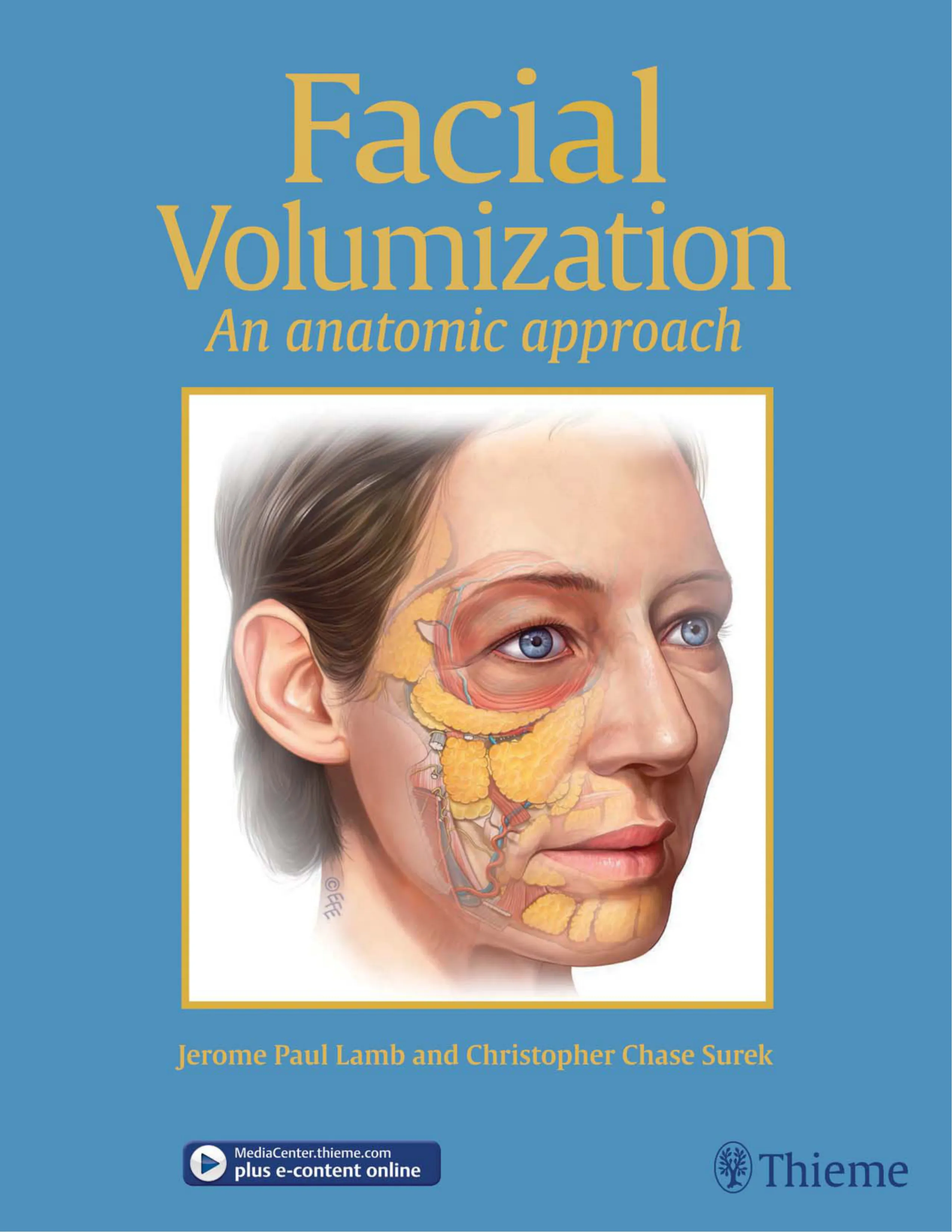


Facial Volumization An anatomic approach Jerome Paul Lamb and Christopher Chase Surek MediaCenter.thieme.com plus e-content online Thieme
Check out these techniques online
with videos on MediaCenter.Thieme.com!
Simply visit MediaCenter.thieme.com and, when prompted during the
registration process, enter the code below to get started today.
U2Q9-4HQU-C739-G64G
Thieme
(Trang không có chữ)
Facial Volumization
An Anatomic Approach
Jerom e Paul Lam b, MD, FACS
Plastic Surgeon Diplomate of the Am erican Board of Plastic Surgery, Inc.
Centerpoint Medical Center, Truman Medical Center
Independence, Missouri, United States
Christopher Chase Surek, DO
Chief Resident
Department of Plastic Surgery
University of Kansas Medical Center
Overland Park, Kansas, United States
58 illustrations
Thieme
New York • Stuttgart • Delhi • Rio de Janeiro
Executive Editor: Sue Hodgson
Developmental Editor: Jennifer Gann
Managing Editor: Elizabeth Palumbo
Director, Editorial Services: Mary Jo Casey
Production Editor: Torsten Scheihagen
International Production Director: Andreas Schabert
International Marketing Director: Fiona Henderson
International Sales Director: Louisa Turrell
Director of Sales, North America: Mike Roseman
President: Brian D. Scanlan
Medical Illustrator: Levent Efe
Library of Congress Cataloging-in-Publication Data
Names: Lamb, Jerome Paul, editor. | Surek, Christopher Chase,
editor.
Title: Facial volumization : an anatomic approach / [edited by]
Jerome Paul Lamb, MD, FACS, Plastic Surgeon Diplomate,
American Board of Plastic Surgery, Inc., Centerpoint Medical
Center, Truman Medical Center, Independence, Missouri,
United States, Christopher Chase Surek, MD, Chief Resident,
Department of Plastic Surgery, University of Kansas Medical
Center, Overland Park, Kansas, United States.
Description: New York : Thieme, [2017]
Identifiers: LCCN 2017021388| ISBN 9781626236943 (print) |
ISBN 9781626238640 (e-book)
Subjects: LCSH: Face--Surgery. | Surgery, Plastic.
Classification: LCC RD119.5.F33 F35 2017 | DDC 617.5/2059--
dc23 LC record available at
Copyright © 2018 by Thieme Medical Publishers, Inc.
Thiem e Publishers New York
333 Seventh Avenue, New York, NY 10001 USA
+1 800 782 3488, custom erservice@thieme.com
Thiem e Publishers Stuttgart
Rüdigerstrasse 14, 70469 Stuttgart, Germ any
+49 [0]711 8931 421, custom erservice@thieme.de
Thieme Publishers Delhi
A-12, Second Floor, Sector-2, Noida-201301
Uttar Pradesh, India
+91 120 45 566 00, customerservice@thieme.in
Thieme Publishers Rio de Janeiro, Thieme Publicações Ltda.
Edifício Rodolpho de Paoli, 25º andar
Av. Nilo Peçanha, 50 – Sala 2508
Rio de Janeiro 20020-906, Brasil
+55 21 3172 2297
Cover design: Thieme Publishing Group
Typesetting by Debra Clark
Printed in China by Everbest Printing Ltd
5 4 3 2 1
ISBN 978-1-62623-694-3
Also available as an e-book:
eISBN 978-1-62623-864-0
This book, including all parts thereof, is legally protected by co-
pyright. Any use, exploitation, or commercialization outside the
narrow limits set by copyright legislation without the publisher’s
consent is illegal and liable to prosecution. This applies in par-
ticular to photostat reproduction, copying, mimeographing or
and electronic data processing and storage.
Im portant note: Medicine is an ever-changing science undergoing
continual developm ent. Research and clinical experience are con-
tinually expanding our knowledge, in particular our knowledge of
proper treatment and drug therapy. Insofar as this book mentions
any dosage or application, readers m ay rest assured that the au-
thors, editors, and publishers have m ade every effort to ensure that
such references are in accordance with the state of knowledge at
the tim e of production of the book.
Nevertheless, this does not involve, imply, or express any
guarantee or responsibility on the part of the publishers in respect
to any dosage instructions and forms of applications stated in the
book. Every user is requested to exam ine carefully the manufac-
in consultation with a physician or specialist, whether the dosage
schedules mentioned therein or the contraindications stated by
the m anufacturers differ from the statements made in the present
book. Such examination is particularly important with drugs that
are either rarely used or have been newly released on the m arket.
Every dosage schedule or every form of application used is enti-
rely at the user’s own risk and responsibility. The authors and
publishers request every user to report to the publishers any di-
screpancies or inaccuracies noticed. If errors in this work are found
after publication, errata will be posted at on the
product description page.
Some of the product nam es, patents, and registered designs
referred to in this book are in fact registered trademarks or proprie-
m ade in the text. Therefore, the appearance of a name without de-
signation as proprietary is not to be construed as a representation
by the publisher that it is in the public dom ain.
This book is dedicated to those colleagues who have encouraged and m entored m e along the way. Most notably,
I thank Glenn Jelks, Bryan Mendelson, and Val Lam bros, who saw the value of our prelim inary research and
encouraged us to publish. During m y form ative years, it was Don Kam inski, George Block, and Gus Colon who
provided that guidance and support in m edical school, general surgery, and plastic surgery residency that has
graced m y life. Lastly, this book is dedicated to m y w ife Carri, m y son Jake, and m y daughter Madi, who have
given up tim e w ith m e that w ill never be able to be returned in order for m e to fulfill a dream .
Jerom e Paul Lam b
I dedicate this book to those who have loved, supported, and inspired m e. I want to thank m y education
m entors who have set great examples for m e to follow. My true companion Krystle Surek, thank you for
your patience during this process and for understanding m y passion for this field. To Sharon Surek and
Christopher L. Surek who raised m e to chase m y dream s and em bodied the values of hum ility and
unconditional love. To m y grandfather Richard Weber who showed m e that life is what you m ake of it
and that giving up is never an option. Lastly, to our patients of past, present, and future—they are the
instrum ents by which we are measured, and it is m y sincere hope that this body of work w ill translate into
renewed happiness and joy in their lives.
Christopher Chase Surek
(Trang không có chữ)
Contents
Video Contents ............................................................................................................................... ix
Foreword ........................................................................................................................................ xi
Preface ........................................................................................................................................... xiii
Contributors ................................................................................................................................... xv
1
The Midface ...................................................................................................................................... 1
Jerome Paul Lamb and Christopher Chase Surek
2
The Lymphatic Anatomy of the Lower Eyelid and the Malar Region of the Face ........................... 25
Sajna Shoukath and Mark Winter Ashton
3
The Perioral Area, the Chin, and the Jowl ...................................................................................... 35
Jerome Paul Lamb, Christopher Chase Surek, and James D. Vargo
4
The Temple and the Brow .............................................................................................................. 59
Jerome Paul Lamb and Christopher Chase Surek
vii
(Trang không có chữ)
ix
Video Contents
1
The Midface
Video 1.1 Pneumatization of the Deep Pyriform Space
Video 1.2 Mid-Face Augmentation
Video 1.3 Vascular Anatomy of the Face
Video 1.4 Deep Pyriform Space Approach to the Tear Trough
Video 1.5 DeMaio V1 and V2
3
The Perioral Area, the Chin, and the Jowl
Video 3.1 The Nasolabial Fold and the Marionette Lines
Video 3.2 Nasolabial Folds with Cannula and Deep Pyriform Space Injection
Video 3.3 Anesthesia
Video 3.4 Building a Fuller Upper Lip: The Retro-Orbicularis Oris Fat Compartment
Video 3.5 Crosshatching
Video 3.6 White Roll and Philtrum
Video 3.8 Red Lip Enhancement
Video 3.9 Augmentation of the Aging Orbicularis Oris Muscle
Video 3.10 Restoring the Lower Lip White Roll
Video 3.11 Lower Lip Enhancement
Video 3.12 The Pre-Jowl
4
The Temple and the Brow
Video 4.1 Lateral Temporal–Cheek Fat Compartment Volumization
Video 4.2 Upper Brow Volumization
Video 4.3 Swift: One Up and One Over
(Trang không có chữ)
Foreword
As a young plastic surgeon, I remember well how the in-
terior of the face looked like a mysterious and dangerous
territory where everything looked like everything else and
only obedience to the empirical ritual of the facelift opera-
tion let m e feel secure. In tim e, increasingly subtle changes
of anatomy became old friends while passing them in sur-
gery. I contemplated the retaining ligaments of the face—
thin and weak individually, but very powerful in aggrega-
te. Further along I began to understand how the anatomy
contributed to both the look of the face and how such an
understanding could be a tool to comprehending the older
face and better ways to rehabilitate it. Knowledge is power.
In replacing or adding volum e to the aging face subtle-
ties of placem ent went from being ignored to being obvi-
ous. At first one is happy that an area can be filled at all.
In tim e, one’s powers of observation and discrimination
increase to the point that earlier attempts look crude and
artless and one sees and strives for more graceful and less
exaggerated results.
Almost all aesthetic modifications of the face are based
on anatomy, the details of which are usually the last con-
sideration of the aesthetic practitioner. Facelifts were
done for approximately 6 decades before the anatomy of
the facial nerve was understood. The first several years of
filler use was for the most part filling wrinkles. Injectors
had very little understanding of the relationship of loca-
tion and depth of placem ent to results until about a de-
cade after their introduction when finally a m ore balanced
and volumetric use of fillers was established. Cosmetic
xi
lasers were accepted very quickly before the occurrence of
late depigmentation was appreciated. One might conclude
that early comm ercial adoption of a technique precludes
thoughtful study of its underlying science.
Things have changed: we have now grown into in-
creased wisdom of many aspects of the face. The study of
facial anatomy has blossomed in the last few years; with
more attention being paid to areas of perceptual interest,
the structures that define faces of different ages can be
seen to have altered in knowable ways, not mysterious and
arbitrary ones.
Drs. Lamb and Surek have given us a remarkable volu-
me of facial anatomy, compiling information from other
authors but also including extensive clean dissections
and descriptions of their own. The greatest of explana-
tory tools, the diagrams, are particularly valuable as they
are concise and correct and address the clinician’s con-
cerns. Anatomic phenomena of the face, hollows, ripples,
mounds, and others are revealed to have their origins deep
in the face in ways that are not intuitive. Treatm ents can
be made more rationally with such understandings and
many are included in this volum e. Knowledge is power.
This book is a m ajor step forward in understanding not
only the dry anatomy of the face, but how it works and
how it can be modified for the good. Any student of the
face will be well advised to study the topic and this book
in particular.
Val Lam bros, MD
(Trang không có chữ)
Preface
Bryan Mendelson said it best: “Human beings are the only
species on the planet whose age shows on their face.”
Treating the aging face is one of the great challenges of
aes thetic surgery, and volume replenishment is a critical
component of the treatment algorithm. Although volumi-
zation techniques are com m only perform ed, they are still
relatively subjective, and they are often based on injector’s
“aesthetic eye.” As anatomists and surgeons, we believe a
strong knowledge of facial anatomy is critical for an injec-
tor to be accurate, safe, and consistent when performing
facial volumization. Although there have been several
published works about the individual components of the
face, there has not been a comprehensive synthesis of fa-
cial anatomy specific to injection techniques. This book is
designed to bridge the gap between the anatomy lab and
your injection clinic. We intend to distill high-yield, cli-
nically relevant anatomical details of commonly injected
regions of the face and to guide the clinician in the safe
conduct of these procedures.
The full-page illustrations will take the reader through
each layer of the face, from deep to superficial, to demon-
strate the scaffold construction of this complex anatomical
landscape. As the reader turns the pages, they will arrive
on new anatomical layers. On the left side of the page, the
xiii
reader will find a written synopsis of the vascular, muscu-
lar, ligament, and adipose components of the specific lay-
er paired with cadaveric photographs. Personally, we find
the synergy between the anatom ical descriptions and the
medical illustration to equip us with a “snapshot” that we
can think about when we are injecting these regions on
our patients. It is our hope that this presentation improves
your understanding of depth and of the plane of injection.
At the end of Chapters 1, 2, and 4, we share our preferred
techniques for volumization in each region; these descrip-
tions are accompanied by a medical illustration depicting
the critical anatomy for the defined technique.
The supplemental video companion of the book will
take the reader step by step through each technique as it
is performed on a patient. These videos contain medical
illustrations in the top corner of the screen for the reader
to visualize the anatomy as the injection is performed. The
gliding nature and dynamic planes of the face create a for-
midable challenge for the injector when treating an aging
face. Please consider this book as a companion, toolbox,
and potential “secret weapon” to assist you with obtaining
optimal aesthetic outcomes for your patients.
Jerom e Paul Lamb and Christopher Chase Surek
(Trang không có chữ)
xv
Contributors
Mark Winter Ashton, MD
Professor
Department of Surgery
University of Melbourne
Parkville, Victoria, Australia
Jerom e Paul Lam b, MD, FACS
Plastic Surgeon Diplomate of the American Board
of Plastic Surgery, Inc.
Centerpoint Medical Center, Truman Medical
Center
Independence, Missouri, United States
Sajna Shoukath, PhD
Student
Department of Surgery
University of Melbourne
Parkville, Victoria, Australia
Christopher Chase Surek, DO
Chief Resident
Department of Plastic Surgery
University of Kansas Medical Center
Overland Park, Kansas, United States
Jam es D. Vargo, MD
Resident Physician
Department of Plastic Surgery
University of Kansas Medical Center
Kansas City, Missouri, United States
(Trang không có chữ)
1
Chapter 1
The Midface
The Orbital Retaining Ligament,
the Zygomaticocutaneous
Ligaments, the Maxillary
Retaining Ligaments, and the Masseteric
Ligaments
3
The Preperiosteal Fat Pad and
the Deep Pyriform Space
6
The Prezygomatic Space
8
The Medial and Lateral
Suborbicularis Oculi Fat
Compartments and the Deep
Medial Cheek Fat Compartment
10
The Facial Nerve
12
The Vascular Anatomy of
the Midface
14
The Premaxillary Space
16
The Infraorbital Fat
Compartment “Malar Bag”
18
The Authors’ Preferred
Techniques for Midfacial
Volumization
20
(Trang không có chữ)
3
1 The Midface
Jerome Paul Lamb and Christopher Chase Surek
The Orbital Retaining Ligament,
the Zygomaticocutaneous
Ligaments, the Maxillary Retaining
Ligaments, and the Masseteric
Ligaments
-
-
-
-
-
-
Fig. 1.1
Fig. 1.2
-
Fig. 1.1 The malar equator (turquoise line) bisects the midface and
anatomically correlates with the zygomaticocutaneous ligaments.
Fig. 1.2 The zygomaticocutaneous ligaments form the lower “ham-
mock” border of the prezygomatic space. The ligaments serve as a
partition of the upper and lower midface, respectively.
4
1 The Midface
The Lower Midface
-
-
Fig. 1.3 -
The Upper Midface
-
-
-
5
1 The Midface
Fig. 1.3 Medical illustration of the key retaining ligaments of the midface: the orbitomalar ligament, the main zygomatic ligament, the zygo-
maticocutaneous ligaments, and the maxillary and masseteric ligaments.
6
1 The Midface
The Preperiosteal Fat Pad and the
Deep Pyriform Space
The Upper Midface
-
-
Fig. 1.4
-
The Lower Midface
Fig.
1.5
Fig. 1.6 -
Fig. 1.5 Medical illustration of the deep pyriform space and impor-
tant adjacent anatomical structures. (From Surek C, Vargo J, Lamb J.
-
tions. Plast Reconstr Surg. 2016;138(1). 2016 with permission)
Fig. 1.6 The deep pyriform space is cradled medially by the pyriform
aperture and depressor nasalis. The angular artery courses between
the space and the deep medial cheek fat compartment. Note that
within the roof of the space. (From Surek C, Vargo J, Lamb J. Deep
Plast Reconstr Surg. 2016;138(1). 2016 with permission)
Fig. 1.7 Demonstration of the mimetic muscle insertions into the
nasolabial fold. (From Surek C, Vargo J, Lamb J. Deep pyriform space:
Surg. 2016;138(1). 2016 with permission)
Fig. 1.4 The preperiosteal fat pad lies within the prezygomatic space
adherent to the maxilla.
-
Video 1.1
Fig.
1.7
-
Fig. 1.8
7
1 The Midface
Fig. 1.8 Medical illustration of the deepest compartments and spaces in the midface.
8
1 The Midface
The Prezygomatic Space
The Upper Midface
Fig. 1.9
-
Fig. 1.10 -
Fig. 1.11; Video
1.2 -
-
Fig. 1.9 Illustration of the anatomical depth relationships in the
upper midface. The prezygomatic space is demonstrated in the
deep suborbicularis plane (blue capsule). The orbitomalar ligament
is demonstrated arborizing through the orbicularis oculi muscle
inserting into the skin forming the tear trough crease. The zygomati-
cocutaneous ligaments arborize through the orbicularis, forming
a partition between the infraorbital “malar” fat compartment
-
ous insertion of the ligaments forms the characteristic skin crease
demonstrated in clinical malar mounds.
Fig. 1.10 Frontal view. The prezygomatic space capsule has been
stained with methylene blue. The infraorbital “malar” fat compart-
oculi. Blunt cannulas placed percutaneously prior to dissection are
found inside the prezygomatic space. (From Surek CC, Beut J, Ste-
phens R, Jelks G, Lamb J. Pertinent anatomy and analysis for midface
volumizing procedures. Plast Reconstr Surg 2015;135(5):818e–829e
with permission).
Fig. 1.11 Demonstration of the “pinch-and-pull” technique for
penetration of a blunt cannula into a suborbicularis plane into the
prezygomatic space. (From Surek C, Beut J, Stephens R, Lamb J, Jelks
Aesthet Surg J 2015;35(2):121–134 with permission)
Injection Pearl
For consistent deep volumization of the cheek, the prezygo-
matic space can be a secret weapon.
Fig. 1.12
9
1 The Midface
Fig. 1.12 Medical illustration of the prezygomatic space.
10
1 The Midface
The Medial and Lateral
Suborbicularis Oculi Fat
Compartments and the Deep
Medial Cheek Fat Compartment
The Upper Midface
-
The Lower Midface
-
-
-
-
-
The Lateral Component of the Deep Medial
Cheek Fat
-
-
The Medial Component of the Deep Medial
Cheek Fat
-
-
-
Figs. 1.13 1.14
Injection Pearl
composition wisely. We recommend medium to large particle
Fig. 1.13
retracted. Zygomaticus major (ZM) and levator labii superioris (LLS)
are labeled. The orbicularis oculi has been resected. Remnants of the
lateral suborbicularis oculi fat compartment (SOOF) are noted on top
of the preperiosteal fat (PPF). The medial SOOF and the infraorbital
(IO) neurovascular bundle are labeled. The loose areolar consistency
of the deep medial cheek fat is noted lateral to the transected levator
anguli oris (LAO). (From Surek CC, Beut J, Stephens R, Jelks G, Lamb J.
Pertinent anatomy and analysis for midface volumizing procedures.
Plast Reconstr Surg 2015;135(5):818e–829e with permission)
Fig. 1.14
-
tion of the deep medial cheek fat (DMCF) and medial suborbicularis
oculi fat compartment (SOOF) stained with methylene blue. The
zygomaticus major (ZM), levator anguli oris (LAO), levator labii
superioris (LLS), and levator labii superioris alaeque nasi (LLSAN)
been injected into the lateral SOOF overlying the preperiosteal fat
compartment. (From Surek CC, Beut J, Stephens R, Jelks G, Lamb J.
Pertinent anatomy and analysis for midface volumizing procedures.
Plast Reconstr Surg 2015;135(5):818e–829e with permission)
Fig. 1.15 -
11
1 The Midface
Fig. 1.15
12
1 The Midface
The Facial Nerve
•
•
•
•
•
The Frontal or Temporal Branch
-
-
-
-
The Zygomatic Branch
-
The Buccal Branch
The Marginal Mandibular Branch
-
-
The Cervical Branch
-
Fig. 1.16 -
13
1 The Midface
Fig. 1.16
14
1 The Midface
Venous Anatomy of the Midface
-
-
-
-
Injection Pearl
The injector must have a keen understanding of the DEPTH
of all structures in the upper midface to avoid unwanted
outcomes.
Fig. 1.17
The Vascular Anatomy of the
Midface
Arterial Anatomy of the Midface
Vid-
eo 1.3
-
-
-
-
-
-
-
15
1 The Midface
Fig. 1.17 Medical illustration of the high-yield venous and arterial anatomy of the face.
16
1 The Midface
The Premaxillary Space
-
-
Figs. 1.18 1.19
Fig.
1.20
Fig. 1.18 Medical illustration of the deep pyriform space and impor-
tant adjacent anatomical structures.
Fig. 1.19 Instruments are placed in the premaxillary space and
the deep pyriform. The instruments were advanced through the
tear trough ligament demonstrating that these structures reside in
-
Reconstr Surg. 2016;138(1). 2016 with permission)
17
1 The Midface
Fig. 1.20 Medical illustration of the premaxillary space and the platysma-auricular fascia.
18
1 The Midface
Injection Pearl
-
animation.
Injection Pearl
Langer’s lines.
Fig. 1.23 -
The Infraorbital Fat Compartment
Cheek Compartments
The Upper Midface
Fig. 1.21
-
Fig. 1.22
Upper Midface Pitfall 1
Iatrogenic malar mound: “The bee sting appearance.”
-
-
-
-
-
Fig. 1.21 Illustration of the anatomical depth relationships in the
upper midface. The prezygomatic space is demonstrated in the
deep suborbicularis plane (blue capsule). The orbitomalar ligament
is demonstrated arborizing through the orbicularis oculi muscle
inserting into the skin forming the tear trough crease. The zygo-
maticocutaneous ligaments arborize through the orbicularis forming
a partition between the infraorbital “malar” fat compartment
-
ous insertion of the ligaments forms the characteristic skin crease
demonstrated in clinical malar mounds.
Fig. 1.22 Photographic illustration of an iatrogenic malar mound
compartment. Note the cutaneous insertions of the orbitomalar liga-
ment superiorly and the zygomaticocutaneous ligaments inferiorly.
19
1 The Midface
Fig. 1.23
20
1 The Midface
-
Anterior Midface (Deep Injection)
-
Video 1.4
Fig. 1.24
The Authors’ Preferred Techniques
for Midfacial Volumization
Deep Volumization
-
Lateral Midface (Deep Injection)
Video 1.2
21
1 The Midface
Fig. 1.24 Medical illustration depicting the authors’ preferred technique for deep volumization in the anterior and lateral midface.
22
1 The Midface
-
Video 1.5
-
Fig. 1.25 -
-
Fig. 1.25
pathway of Langer’s lines.
-
-
Fig. 1.26
23
1 The Midface
Fig. 1.26
deep bolus injections per DeMaio.
24
1 The Midface
-
-
-
-
-
-
Suggested Reading
-
-
-
-
2
Chapter 2
The Lymphatic Anatomy
of the Lower Eyelid
and the Malar Region
of the Face
Introduction
27
Facial Lymphatics
28
32
(Trang không có chữ)
27
2 The Lymphatic Anatomy of the Lower Eyelid
and the Malar Region of the Face
Sajna Shoukath and Mark Winter Ashton
A keen understanding of facial lymphatics is the m issing link in
the anatom y of facial injections. This knowledge equips the injec-
tor w ith a secret weapon in their arm amentarium to prevent sub-
optim al aesthetic outcom es secondary to prolonged lymphedem a.
In particular, this emphasizes the importance of injection depth
in the m idface.
We would like to thank Dr. Shoukath and Professor Ashton
relevant to the injector and fat grafting surgeon. Iatrogenic per-
sistent periorbital edem a m ay be the result of lymphatic injury,
lymphatic fouling, or possibly particle cohesivity in the case of
in the prezygom atic space, w hen entered laterally in a m obile
segment of the periorbital, should provide safety as the cannula
is directed toward m alar periosteum and then advanced m edi-
ally. Sharp needle injection in the region of the bilam ellar orbital
retaining ligam ent poses som e risk of injury or impingem ent of
the deep lateral lymphatic channel. Based upon anatom ic dis-
sections in the region of the zygom aticom axillary retaining liga-
m ent, the caudal border of the prezygom atic space is a series of
osteocutaneous ligam ents, rather than a m em branous structure
as is present cephalad in the orbital retaining ligam ent. As such,
volum e-based procedures near the caudal aspect of the prezygo-
m atic space would theoretically be less prone to an impingement
since the lymphatics do not pierce a dense anatom ic structure.
—Jerom e P. Lam b and Christopher C. Surek
Introduction
Our knowledge of the human lymphatic system is predomi-
nantly based on the pioneering work of Sappey1 and his suc-
cessors2 in the 19th and early 20th centuries. This is because
-
ysis impossible.
possible.3 This is important because many of the questions con-
cerning lymphatic anatomy are not answered by the existing
literature.4
-
ameter.5 This network subsequently drains into larger “precol-
-
6
28
-
tible to lymphedema and chemosis. Indeed, the recent trend to
complication rates of persistent chemosis beyond 2 to 3 weeks
in one series recently published.9 In each published series, the
Facial Lymphatics
-
rotid and submandibular lymph nodes. -
predictable in their location. Knowledge of the location of these
as the fragile and compressible nature of the collecting lym-
Fig. 2.1
29
Fig. 2.1 Illustration of the lymphatic channels of the midface and the lower eyelid.
30
Fig. 2.2
-
sistent.10 Elsewhere in the body, the precollecting lymphatic
2C.5 5 The face is no
lower eyelid and eye drains into precollectors in the deep der-
precollectors fall into two groups: those orientated toward the
-
collectors coalesces to form a collecting trunk, which turns
continues within the nasolabial fat compartment11 to drain into
the submandibular lymph node. The lateral group of precollec-
-
ally within the lateral orbital fat compartment11 to drain into
the preauricular lymph nodes.12 These two medial and lateral
As would be predicted, there is an additional system of lym-
is, in layer 4. This deep system drains the lower eyelid and up-
tarsal plate and Meibomian glands in the lateral third of the
lower eyelid. This deep lymphatic system is joined by connec-
-
-
-
-
probable that this system also exists but may be smaller or less
Thus, there are three main lymphatic channels of the lower
a deep lateral system of lymphatics. The medial system drains
to the submandibular gland, and the lateral system drains to
the parotid.
As would be predicted from elsewhere in the body, the capil-
lary network and the precollectors located within the conjunc-
medial and lateral eye. There is also a free communication be-
tween precollectors of the lower and upper eyelids around the
lateral canthus. Our studies would suggest that the majority of
lymphatic drainage from both the upper and lower eyelids is
through the lateral system.
31
Fig. 2.2 Illustration of the facial lymphatic architecture.
32
13 in
his composite rhytidectomy technique in which much of the dis-
-
14
been described15
more caudal on the face. Mendelson et al16 noted that excision of
subcutaneous fat in malar mounds lead to prolonged swelling.
New studies into lymphatic anatomy of the lower eyelid and
12 -
orbicularis muscle approximately 10 mm from the lid margin in
precisely the region of the infraorbital incisions described ear-
-
cutaneous fat compartments.11
be reformed after injury, and hence, it can be expected that
cheek edema while they are being repaired.
-
-
ing through the tarsal plate laterally. The deep lymphatic collec-
tors subsequently formed are deep to the preseptal orbicularis
oculi and are located at the junction of the orbicularis retaining
10 the collectors descend to the preperiosteal fat around the
the parotid.
Table 2.1
that the incidence of chemosis increases as the dissection in the
attributable to the minimal amount of dissection around the
lateral canthus and the orbicularis retaining ligament. Trans-
not injured. It is notable that in both of these techniques, only
one of the lymphatic systems of the face is potentially damaged.
interconnections through the preseptal orbicularis, it is prob-
able that damage to one system will be compensated for by the
other without chemosis occurring.
compromised and relies on cross-connections with a poten-
proposed that damage to the deep system, in addition to the
-
eral canthal support and re-draping of the orbicularis retaining
ligament.9,20,21
This is particularly important when surgical procedures on
in the malar region or nasojugal fold. Our studies would suggest
that the key lymphatic drainage pathways for the lower eyelid
are in close proximity to the nasojugal fold medially and the
extreme care should concomitant lower eyelid surgery also be
proposed.
Table 2.1
Author (Year)
No. of patients
Chemosis rates
Seitz et al (2012)18
Transconjunctival deep midface lift
124
0.8%
Honrado et al (2004)8
3988
1%
Prischmann et al (2013)9
Transconjunctival
39
5.1%
Undavia et al (2015)19
Transconjunctival
66
7.6%
Weinfeld et al (2008)20
Release ORL
Lateral canthopexy/plasty
312
11.5%
Codner et al (2008)21
Release ORL
Lateral canthopexy/plasty
264
12.1%
Prischmann et al (2013)9
694
34.5%