
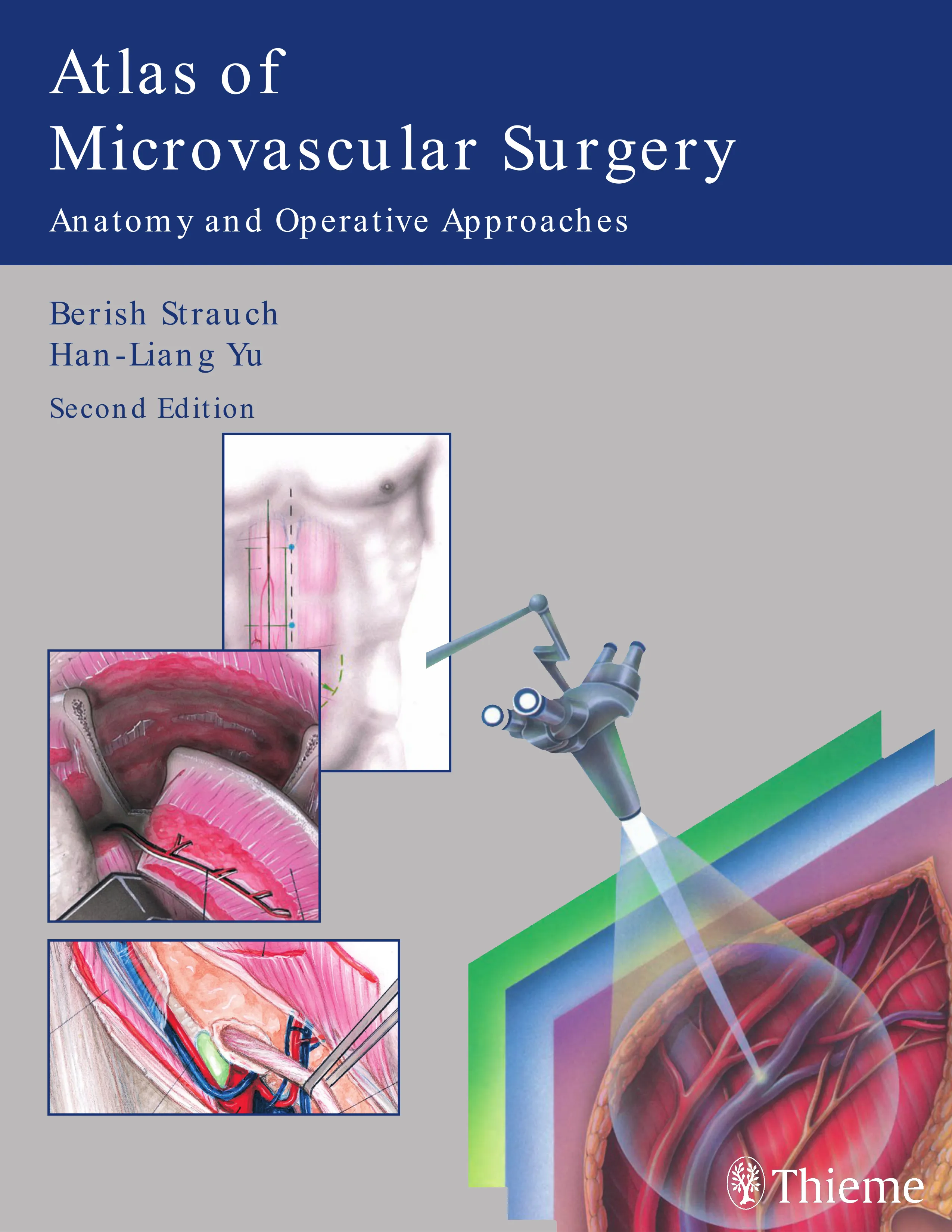

Atlas of
Microvascular Surgery
Berish Strauch
Han-Liang Yu
Second Edition
Anatomy and Operative Approaches
Atlas of Microvascular Surgery
Anatomy and Operative Techniques
Second Edition
Thiem e
(Trang không có chữ)
Atlas of Microvascular Surgery
Anatomy and Operative Techniques
Second Edition
Berish Strauch, M.D.
Professor and Chair, Department of Plastic and Reconstructive Surgery
Albert Einstein College of Medicine of Yeshiva University and
Monte ore Medical Center
Bronx, New York
Han-Liang Yu, M.D.
Associate Clinical Professor, Department of Plastic and Reconstructive Surgery
Albert Einstein College of Medicine of Yeshiva University and
Monte ore Medical Center
Bronx, New York
Liugo Liang
Medical Illustrator
Thieme
New York • Stuttgart
Thieme Medical Publishers, Inc.
333 Seventh Ave.
New York, NY 10001
Executive Editor: Timothy Hiscock
Vice-President, Production and Electronic Publishing: Anne T. Vinnicombe
Sales Director: Ross Lumpkin
Chief Financial Of cer: Peter van Woerden
President: Brian D. Scanlan
Compositor: Compset Inc.
Printer: Everbest Printing Company
Library of Congress Cataloging-in-Publication Data
Library of Congress Cataloging-in-Publication Data
Strauch, Berish, 1933–
Atlas of microvascular surgery : anatomy and operative techniques /
Berish Strauch, Han-Liang Yu. — 2nd ed.
p. ; cm.
Includes bibliographical references and index.
ISBN 1-58890-466-0 (TMP : alk. paper) — ISBN 3-13-783002-8 (GTV
: alk. paper)
1. Blood-vessels—Surgery—Atlases. 2. Blood-vessels—Anatomy—Atlases.
3. Microsurgery—Atlases. 4. Flaps (Surgery)—Atlases. I. Yu,
Han-Liang.
II. Title.
[DNLM: 1. Blood Vessels--anatomy & histology—Atlases. 2. Micro-
surgery—methods—Atlases. 3. Vascular Surgical Procedures—methods
—Atlases. WG 17 S912a 2006]
RD598.5.S79 2006
617.4'13—dc22
2005032830
Copyright ©2006 by Thieme Medical Publishers, Inc. This book, including all parts thereof, is legally protected by copyright. Any use, exploitation,
or commercialization outside the narrow limits set by copyright legislation without the publisher's consent is illegal and liable to prosecution. This
applies in particular to photostat reproduction, copying, mimeographing or duplication of any kind, translating, preparation of micro lms, and
electronic data processing and storage.
Important note: Medical knowledge is ever-changing. As new research and clinical experience broaden our knowledge, changes in treatment and
drug therapy may be required. The authors and editors of the material herein have consulted sources believed to be reliable in their efforts to pro-
vide information that is complete and in accord with the standards accepted at the time of publication. However, in the view of the possibility of hu-
man error by the authors, editors, or publisher of the work herein or changes in medical knowledge, neither the authors, editors, or publisher, nor
any other party who has been involved in the preparation of this work, warrants that the information contained herein is in every respect accurate
or complete, and they are not responsible for any errors or omissions or for the results obtained from use of such information. Readers are encour-
aged to con rm the information contained herein with other sources. For example, readers are advised to check the product information sheet in-
cluded in the package of each drug they plan to administer to be certain that the information contained in this publication is accurate and that
changes have not been made in the recommended dose or in the contraindications for administration. This recommendation is of particular impor-
tance in connection with new or infrequently used drugs.
Some of the product names, patents, and registered designs referred to in this book are in fact registered trademarks or proprietary names even
though speci c reference to this fact is not always made in the text. Therefore, the appearance of a name without designation as proprietary is not
to be construed as a representation by the publisher that it is in the public domain.
Printed in China
5 4 3 2 1
TMP ISBN 1-58890-466-0
GTV ISBN 3 13 783002 8
In Memoriam, Dr. Zhong-Wei Chen . . . . . . . . . . . . . . . . . . . . . . . . . . . . . . . . . . . . . . . . . . . . . . . . . . . . . . . . . . . . . .xi
Preface to the Second Edition . . . . . . . . . . . . . . . . . . . . . . . . . . . . . . . . . . . . . . . . . . . . . . . . . . . . . . . . . . . . . . . . . . .xv
Preface to the First Edition . . . . . . . . . . . . . . . . . . . . . . . . . . . . . . . . . . . . . . . . . . . . . . . . . . . . . . . . . . . . . . . . . . . .xvii
Introduction . . . . . . . . . . . . . . . . . . . . . . . . . . . . . . . . . . . . . . . . . . . . . . . . . . . . . . . . . . . . . . . . . . . . . . . . . . . . . . . . . .I1
De nitions . . . . . . . . . . . . . . . . . . . . . . . . . . . . . . . . . . . . . . . . . . . . . . . . . . . . . . . . . . . . . . . . . . . . . . . . . . . . . . . . . . . . . . . . . .I1
Training in Basic Microsurgical Techniques . . . . . . . . . . . . . . . . . . . . . . . . . . . . . . . . . . . . . . . . . . . . . . . . . . . . . . . . . . . . . .I1
Exposure of Recipient Vessels . . . . . . . . . . . . . . . . . . . . . . . . . . . . . . . . . . . . . . . . . . . . . . . . . . . . . . . . . . . . . . . . . . . . . . . . . .I2
Microsurgical Anatomy and its Variations . . . . . . . . . . . . . . . . . . . . . . . . . . . . . . . . . . . . . . . . . . . . . . . . . . . . . . . . . . . . . . .I3
Donor Sites . . . . . . . . . . . . . . . . . . . . . . . . . . . . . . . . . . . . . . . . . . . . . . . . . . . . . . . . . . . . . . . . . . . . . . . . . . . . . . . . . . . . . . . . . .I4
Flap Design . . . . . . . . . . . . . . . . . . . . . . . . . . . . . . . . . . . . . . . . . . . . . . . . . . . . . . . . . . . . . . . . . . . . . . . . . . . . . . . . . . . . . . . . . .I4
Harvesting Techniques . . . . . . . . . . . . . . . . . . . . . . . . . . . . . . . . . . . . . . . . . . . . . . . . . . . . . . . . . . . . . . . . . . . . . . . . . . . . . . . .I5
Comments and Insights . . . . . . . . . . . . . . . . . . . . . . . . . . . . . . . . . . . . . . . . . . . . . . . . . . . . . . . . . . . . . . . . . . . . . . . . . . . . . . .I5
PART I
UPPER EXTREMITY
Chapter 1
Shoulder, Arm, and Axilla . . . . . . . . . . . . . . . . . . . . . . . . . . . . . . . . . . . . . . . . . . . . . . . . . . . . . . . . . . . . . . . . . . . . . .3
Deltoid Flap . . . . . . . . . . . . . . . . . . . . . . . . . . . . . . . . . . . . . . . . . . . . . . . . . . . . . . . . . . . . . . . . . . . . . . . . . . . . . . . . . . . . . . . . . .3
Medial Arm Flap . . . . . . . . . . . . . . . . . . . . . . . . . . . . . . . . . . . . . . . . . . . . . . . . . . . . . . . . . . . . . . . . . . . . . . . . . . . . . . . . . . . . . .8
Lateral Arm Flap . . . . . . . . . . . . . . . . . . . . . . . . . . . . . . . . . . . . . . . . . . . . . . . . . . . . . . . . . . . . . . . . . . . . . . . . . . . . . . . . . . . . .12
Posterior Arm Flap . . . . . . . . . . . . . . . . . . . . . . . . . . . . . . . . . . . . . . . . . . . . . . . . . . . . . . . . . . . . . . . . . . . . . . . . . . . . . . . . . . .18
Recipient Site Exposures . . . . . . . . . . . . . . . . . . . . . . . . . . . . . . . . . . . . . . . . . . . . . . . . . . . . . . . . . . . . . . . . . . . . . . . . . . . . . .22
Bibliography . . . . . . . . . . . . . . . . . . . . . . . . . . . . . . . . . . . . . . . . . . . . . . . . . . . . . . . . . . . . . . . . . . . . . . . . . . . . . . . . . . . . . . . .39
Chapter 2
Forearm Region . . . . . . . . . . . . . . . . . . . . . . . . . . . . . . . . . . . . . . . . . . . . . . . . . . . . . . . . . . . . . . . . . . . . . . . . . . . . .40
Forearm Flaps . . . . . . . . . . . . . . . . . . . . . . . . . . . . . . . . . . . . . . . . . . . . . . . . . . . . . . . . . . . . . . . . . . . . . . . . . . . . . . . . . . . . . . .40
Radial Forearm Flap . . . . . . . . . . . . . . . . . . . . . . . . . . . . . . . . . . . . . . . . . . . . . . . . . . . . . . . . . . . . . . . . . . . . . . . . . . . . . . . . . .48
Radial Forearm Flap for Penile Reconstruction . . . . . . . . . . . . . . . . . . . . . . . . . . . . . . . . . . . . . . . . . . . . . . . . . . . . . . . . . . .55
Reversed Radial Forearm Island Flap . . . . . . . . . . . . . . . . . . . . . . . . . . . . . . . . . . . . . . . . . . . . . . . . . . . . . . . . . . . . . . . . . . .58
Radial Forearm Osteocutaneous Flap . . . . . . . . . . . . . . . . . . . . . . . . . . . . . . . . . . . . . . . . . . . . . . . . . . . . . . . . . . . . . . . . . .59
Radial Forearm Tendinocutaneous Flap . . . . . . . . . . . . . . . . . . . . . . . . . . . . . . . . . . . . . . . . . . . . . . . . . . . . . . . . . . . . . . . . .63
Hybrid Radial Forearm Flap . . . . . . . . . . . . . . . . . . . . . . . . . . . . . . . . . . . . . . . . . . . . . . . . . . . . . . . . . . . . . . . . . . . . . . . . . . .65
Contents
vi
CONTENTS
Ulnar Forearm Flap . . . . . . . . . . . . . . . . . . . . . . . . . . . . . . . . . . . . . . . . . . . . . . . . . . . . . . . . . . . . . . . . . . . . . . . . . . . . . . . . . .67
Interosseous Forearm Flap . . . . . . . . . . . . . . . . . . . . . . . . . . . . . . . . . . . . . . . . . . . . . . . . . . . . . . . . . . . . . . . . . . . . . . . . . . . .72
Posterior Interosseous Flap (Posterior Forearm Flap) . . . . . . . . . . . . . . . . . . . . . . . . . . . . . . . . . . . . . . . . . . . . . . . . . . . . .77
Reversed Posterior Interosseous Flap . . . . . . . . . . . . . . . . . . . . . . . . . . . . . . . . . . . . . . . . . . . . . . . . . . . . . . . . . . . . . . . . . . .82
Reversed Posterior Interosseous Osteocutaneous Flap . . . . . . . . . . . . . . . . . . . . . . . . . . . . . . . . . . . . . . . . . . . . . . . . . . .87
Anterior Interosseous Flap (Dorsal Wrist Flap) . . . . . . . . . . . . . . . . . . . . . . . . . . . . . . . . . . . . . . . . . . . . . . . . . . . . . . . . . . .92
Recipient Site Exposure . . . . . . . . . . . . . . . . . . . . . . . . . . . . . . . . . . . . . . . . . . . . . . . . . . . . . . . . . . . . . . . . . . . . . . . . . . . . . .94
Bibliography . . . . . . . . . . . . . . . . . . . . . . . . . . . . . . . . . . . . . . . . . . . . . . . . . . . . . . . . . . . . . . . . . . . . . . . . . . . . . . . . . . . . . . .103
Chapter 3
Hand and Wrist . . . . . . . . . . . . . . . . . . . . . . . . . . . . . . . . . . . . . . . . . . . . . . . . . . . . . . . . . . . . . . . . . . . . . . . . . . . .109
Anatomy of the Radial Artery—Deep Palmar Arch System . . . . . . . . . . . . . . . . . . . . . . . . . . . . . . . . . . . . . . . . . . . . . . .109
Anatomy of the Ulnar Artery—Super cial Palmar Arch System . . . . . . . . . . . . . . . . . . . . . . . . . . . . . . . . . . . . . . . . . . .110
Recipient Site Exposures . . . . . . . . . . . . . . . . . . . . . . . . . . . . . . . . . . . . . . . . . . . . . . . . . . . . . . . . . . . . . . . . . . . . . . . . . . . .113
PART II
LOWER EXTREMITY
Chapter 4
Gluteal Region . . . . . . . . . . . . . . . . . . . . . . . . . . . . . . . . . . . . . . . . . . . . . . . . . . . . . . . . . . . . . . . . . . . . . . . . . . . . .129
Gluteus Maximus Flap . . . . . . . . . . . . . . . . . . . . . . . . . . . . . . . . . . . . . . . . . . . . . . . . . . . . . . . . . . . . . . . . . . . . . . . . . . . . . . .129
Superior Gluteus Myocutaneous Flap . . . . . . . . . . . . . . . . . . . . . . . . . . . . . . . . . . . . . . . . . . . . . . . . . . . . . . . . . . . . . . . . .133
Gluteal Thigh Flap (Based on the Inferior Gluteal Vessels) . . . . . . . . . . . . . . . . . . . . . . . . . . . . . . . . . . . . . . . . . . . . . . . .136
Vascularized Iliac Bone Graft (Based on the Superior Gluteal Vessels) . . . . . . . . . . . . . . . . . . . . . . . . . . . . . . . . . . . . . .140
Superior Gluteal Artery Perforator Flap (S-GAP Flap) . . . . . . . . . . . . . . . . . . . . . . . . . . . . . . . . . . . . . . . . . . . . . . . . . . . .145
Inferior Gluteal Artery Perforator Flap (I-GAP Flap) . . . . . . . . . . . . . . . . . . . . . . . . . . . . . . . . . . . . . . . . . . . . . . . . . . . . . .148
Recipient Site Exposure . . . . . . . . . . . . . . . . . . . . . . . . . . . . . . . . . . . . . . . . . . . . . . . . . . . . . . . . . . . . . . . . . . . . . . . . . . . . .151
Bibliography . . . . . . . . . . . . . . . . . . . . . . . . . . . . . . . . . . . . . . . . . . . . . . . . . . . . . . . . . . . . . . . . . . . . . . . . . . . . . . . . . . . . . . .154
Chapter 5
Groin Region . . . . . . . . . . . . . . . . . . . . . . . . . . . . . . . . . . . . . . . . . . . . . . . . . . . . . . . . . . . . . . . . . . . . . . . . . . . . . . .155
Groin Flaps . . . . . . . . . . . . . . . . . . . . . . . . . . . . . . . . . . . . . . . . . . . . . . . . . . . . . . . . . . . . . . . . . . . . . . . . . . . . . . . . . . . . . . . .155
Super cial Circum ex Iliac (SCIA) Flap (or Iliofemoral Flap) . . . . . . . . . . . . . . . . . . . . . . . . . . . . . . . . . . . . . . . . . . . . . .158
Iliac Flap . . . . . . . . . . . . . . . . . . . . . . . . . . . . . . . . . . . . . . . . . . . . . . . . . . . . . . . . . . . . . . . . . . . . . . . . . . . . . . . . . . . . . . . . . . .164
Osteocutaneous Groin Flap (Based on the SCIA) . . . . . . . . . . . . . . . . . . . . . . . . . . . . . . . . . . . . . . . . . . . . . . . . . . . . . . . .166
Composite Groin Flap with “Tendon” Transfer . . . . . . . . . . . . . . . . . . . . . . . . . . . . . . . . . . . . . . . . . . . . . . . . . . . . . . . . . .168
Super cial Inferior Epigastric (SIEA) Flap (or Lower Abdominal Flap) . . . . . . . . . . . . . . . . . . . . . . . . . . . . . . . . . . . . . .172
Fourth Lumbar Artery Iliac Flap . . . . . . . . . . . . . . . . . . . . . . . . . . . . . . . . . . . . . . . . . . . . . . . . . . . . . . . . . . . . . . . . . . . . . .175
Vascularized Iliac Bone Graft . . . . . . . . . . . . . . . . . . . . . . . . . . . . . . . . . . . . . . . . . . . . . . . . . . . . . . . . . . . . . . . . . . . . . . . . .179
Vascularized Illiac Bone Graft (Based on the Deep Circum ex Iliac Artery) . . . . . . . . . . . . . . . . . . . . . . . . . . . . . . . . .182
Iliac Osteocutaneous Flap (Based on the Deep Circum ex Iliac Artery) . . . . . . . . . . . . . . . . . . . . . . . . . . . . . . . . . . . .191
Internal Oblique Flap . . . . . . . . . . . . . . . . . . . . . . . . . . . . . . . . . . . . . . . . . . . . . . . . . . . . . . . . . . . . . . . . . . . . . . . . . . . . . . . .196
Internal Oblique Muscle Flap . . . . . . . . . . . . . . . . . . . . . . . . . . . . . . . . . . . . . . . . . . . . . . . . . . . . . . . . . . . . . . . . . . . . . . . . .197
Internal Oblique Osteomuscular Flap . . . . . . . . . . . . . . . . . . . . . . . . . . . . . . . . . . . . . . . . . . . . . . . . . . . . . . . . . . . . . . . . .200
Bibliography . . . . . . . . . . . . . . . . . . . . . . . . . . . . . . . . . . . . . . . . . . . . . . . . . . . . . . . . . . . . . . . . . . . . . . . . . . . . . . . . . . . . . . .201
Chapter 6
Thigh Region . . . . . . . . . . . . . . . . . . . . . . . . . . . . . . . . . . . . . . . . . . . . . . . . . . . . . . . . . . . . . . . . . . . . . . . . . . . . . .203
Gracilis Flap . . . . . . . . . . . . . . . . . . . . . . . . . . . . . . . . . . . . . . . . . . . . . . . . . . . . . . . . . . . . . . . . . . . . . . . . . . . . . . . . . . . . . . . .203
Gracilis Muscle Flap . . . . . . . . . . . . . . . . . . . . . . . . . . . . . . . . . . . . . . . . . . . . . . . . . . . . . . . . . . . . . . . . . . . . . . . . . . . . . . . . .205
Gracilis Myocutaneous Flap . . . . . . . . . . . . . . . . . . . . . . . . . . . . . . . . . . . . . . . . . . . . . . . . . . . . . . . . . . . . . . . . . . . . . . . . . .209
Tensor Fasciae Latae (TFL) Flap . . . . . . . . . . . . . . . . . . . . . . . . . . . . . . . . . . . . . . . . . . . . . . . . . . . . . . . . . . . . . . . . . . . . . . .212
TFL Myocutaneous Flap . . . . . . . . . . . . . . . . . . . . . . . . . . . . . . . . . . . . . . . . . . . . . . . . . . . . . . . . . . . . . . . . . . . . . . . . . . . . .214
TFL Osteomyocutaneous Flap . . . . . . . . . . . . . . . . . . . . . . . . . . . . . . . . . . . . . . . . . . . . . . . . . . . . . . . . . . . . . . . . . . . . . . . .218
CONTENTS
vii
Rectus Femoris Flap . . . . . . . . . . . . . . . . . . . . . . . . . . . . . . . . . . . . . . . . . . . . . . . . . . . . . . . . . . . . . . . . . . . . . . . . . . . . . . . .219
Rectus Femoris Muscle Flap . . . . . . . . . . . . . . . . . . . . . . . . . . . . . . . . . . . . . . . . . . . . . . . . . . . . . . . . . . . . . . . . . . . . . . . . . .220
Rectus Femoris Myocutaneous Flap . . . . . . . . . . . . . . . . . . . . . . . . . . . . . . . . . . . . . . . . . . . . . . . . . . . . . . . . . . . . . . . . . . .222
Medial Thigh Flap . . . . . . . . . . . . . . . . . . . . . . . . . . . . . . . . . . . . . . . . . . . . . . . . . . . . . . . . . . . . . . . . . . . . . . . . . . . . . . . . . .223
Lateral Thigh Flap . . . . . . . . . . . . . . . . . . . . . . . . . . . . . . . . . . . . . . . . . . . . . . . . . . . . . . . . . . . . . . . . . . . . . . . . . . . . . . . . . .226
Anteromedial Thigh Flap . . . . . . . . . . . . . . . . . . . . . . . . . . . . . . . . . . . . . . . . . . . . . . . . . . . . . . . . . . . . . . . . . . . . . . . . . . . .231
Anterolateral Thigh (ALT) Flap . . . . . . . . . . . . . . . . . . . . . . . . . . . . . . . . . . . . . . . . . . . . . . . . . . . . . . . . . . . . . . . . . . . . . . . .234
Anterolateral Lateral Thigh Fasciocutaneous Flap . . . . . . . . . . . . . . . . . . . . . . . . . . . . . . . . . . . . . . . . . . . . . . . . . . . . . . .238
ALT Septofasciocutaneous Flap . . . . . . . . . . . . . . . . . . . . . . . . . . . . . . . . . . . . . . . . . . . . . . . . . . . . . . . . . . . . . . . . . . . . . . .239
ALT Perforator Fasciocutaneous Flap . . . . . . . . . . . . . . . . . . . . . . . . . . . . . . . . . . . . . . . . . . . . . . . . . . . . . . . . . . . . . . . . . .241
ALT Cutaneous Flap . . . . . . . . . . . . . . . . . . . . . . . . . . . . . . . . . . . . . . . . . . . . . . . . . . . . . . . . . . . . . . . . . . . . . . . . . . . . . . . . .243
Vastus Lateralis Myocutaneous Flap . . . . . . . . . . . . . . . . . . . . . . . . . . . . . . . . . . . . . . . . . . . . . . . . . . . . . . . . . . . . . . . . . .245
Composite ALT Flap with “Tendon” Transfer . . . . . . . . . . . . . . . . . . . . . . . . . . . . . . . . . . . . . . . . . . . . . . . . . . . . . . . . . . .246
Chimeric ALT Flap . . . . . . . . . . . . . . . . . . . . . . . . . . . . . . . . . . . . . . . . . . . . . . . . . . . . . . . . . . . . . . . . . . . . . . . . . . . . . . . . . .249
“Flow-Through” ALT Flap . . . . . . . . . . . . . . . . . . . . . . . . . . . . . . . . . . . . . . . . . . . . . . . . . . . . . . . . . . . . . . . . . . . . . . . . . . . .251
Sensate ALT Flap . . . . . . . . . . . . . . . . . . . . . . . . . . . . . . . . . . . . . . . . . . . . . . . . . . . . . . . . . . . . . . . . . . . . . . . . . . . . . . . . . . .253
Recipient Site Exposures . . . . . . . . . . . . . . . . . . . . . . . . . . . . . . . . . . . . . . . . . . . . . . . . . . . . . . . . . . . . . . . . . . . . . . . . . . . .255
Bibliography . . . . . . . . . . . . . . . . . . . . . . . . . . . . . . . . . . . . . . . . . . . . . . . . . . . . . . . . . . . . . . . . . . . . . . . . . . . . . . . . . . . . . . .271
Chapter 7
Lower Leg and Knee . . . . . . . . . . . . . . . . . . . . . . . . . . . . . . . . . . . . . . . . . . . . . . . . . . . . . . . . . . . . . . . . . . . . . . . .275
Fibula and Adjacent Tissue Transfer . . . . . . . . . . . . . . . . . . . . . . . . . . . . . . . . . . . . . . . . . . . . . . . . . . . . . . . . . . . . . . . . . . .275
Vascularized Fibula Bone Graft . . . . . . . . . . . . . . . . . . . . . . . . . . . . . . . . . . . . . . . . . . . . . . . . . . . . . . . . . . . . . . . . . . . . . . .279
Peroneal Flap (Lateral Leg Skin Flap) . . . . . . . . . . . . . . . . . . . . . . . . . . . . . . . . . . . . . . . . . . . . . . . . . . . . . . . . . . . . . . . . . . 285
Fibular Osteocutaneous Flap . . . . . . . . . . . . . . . . . . . . . . . . . . . . . . . . . . . . . . . . . . . . . . . . . . . . . . . . . . . . . . . . . . . . . . . . 289
Fibular Osteomuscular Transfer (with the Soleus Muscle) . . . . . . . . . . . . . . . . . . . . . . . . . . . . . . . . . . . . . . . . . . . . . . . .293
Gastrocnemius Flap . . . . . . . . . . . . . . . . . . . . . . . . . . . . . . . . . . . . . . . . . . . . . . . . . . . . . . . . . . . . . . . . . . . . . . . . . . . . . . . . .297
Medial Gastrocnemius Muscle Flap . . . . . . . . . . . . . . . . . . . . . . . . . . . . . . . . . . . . . . . . . . . . . . . . . . . . . . . . . . . . . . . . . . .299
Lateral Gastrocnemius Muscle Flap . . . . . . . . . . . . . . . . . . . . . . . . . . . . . . . . . . . . . . . . . . . . . . . . . . . . . . . . . . . . . . . . . . .303
Saphenous Flap . . . . . . . . . . . . . . . . . . . . . . . . . . . . . . . . . . . . . . . . . . . . . . . . . . . . . . . . . . . . . . . . . . . . . . . . . . . . . . . . . . . .305
Anterior Tibial Flap . . . . . . . . . . . . . . . . . . . . . . . . . . . . . . . . . . . . . . . . . . . . . . . . . . . . . . . . . . . . . . . . . . . . . . . . . . . . . . . . .311
Reverse Anterior Tibial Island Flap . . . . . . . . . . . . . . . . . . . . . . . . . . . . . . . . . . . . . . . . . . . . . . . . . . . . . . . . . . . . . . . . . . . .316
Medial Leg Flap . . . . . . . . . . . . . . . . . . . . . . . . . . . . . . . . . . . . . . . . . . . . . . . . . . . . . . . . . . . . . . . . . . . . . . . . . . . . . . . . . . . .317
Posterior Leg Flap . . . . . . . . . . . . . . . . . . . . . . . . . . . . . . . . . . . . . . . . . . . . . . . . . . . . . . . . . . . . . . . . . . . . . . . . . . . . . . . . . .323
Recipient Site Exposures . . . . . . . . . . . . . . . . . . . . . . . . . . . . . . . . . . . . . . . . . . . . . . . . . . . . . . . . . . . . . . . . . . . . . . . . . . . .328
Bibliography . . . . . . . . . . . . . . . . . . . . . . . . . . . . . . . . . . . . . . . . . . . . . . . . . . . . . . . . . . . . . . . . . . . . . . . . . . . . . . . . . . . . . . .367
Chapter 8
Ankle and Foot . . . . . . . . . . . . . . . . . . . . . . . . . . . . . . . . . . . . . . . . . . . . . . . . . . . . . . . . . . . . . . . . . . . . . . . . . . . . .372
Dorsalis Pedis Flap . . . . . . . . . . . . . . . . . . . . . . . . . . . . . . . . . . . . . . . . . . . . . . . . . . . . . . . . . . . . . . . . . . . . . . . . . . . . . . . . . .372
Dorsalis Pedis Skin Flap . . . . . . . . . . . . . . . . . . . . . . . . . . . . . . . . . . . . . . . . . . . . . . . . . . . . . . . . . . . . . . . . . . . . . . . . . . . . .377
Dorsalis Pedis Osteocutaneous Flap with Second Metatarsal . . . . . . . . . . . . . . . . . . . . . . . . . . . . . . . . . . . . . . . . . . . . .381
Dorsalis Pedis Tendinocutaneous Flap with Extensor Tendons . . . . . . . . . . . . . . . . . . . . . . . . . . . . . . . . . . . . . . . . . . . .385
Extensor Digitorum Brevis Flap . . . . . . . . . . . . . . . . . . . . . . . . . . . . . . . . . . . . . . . . . . . . . . . . . . . . . . . . . . . . . . . . . . . . . . .388
Medial Plantar Flap . . . . . . . . . . . . . . . . . . . . . . . . . . . . . . . . . . . . . . . . . . . . . . . . . . . . . . . . . . . . . . . . . . . . . . . . . . . . . . . . .393
Free Toe and Toe Tissue Transfers . . . . . . . . . . . . . . . . . . . . . . . . . . . . . . . . . . . . . . . . . . . . . . . . . . . . . . . . . . . . . . . . . . . . .399
Free Big Toe Transfer . . . . . . . . . . . . . . . . . . . . . . . . . . . . . . . . . . . . . . . . . . . . . . . . . . . . . . . . . . . . . . . . . . . . . . . . . . . . . . . .403
Free Second Toe Transfer . . . . . . . . . . . . . . . . . . . . . . . . . . . . . . . . . . . . . . . . . . . . . . . . . . . . . . . . . . . . . . . . . . . . . . . . . . . .409
First Web Space Skin Flap . . . . . . . . . . . . . . . . . . . . . . . . . . . . . . . . . . . . . . . . . . . . . . . . . . . . . . . . . . . . . . . . . . . . . . . . . . . .416
viii
CONTENTS
Wrap-Around Flap . . . . . . . . . . . . . . . . . . . . . . . . . . . . . . . . . . . . . . . . . . . . . . . . . . . . . . . . . . . . . . . . . . . . . . . . . . . . . . . . . .419
Modi ed Wrap-Around Flap . . . . . . . . . . . . . . . . . . . . . . . . . . . . . . . . . . . . . . . . . . . . . . . . . . . . . . . . . . . . . . . . . . . . . . . . .422
Free Pulp Flap . . . . . . . . . . . . . . . . . . . . . . . . . . . . . . . . . . . . . . . . . . . . . . . . . . . . . . . . . . . . . . . . . . . . . . . . . . . . . . . . . . . . . .430
Vascularized Joint Transfer . . . . . . . . . . . . . . . . . . . . . . . . . . . . . . . . . . . . . . . . . . . . . . . . . . . . . . . . . . . . . . . . . . . . . . . . . . .431
Recipient Site Exposures . . . . . . . . . . . . . . . . . . . . . . . . . . . . . . . . . . . . . . . . . . . . . . . . . . . . . . . . . . . . . . . . . . . . . . . . . . . .438
Bibliography . . . . . . . . . . . . . . . . . . . . . . . . . . . . . . . . . . . . . . . . . . . . . . . . . . . . . . . . . . . . . . . . . . . . . . . . . . . . . . . . . . . . . . .444
PART III
TRUNK
Chapter 9
Thorax . . . . . . . . . . . . . . . . . . . . . . . . . . . . . . . . . . . . . . . . . . . . . . . . . . . . . . . . . . . . . . . . . . . . . . . . . . . . . . . . . . . .453
Pectoralis Major Flap . . . . . . . . . . . . . . . . . . . . . . . . . . . . . . . . . . . . . . . . . . . . . . . . . . . . . . . . . . . . . . . . . . . . . . . . . . . . . . . .453
Pectoralis Major Muscle Flap . . . . . . . . . . . . . . . . . . . . . . . . . . . . . . . . . . . . . . . . . . . . . . . . . . . . . . . . . . . . . . . . . . . . . . . . .456
Pectoralis Major Myocutaneous Flap . . . . . . . . . . . . . . . . . . . . . . . . . . . . . . . . . . . . . . . . . . . . . . . . . . . . . . . . . . . . . . . . . .461
Pectoralis Major Osteomyocutaneous Flap . . . . . . . . . . . . . . . . . . . . . . . . . . . . . . . . . . . . . . . . . . . . . . . . . . . . . . . . . . . .463
Clavicular Segment of the Pectoralis Major Muscle Flap . . . . . . . . . . . . . . . . . . . . . . . . . . . . . . . . . . . . . . . . . . . . . . . . .465
Pectoralis Minor Muscle Flap . . . . . . . . . . . . . . . . . . . . . . . . . . . . . . . . . . . . . . . . . . . . . . . . . . . . . . . . . . . . . . . . . . . . . . . . .471
Serratus Anterior Flap . . . . . . . . . . . . . . . . . . . . . . . . . . . . . . . . . . . . . . . . . . . . . . . . . . . . . . . . . . . . . . . . . . . . . . . . . . . . . . .476
Serratus Anterior Muscle Flap . . . . . . . . . . . . . . . . . . . . . . . . . . . . . . . . . . . . . . . . . . . . . . . . . . . . . . . . . . . . . . . . . . . . . . . .478
Serratus Anterior Myocutaneous Flap . . . . . . . . . . . . . . . . . . . . . . . . . . . . . . . . . . . . . . . . . . . . . . . . . . . . . . . . . . . . . . . . .483
Composite Serratus–Latissimus Muscle Flap . . . . . . . . . . . . . . . . . . . . . . . . . . . . . . . . . . . . . . . . . . . . . . . . . . . . . . . . . . .485
Serratus Anterior Costo-Osteomuscular Flap . . . . . . . . . . . . . . . . . . . . . . . . . . . . . . . . . . . . . . . . . . . . . . . . . . . . . . . . . . .487
Revascularized Costal Bone Graft and Intercostal Flap . . . . . . . . . . . . . . . . . . . . . . . . . . . . . . . . . . . . . . . . . . . . . . . . . . .489
Vascularized Rib Graft (Based on the Posterior Intercostal Artery) . . . . . . . . . . . . . . . . . . . . . . . . . . . . . . . . . . . . . . . .490
Lateral Intercostal Skin Flap . . . . . . . . . . . . . . . . . . . . . . . . . . . . . . . . . . . . . . . . . . . . . . . . . . . . . . . . . . . . . . . . . . . . . . . . . .495
Posterior Costal Osteocutaneous Flap . . . . . . . . . . . . . . . . . . . . . . . . . . . . . . . . . . . . . . . . . . . . . . . . . . . . . . . . . . . . . . . . .500
Vascularizxed Rib Graft (Based on the Anterior Intercostal Artery) . . . . . . . . . . . . . . . . . . . . . . . . . . . . . . . . . . . . . . . .503
Anterior Costal Osteocutaneous Flap . . . . . . . . . . . . . . . . . . . . . . . . . . . . . . . . . . . . . . . . . . . . . . . . . . . . . . . . . . . . . . . . .506
Lateral Thoracic Flap (Axillary Flap) . . . . . . . . . . . . . . . . . . . . . . . . . . . . . . . . . . . . . . . . . . . . . . . . . . . . . . . . . . . . . . . . . . .509
Bibliography . . . . . . . . . . . . . . . . . . . . . . . . . . . . . . . . . . . . . . . . . . . . . . . . . . . . . . . . . . . . . . . . . . . . . . . . . . . . . . . . . . . . . . .515
Chapter 10
Abdominal Wall and Cavity . . . . . . . . . . . . . . . . . . . . . . . . . . . . . . . . . . . . . . . . . . . . . . . . . . . . . . . . . . . . . . . . .518
Rectus Abdominis Flap . . . . . . . . . . . . . . . . . . . . . . . . . . . . . . . . . . . . . . . . . . . . . . . . . . . . . . . . . . . . . . . . . . . . . . . . . . . . . .518
Rectus Abdominis Muscle Flap (Based on the Deep Inferior Epigastric Artery) . . . . . . . . . . . . . . . . . . . . . . . . . . . . . .522
Rectus Abdominis Myocutaneous Flap (Based on the Inferior Epigastric Artery) . . . . . . . . . . . . . . . . . . . . . . . . . . . .524
Lower Transverse Rectus Abdominis Myocutaneous Flap (Lower TRAM Flap) . . . . . . . . . . . . . . . . . . . . . . . . . . . . . . .526
Modi ed Lower TRAM Flap . . . . . . . . . . . . . . . . . . . . . . . . . . . . . . . . . . . . . . . . . . . . . . . . . . . . . . . . . . . . . . . . . . . . . . . . . .530
Thoracoumbilical Flap . . . . . . . . . . . . . . . . . . . . . . . . . . . . . . . . . . . . . . . . . . . . . . . . . . . . . . . . . . . . . . . . . . . . . . . . . . . . . .532
Deep Inferior Epigastric Perforator (DIEP) Flap . . . . . . . . . . . . . . . . . . . . . . . . . . . . . . . . . . . . . . . . . . . . . . . . . . . . . . . . .535
Modi ed DIEP Flap . . . . . . . . . . . . . . . . . . . . . . . . . . . . . . . . . . . . . . . . . . . . . . . . . . . . . . . . . . . . . . . . . . . . . . . . . . . . . . . . .541
Greater Omentum Transfer . . . . . . . . . . . . . . . . . . . . . . . . . . . . . . . . . . . . . . . . . . . . . . . . . . . . . . . . . . . . . . . . . . . . . . . . . .543
Free Omentum Transfer . . . . . . . . . . . . . . . . . . . . . . . . . . . . . . . . . . . . . . . . . . . . . . . . . . . . . . . . . . . . . . . . . . . . . . . . . . . . .545
Extending Greater Omentum . . . . . . . . . . . . . . . . . . . . . . . . . . . . . . . . . . . . . . . . . . . . . . . . . . . . . . . . . . . . . . . . . . . . . . . .548
Composite Flap of Stomach and Omentum . . . . . . . . . . . . . . . . . . . . . . . . . . . . . . . . . . . . . . . . . . . . . . . . . . . . . . . . . . . .550
Free Jejunum Transfer . . . . . . . . . . . . . . . . . . . . . . . . . . . . . . . . . . . . . . . . . . . . . . . . . . . . . . . . . . . . . . . . . . . . . . . . . . . . . . .555
Recipient Site Exposures . . . . . . . . . . . . . . . . . . . . . . . . . . . . . . . . . . . . . . . . . . . . . . . . . . . . . . . . . . . . . . . . . . . . . . . . . . . .561
Bibliography . . . . . . . . . . . . . . . . . . . . . . . . . . . . . . . . . . . . . . . . . . . . . . . . . . . . . . . . . . . . . . . . . . . . . . . . . . . . . . . . . . . . . . .564
CONTENTS
ix
Chapter 11
Back . . . . . . . . . . . . . . . . . . . . . . . . . . . . . . . . . . . . . . . . . . . . . . . . . . . . . . . . . . . . . . . . . . . . . . . . . . . . . . . . . . . . .571
Latissimus Dorsi Flap . . . . . . . . . . . . . . . . . . . . . . . . . . . . . . . . . . . . . . . . . . . . . . . . . . . . . . . . . . . . . . . . . . . . . . . . . . . . . . . .571
Latissimus Dorsi Muscle Flap . . . . . . . . . . . . . . . . . . . . . . . . . . . . . . . . . . . . . . . . . . . . . . . . . . . . . . . . . . . . . . . . . . . . . . . . .579
Latissimus Dorsi Myocutaneous Flap . . . . . . . . . . . . . . . . . . . . . . . . . . . . . . . . . . . . . . . . . . . . . . . . . . . . . . . . . . . . . . . . . .585
Bilobed Split Latissimus Dorsi Flap . . . . . . . . . . . . . . . . . . . . . . . . . . . . . . . . . . . . . . . . . . . . . . . . . . . . . . . . . . . . . . . . . . . .588
Lateral Split Latissimus Dorsi Flap . . . . . . . . . . . . . . . . . . . . . . . . . . . . . . . . . . . . . . . . . . . . . . . . . . . . . . . . . . . . . . . . . . . .590
Rib–Latissimus Dorsi Osteomyocutaneous Flap . . . . . . . . . . . . . . . . . . . . . . . . . . . . . . . . . . . . . . . . . . . . . . . . . . . . . . . .593
Scapular Bone–Latissimus Dorsi Osteomuscular Flap . . . . . . . . . . . . . . . . . . . . . . . . . . . . . . . . . . . . . . . . . . . . . . . . . . .596
Thoracodorsal Artery Perforator (TAP) Flap . . . . . . . . . . . . . . . . . . . . . . . . . . . . . . . . . . . . . . . . . . . . . . . . . . . . . . . . . . . .600
Scapular and Parascapular Flaps . . . . . . . . . . . . . . . . . . . . . . . . . . . . . . . . . . . . . . . . . . . . . . . . . . . . . . . . . . . . . . . . . . . . . .605
Scapular Osteocutaneous Flap . . . . . . . . . . . . . . . . . . . . . . . . . . . . . . . . . . . . . . . . . . . . . . . . . . . . . . . . . . . . . . . . . . . . . . .613
Scapular and Parascapular Combined Flap . . . . . . . . . . . . . . . . . . . . . . . . . . . . . . . . . . . . . . . . . . . . . . . . . . . . . . . . . . . . .617
Scapular and Latissimus Dorsi Combined Flap . . . . . . . . . . . . . . . . . . . . . . . . . . . . . . . . . . . . . . . . . . . . . . . . . . . . . . . . . .619
Bibliography . . . . . . . . . . . . . . . . . . . . . . . . . . . . . . . . . . . . . . . . . . . . . . . . . . . . . . . . . . . . . . . . . . . . . . . . . . . . . . . . . . . . . . .623
PART IV
HEAD AND NECK
Chapter 12
Head and Neck . . . . . . . . . . . . . . . . . . . . . . . . . . . . . . . . . . . . . . . . . . . . . . . . . . . . . . . . . . . . . . . . . . . . . . . . . . . .631
Temporoparietal Flap . . . . . . . . . . . . . . . . . . . . . . . . . . . . . . . . . . . . . . . . . . . . . . . . . . . . . . . . . . . . . . . . . . . . . . . . . . . . . . .631
Temporoparietal Fascia Flap . . . . . . . . . . . . . . . . . . . . . . . . . . . . . . . . . . . . . . . . . . . . . . . . . . . . . . . . . . . . . . . . . . . . . . . . .633
Bilobed Temporal Fascia Flap . . . . . . . . . . . . . . . . . . . . . . . . . . . . . . . . . . . . . . . . . . . . . . . . . . . . . . . . . . . . . . . . . . . . . . . . .637
Temporocalvarial Osteofascial Flap . . . . . . . . . . . . . . . . . . . . . . . . . . . . . . . . . . . . . . . . . . . . . . . . . . . . . . . . . . . . . . . . . . .639
Free Scalp Skin Flap . . . . . . . . . . . . . . . . . . . . . . . . . . . . . . . . . . . . . . . . . . . . . . . . . . . . . . . . . . . . . . . . . . . . . . . . . . . . . . . . .641
Super cial Cervical Flap . . . . . . . . . . . . . . . . . . . . . . . . . . . . . . . . . . . . . . . . . . . . . . . . . . . . . . . . . . . . . . . . . . . . . . . . . . . . .644
Submental Flap . . . . . . . . . . . . . . . . . . . . . . . . . . . . . . . . . . . . . . . . . . . . . . . . . . . . . . . . . . . . . . . . . . . . . . . . . . . . . . . . . . . .650
Recipient Site Exposures . . . . . . . . . . . . . . . . . . . . . . . . . . . . . . . . . . . . . . . . . . . . . . . . . . . . . . . . . . . . . . . . . . . . . . . . . . . .656
Bibliography . . . . . . . . . . . . . . . . . . . . . . . . . . . . . . . . . . . . . . . . . . . . . . . . . . . . . . . . . . . . . . . . . . . . . . . . . . . . . . . . . . . . . . .660
Index . . . . . . . . . . . . . . . . . . . . . . . . . . . . . . . . . . . . . . . . . . . . . . . . . . . . . . . . . . . . . . . . . . . . . . . . . . . . . . . . . . . . . . . . . . . . .663
(Trang không có chữ)
In Memoriam
Zhong-Wei Chen, M.D.
(1929–2004)
In memory of Dr. Zhong-Wei Chen, coauthor of the
rst edition of this book, father of replantation, and
a founder of microvascular surgery.
Intense sorrow has been our reaction to the death
of Dr. Chen, with our recollections of working with him
for more than 30 years. He died March 23, 2004, in an
accidental fall. We deeply regret the loss of an innovative
and brilliant surgeon in orthopedic, hand, and microvas-
cular surgery.
Dr. Chen was born in Ningbo, Zhejiang Province,
China, on October 1, 1929. He was an only son with
seven sisters. His father was the founder of one of the
earliest Western hospitals in his hom etown, and his
mother was a pharmacist. In 1948, he was admitted to
the Shanghai Tongde Medical College, a Germ an-
funded institute, which was later m erged with the
Shanghai Second Medical College. During his medical
college years, he worked as a teacher assistant in the
Departm ent of Anatomy, where he did a great number
of cadaver dissections. This special experience, as he
told us, becam e a strong foundation for building his
career in surgery.
After graduating from m edical school in 1954,
Dr. Chen was selected for a fellowship program in
orthopedic surgery at the Shanghai Lister Hospital, and
he was trained there by Dr. Yan-Qing Ye, one of the
founders of m odern orthopedics in China. After spe-
cialty training, Dr. Chen was chosen to establish the
Departm ent of Orthopedics at the Shanghai Sixth
People’s Hospital. Because of his outstanding achieve-
m ents in both the national and international spheres,
he was appointed professor and chairm an of the
Departm ent of Orthopedics at Zhong-Shan Hospital in
March 1982. This is a teaching institution af liated with
the Shanghai Medical University (recently nam ed
Fudan University Medical School).
In January 1963, Dr. Chen and his colleagues
successfully performed a replantation, which reattached
an am putated hand at the level above the w rist in
a young factory worker. After functional recovery,
this clinical case was published as the rst replantation
case in the literature. This achievem ent was recog-
nized internationally, and Dr. Chen, as a national
m edical hero, was recognized for his achievem ent
throughout China.
In 1964, about 6 months after the successful Chinese
case report, R. A. Malt and colleagues in America
reported another successful replantation of an ampu-
tated arm in a 12-year-old boy, performed in 1962.
Before these milestones in Chinese and American sur-
gery, amputated limbs were discarded and stumps were
debrided and treated with closure or as open wounds.
In 1966, Dr. Chen achieved successful nger replan-
tation, and he also perform ed limb replantation after
segmental resection of low-grade malignant tumor. By
1983, his group at the Shanghai Sixth People’s Hospital
had accumulated 1100 cases of replantation of ampu-
tated limbs and digits in various types of injury. With
this abundant experience, Dr. Chen created a set of
standards for evaluation of functional recovery after
replantation, known as “Chen’s criteria” for extremity
replantation.
For his enorm ous contributions to the develop-
m ent of replantation, Dr. Chen was recognized as “The
Father of Replantation” at the International Hand
Surgery Conference in Rotterdam, the Netherlands, in
August 1978.
With his achievem ents in replantation, consum -
m ate m icrovascular surgical technique, and m aturing
experience, Dr. Chen began exploring revascularized
tissue transfer for reconstructive microsurgery. In July
1973, he was the rst to use a revascularized pectoralis
m ajor muscle to restore function to the exor muscle of
the forearm in a case of Volkman’s ischemic contrac-
ture. Dealing with congenital pseudarthrosis of the
tibia, one of orthopedic surgery’s most dif cult prob-
lems, he rst used a revascularized bula bone transfer
in July 1977, with successful results. In 1980, he devel-
oped a thum b reconstruction technique using a free
second toe and wrap-around ap from the big toe,
almost at the same time that W. A. Morrison reported
the sam e procedure. The reconstructed thum b m im -
icked the appearance and function of a normal thumb.
In addition, he introduced (1981) the clinical use
of a vascularized nerve graft for a long segm ent nerve
defect in a scarred recipient bed. In 1982, he also de-
veloped the procedure of pedicled vascularized iliac
crest bone transfer based on the deep circum ex iliac
artery, to treat aseptic necrosis of the fem oral head or
osteochondrosis in children.
Dr. Chen’s 125 articles in medical journals and 11
books emphasized his contributions to m icrosurgery,
hand surgery, and orthopedic surgery in both clinical
and research endeavors. Am ong his contributions, the
rst edition of the Atlas of Microvascular Surgery,
Anatomy and Operative Approaches, in which we coop-
erated with him, contained many of his innovations and
experiences.
During the period of China’s “open door” in the
1970s, Dr. Chen, as a sym bolic gure in the Chinese
delegation of m icrosurgeons, was invited to visit
North Am erica in 1972 for an academ ic exchange. In
January 1974, he gave the Founder’s Lecture at the
29th annual m eeting of the Am erican Society for
Surgery of the Hand in Dallas, Texas. In 1979, he was
elected a founding m em ber of the International Soci-
ety of Reconstructive Microsurgery at the fth annual
m eeting in Brazil.
Continuing with his recognition and honors, in
1983, he was invited to New York University Medical
Center as Kazanjian Memorial Lecturer. He was elected
president of the International Society of Reconstructive
Microsurgery in Paris in 1985. Dr. Chen was one of three
recipients of the Millennium Award of the International
Society of Reconstructive Microsurgery in Los Angeles
in 1999. He was named an outstanding scientist by the
Journal of Long-Term Effects of Medical Implants in 2003.
He attended about 40 sem inars abroad relating to
replantation and m icrosurgery in 20 different coun-
tries, and he was honored as a visiting professor at
14 universities or institutes, including Harvard, the
University of Southern California, Tokyo University, the
Australian Royal Society of Surgery, and the University
of Zurich, among others.
In China, he received even m ore honors from the
governm ent. He was selected as a representative of
the fourth and fth sessions of the Chinese National
Congress, serving between 1969 and 1979. In 1980, he
was elected to the Chinese Academy of Science, w hich
is the suprem e academ ic authority in China. He was
also elected as a m em ber of the Third World Academy
of Science in 1986. In 1994, he received the Chinese
national Qiu-Shi Award, funded by Hong Kong entre-
preneurs for the top 10 innovative scientists m aking
the greatest national contributions. Dr. Chen was
the only m edical scientist in this group. At the tim e
of his death, he was the honorary president of the
Chinese Society of
Reconstructive Microsurgery,
vice president of the National Society of Surgery of
the Chinese Medical Association, and chairm an of the
Chinese Society for Research in Rehabilitation of
Neural Disability.
In addition to his enormous reputation in China and
internationally as a clinician and researcher, Dr. Chen was
xii
IN MEMORIAM
an outstanding teacher. To popularize the techniques of
replantation and microvascular surgery, he established a
1-year training program for orthopedic surgeons from
the cities and provinces in China, so that most of the
county hospitals, even in remote areas, could perform the
replantation of amputated extremities on site. He also
created a short-term program of academic exchange for
foreign surgeons from countries including the United
States, Canada, Finland, Greece, Germany, and others. He
was a mentor in training and guidance for about 40 Ph.D.
candidates in the specialty of microsurgery.
It is gratifying that his daughter, Dr. Lilly Chen, with
her father’s guidance, has becom e an excellent plastic
surgeon in New York City, and continues in the eld of
replantation and microvascular surgery.
Dr. Zhong-Wei Chen is survived by his w ife,
Dr. Hui-Zhu Yin, an otolaryngologist; his daughter,
Dr. Lilly Chen; his son, Li-Jian Chen; and his three
grandchildren, Joshua, Alexander, and Yuan-m in.
Berish Strauch, M.D.
Han-Liang Yu, M.D.
IN MEMORIAM
xiii
REFERENCES
American Replantation Mission to China: replantation
in China. Plast Reconstr Surg 1973;52:474
Chen L, Zhang F. My father, Zhong-Wei Chen. J Reconstr
Microsurg 2005;21:3
Chen Z-W, Chien YQ, Bao YS. Salvage of the forearm
following complete traumatic amputation: report of a
case. Chin Med J 1963;11:767
Chen Z-W, Yang DY, Chang DS. Microsurgery. Berlin:
Springer-Verlag; 1982
Chen Z-W, Yu ZJ, Wang Y. Vascularized free bula graft:
report of 30 cases. Shanghai Med J 1979;2:1
Jobe MT. Microsurgery. In: Canale ST, ed. Operative
Orthopaedics. 10th ed. St. Louis: Mosby; 2003
Malt RA, McKhann C. Replantation of several arm s.
JAMA 1964;189:716
Shanghai Sixth People’s Hospital. Free muscle trans-
plantation by microsurgical neurovascular anastomosis:
report of a case. Chin Med J 1975;55:562
Wang YC. The Biography of Zhong-Wei Chen. Ningbo,
China: Ningbo Publishers; 2003
(Trang không có chữ)
Preface to the Second Edition
This atlas, when originally published, was the rst book to provide a comprehensive approach to microsurgical
anatomy and to the details involved in harvesting vascularized tissue from multiple body donor sites. The book’s spe-
cial features included concise descriptions, beautiful colored drawings, anatomic and clinical applications, and step-
by-step harvesting procedures. Since its publication, microsurgeons have found it convenient, a pleasure to read, and
helpful. A common com ment from most readers has been: “Give it 20 minutes of reading, and you’ll have m ore con-
dence during surgery.”
It has been more than 10 years since this book was rst published in 1992. Microsurgery, then a relatively new
specialty, has continued its development in the discovery of donor sites, surgical harvesting techniques, anatomic in-
vestigations, and clinical applications. It is very gratifying to see that microsurgical techniques have become so pop-
ular: Many new applications have been reported using revascularized tissue transfer, and training in microsurgical
techniques has becom e a fairly regular course for surgical residents or even for some medical students in many teach-
ing hospitals.
In this new edition, we have provided 22 recently developed donor sites and harvesting procedures. Among
them are perforator aps based on the deep inferior epigastric artery, superior and inferior gluteal arteries, thora-
codorsal artery, lateral circum ex femoral artery, and others. These skin aps leave the muscles intact, the aps are
less bulky, and donor sites have less postoperative morbidity.
We have reviewed and refreshed all the free aps in the rst edition. Many have been updated with our accu-
mulated further experiences and recent references in anatom ic knowledge. Comments and insights into clinical
applications or harvesting techniques re ect this. The anatomic features of the vessels and nerves, with their sup-
plying tissues, are the foundations for the donor sites involved in free tissue transfer. Anatomic variations are of most
concern for experienced microsurgeons. We have included many references and drawings with respect to anatomic
variations, which continually make these donor sites more dependable. Most of the references we have added are
concerned with indications, complications, and harvesting techniques for each donor site.
To improve the quality of some older anatomic drawings, we have replaced them or added more color and detail
in the artwork. In total, and including newly developed donor sites and harvesting techniques, there are 115 new
drawings and related text added to this second edition.
This book is dedicated to those who have extended and perfected more recent donor sites in the last decade. We
also wish to continually dedicate our work to those pioneers whose earlier discoveries have been widely applied.
For making this atlas possible, we especially acknowledge two people:
Mr. Li-Guo Liang, an outstanding m edical illustrator and ne artist, for his magni cent illustrations; and
Dr. R. D. Landres, Director of Academic Affairs, Departm ent of Plastic and Reconstructive Surgery, Albert
Einstein College of Medicine and Monte ore Medical Center, for her invaluable editorial assistance in the preparation
of the text.
Berish Strauch, M.D.
Han-Liang Yu, M.D.
(Trang không có chữ)
Preface to the First Edition
Donor sites are as important for m icrovascular surgeons as fabric materials are for tailors.
It has been almost three decades since microvascular surgical techniques have been clinically applied. In the initial
period of the 1960s, successful replantations of the upper extremities were reported in the United States and China.
The development of replantation spurred the improvement of optical equipment, suture materials, and instruments,
as well as the re nement of anastomotic techniques for microvascular surgery. These developments also set the stage
for attempts at free tissue transfer in the early 1970s. Limited series of free toe transfers and free skin aps were
subsequently carried out.
In the 1970s, in addition to free toe transfers and free skin aps, revascularized muscle, bone, and nerve grafts,
and composite tissue transfers were developed, and many of the useful donor sites were discovered in this decade,
based on anatomic investigations and clinical trials. Microvascular surgery grew as an ancillary developm ent of
general surgical, plastic surgical, and orthopaedic techniques and obtained a rm foothold in m edical science.
In the 1980s, the techniques m atured with further anatom ic investigations and clinical applications. Many
surgeons m astered these m icrosurgical techniques, which were then popularized and became more effective in
patient management.
Seeking to make the techniques of free tissue transfer convenient for surgeons to approach rather than hiding
them in relatively obscure references, this Atlas of Microvascular Surgery: Anatomy and Operative Approaches has the
following goals.
1. This is a comprehensive work and includes 43 donor sites with detailed anatomy and 98 basic and varied techniques
of ap design, harvesting, or fabrication, as well as 39 procedures for exposure of the recipient vessels. Almost all
of the necessary anatomic information and surgical techniques for reconstructive microsurgery are described and
illustrated.
2. This book is concise and precise. An enormous amount of literature has been collected and reviewed relating to
reconstructive microsurgery. The essentials have been selected and com bined with our anatom ic investigations
and clinical experiences, and the results have been condensed into short, clear, and succinct descriptions.
3. The Atlas is relatively easy to peruse and to understand. There are approxim ately 570 illustrations that present
clear anatomy and variations thereof for step-by-step operative procedures. We have avoided the use of
photographs as we have not generally found them as instructive as detailed draw ings. Each ap is preceded by
a description of the relevant anatomy. The rst of each set of draw ings for every ap described lists all of the
anatom ic structures that w ill be discussed or indicated for that particular ap. All gures follow ing list only
the structures speci c for that draw ing.
In addition to describing the anatomy and detailed harvesting of all the flaps, we have also attempted to
describe the anatomic approaches to the major vascular recipient sites. These sections are found at the end of each
anatomic area.
4. We hope that the work will prove to be a valuable reference for those who are actively involved in microsurgery.
In addition to general anatomy and operative techniques, variations in arterial anatomy are emphasized, and clinical
comments are added for each donor site. Even for the experienced surgeon, brie y reviewing related chapters
before attempting a speci c microvascular transfer should prove very helpful: details of the related anatomy and
speci c procedure will thereby be recalled, and reminders of clinical advantages and disadvantages should allow
a review of the clinical applications. Even a quarter of an hour spent on the relevant information will increase the
con dence of the microsurgeon.
We consider this work as an international collaboration and extension of knowledge between the United States
and China, as well as a collaborative effort between plastic and orthopaedic surgeons in whose specialties a great deal
of microvascular surgery is performed.
Our special thanks for making this book possible go to Dr. R. D. Landres, Director of Academic Affairs, Department
of Plastic and Reconstructive Surgery, Albert Einstein College of Medicine and Monte ore Medical Center. She has
provided invaluable editorial assistance in the preparation of the text. We also gratefully acknowledge the extensive
drawings of Li-Guo Liang, the artist and coordinator of the medical illustrations.
This book is dedicated to all the pioneers in microvascular surgery whose tireless and brilliant efforts have
resulted in the disclosure of donor sites for wide clinical application.
Berish Strauch, M.D.
Han-Liang Yu, M.D.
xviii
PREFACE TO THE FIRST EDITION
DEFINITIONS
In certain operative procedures the naked eye is not
suf cient for the m ost precise and accurate results.
Surgeons need im proved visualization under m icro-
scopic m agni cation, along with the utilization of
accompanying microsurgical instruments.
Microsurgery is a surgical territory in w hich
incisions, sutures, dissections, ligations, osteotom ies,
etc., are perform ed under the m icroscope. Within
this
territory are included, for exam ple, techn-
iques for suturing the cornea in ophthalm ologic
procedures and fenestration for otosclerosis in ear
procedures.
Microvascular surgery is a particular area of m i-
crosurgery in w hich sm all vessels w ith diam eters of
less than 2.5 m m can be repaired for lim b or tissue
revascularization. This area of techniques includes
both replantation and reconstructive m icrosurgery.
Reconstructive m icrosurgery com prises a surgical
category utilizing revascularized tissue transfer to
repair acquired or congenital defects or m alform a-
tions. The objective of these reparative procedures is
the restoration of function or the im provem ent of
appearance, or both. Acquired defects or m alform a-
tions are usually the result of traum atic injury, burns,
or m alfunction due to disease or the surgical resection
of tum or or other affected tissues.
Reconstructive microsurgical procedures generally
consist of the following steps:
1. Preparation of the recipient site, including resection
of tum or or other diseased tissues, and exposure of
the recipient artery and veins, as well as nerves, ten-
dons, or bony structures, as required.
2. Elevation of donor tissues on a vascular pedicle,
keeping the circulation intact.
3. Transfer of the elevated tissues from the donor to the
recipient site for replantation by vessel anastomoses.
Sometimes, bony xation and repair of nerves or ten-
dons may be needed as, for example, in a toe-to-hand
transfer.
In reference to the measures just mentioned, harvest-
ing of donor tissues and the exposure of recipient ves-
sels are key techniques for any individual case involving
reconstructive microsurgery. For this reason, this book
will focus particularly on these two issues, especially on
the harvesting of donor tissues.
In textual descriptions and gure legends, the
anatomic terminology follows Gray’s Anatomy; numbers
in the text following anatom ic term s represent and
parallel the structures illustrated, so that the reader can
easily match the text with the gures.
Introduction
TRAINING IN BASIC MICROSURGICAL TECHNIQUES
We emphasize that the surgeon must be well trained in
m icrovascular techniques on anim al m odels in the
laboratory before attempting clinical trials. A goal of
crucial im portance to the success of revascularized
tissue transfer is the patency of vessel anastomoses. To
this end, we suggest training in the following areas.
MASTERING MICROSURGICAL INSTRUMENTS
The surgeon should be com fortable in handling all
microsurgical instruments under the operating micro-
scope, with hands and eyes cooperating adroitly.
ADJUSTING THE OPERATING MICROSCOPE
Eye pieces for both eyes should be adjusted to obtain a
stereopsis that facilitates operative maneuvers. The sur-
geon should also be skilled in controlling the foot pedals to
keep the focus sharp and should be able to change magni-
cation at will, whenever required during the procedure.
Different operative maneuvers require varying mag-
ni cations. We suggest that low magni cation (5
to
9 ) is best used for vessel preparation (dissection, clamp
application, trimming of adventitia, irrigation) and knot
tying in a wide-view eld. Moderate magni cation (10
to 17 ) is suggested for suture placement. High magni -
cation (18
or more) should be used for inspection.
SUTURE SELECTION
Depending on the diam eter of the vessel used for
anastomosis, a proper sized suture should be selected.
Commonly, 10-0 mono lament nylon is used for vessels
with diameters of 0.8 to 1.5 mm; 9-0 is used for vessels
with diameters of 1.6 to 2.5 mm; and 11-0 is used for
vessels with diam eters of less than 0.7 mm .
COURSE OF TRAINING
Training should proceed in an orderly, step-by-step fash-
ion. When beginning practice in suturing and knot tying
on a silicone sheet, end-to-end anastomoses are per-
formed on the carotid artery of rats with gradual
advancement to the rat femoral artery and vein. Bypasses
can then be undertaken in the femoral artery with grafts
of the saphenous artery to practice end-to-side anasto-
moses. Finally, successful executions of ear replantation
in the rabbit and free epigastric ap transfer to the neck in
a rat model should produce expertise and con dence.
To reach a desired level of training, the surgeon
needs at least 3 months of practice as well as patience
and focused effort.
PRODUCING SECURE ANASTOMOSES
To attain a high patency rate subsequent to performing
anastomoses, the surgeon should attend consistently to
the following requirements:
1. An atraum atic technique is em ployed with gentle
handling of vessels with adventitia; avoiding damage
to the intim a is crucial.
2. Proper trim m ing of the adventitia is important so
that there is no interference with the anastomosis.
3. An appropriate tension is bene cial for anastomotic
patency. If the tension is excessive, an interpositional
vein graft should be considered.
4. Depending on vessel diam eter, appropriate and
equal intervals between stitches are required, and,
according to vessel-wall thickness, appropriate
m arginal distances between the point of suture
insertion and the transected vessel edge m ust be
determined.
5. During the anastomosis, all possible effort should be
directed toward the successful eversion of the intima
of both transected vessel edges, and inversion should
be avoided.
Since descriptions and illustrations of details of basic
m icrosurgical techniques in both experim ental and
clinical microsurgery have been published, we recom-
mend some of these works for the novice microsurgeon
rather than repeating this inform ation in this book.
Acland RD. Microsurgery Practice Manual. Louisville,
KY: University of Louisville Microsurgery Laboratory,
Price Institute of Surgical Research, Department of
Surgery, Health Sciences Center; 1977.
Ballantyne DL, Razabone RM, Harper AD. Microvas-
cular Surgery: A Laboratory Manual. New York: Insti-
tute of Reconstructive Plastic Surgery, New York
University Medical Center; 1980.
Berger A, Tizian C. Atlas of Microsurgical Technique.
Norfolk, VA: Hampton Press Publishing; 1987.
Chen ZW, Yang TY, Chang TS. Microsurgery. Berlin,
Heidelberg, New York: Shanghai Scienti c and
Technical Publishers and Springer-Verlag; 1982.
Moy OJ, Ablover RH. Microsurgical method and re-
plantation. In: Peimer CA, ed. Surgery of the Upper
Hand and Upper Extrem ity. New York: McGraw-
Hill;1996:1845–1874
Sanders WE. Principles of microvascular surgery. In:
Green DP, ed. Operative Hand Surgery. New York:
Churchill-Livingston;1993:1039–1083.
I2
INTRODUCTION
EXPOSURE OF RECIPIENT VESSELS
As was m entioned before, the exposure of recipient
vessels is signi cant in reconstructive m icrosurgery,
and in replantation m icrosurgery for determ ining
the m ain vessels. However, as far as we know, there is a
dearth of published information concerning recipient-
vessel exposure. For this reason, our book will describe
the relevant techniques for the surgeon’s reference.
For general use, incisions and procedures will be de-
scribed and illustrated in normal vessels: in reality, there
are commonly defects and deformities at the recipient
A know ledge of the anatomy of donor-tissue blood
supply is absolutely indispensable for harvesting
procedures. In addition, anatom ic investigations have
always been prerequisites for clinical applications of
every revascularized tissue transfer. In our literature
searches, we found that arterial anatomy and its varia-
tions have been the m ost com plicated and difficult
subjects in donor-site descriptions. Such descriptions
have been complex and confusing, with insuf cient in-
form ation and often even with disagreem ent am ong
authors.
According to our investigations and experience, we
have tried to m ake such descriptions well-organized
and accurate as well as concise and practical. We also
have attempted more detailed anatomic descriptions of
commonly applicable donor sites than have appeared
previously. Vessel diameter and pedicle length m eas-
urements as provided in the text or tables refer to the
average in adults; variations in children are also pro-
vided. In addition to the normal, variations in arterial
anatomy are emphasized so that the surgeon will have
some idea of proper management of these when they
are identi ed intraoperatively.
The m orphology of relevant m uscles, bones, and
nerves as well as of venous drainage is clearly described
for each speci c donor site.
Revascularized tissue transfer consists m ainly of
the free skin ap, free muscle ap, revascularized bone
graft, and free toe transfer, as well as revascularized
nerve graft, etc.
The free skin ap comprises three patterns accord-
ing to the nature of the blood supply:
1. The axial skin ap is supplied by its direct cutaneous
vessels, for example, the groin ap.
2. The musculocutaneous ap is supplied by its muscu-
locutaneous perforators, for example, the latissimus
dorsi musculocutaneous ap.
3. The fasciocutaneous ap is supplied by its septofas-
ciocutaneous perforators, for exam ple, the radial
forearm ap.
The revascularized bone graft usually needs nour-
ishment from the nutrient artery included in its vascu-
lar pedicle; it can also be nourished by the vascular
network on the periosteum only.
FLAP DESIGN
I3
MICROSURGICAL ANATOMY AND ITS VARIATIONS
DONOR SITES
It is w idely know n that there are num erous donor
sites in the body that can provide m aterials for
restoration of function or appearance at other sites
w ith little or no m orbidity. The descriptions of donor
sites in this atlas concentrate on tissue transfer
involving revascularization by m icrovascular anasto-
m osis. Pedicle tissue transfers (even those w ith
vascular pedicles) are not included. However, m ost
of the donor sites described in this book can be used
w ith vascular pedicles for local transfers in indicated
cases.
Tissues for m icrovascular reconstruction include
the free skin ap, free muscle ap, revascularized bone
graft, revascularized joint graft, and free toe transfer, as
well as free omentum and free jujunum transfer, among
others. Tendons, fascia, nerves, or nail and hair can be
included in composite aps used as vascularized grafts
with certain indications.
There may be several techniques for harvesting the
various tissues supplied by a donor site. Following
anatomic description, the rst tissue-harvesting proce-
dure, which is usually the basic technique at this donor
site, is described in detail.
The term s “free” and “revascularized” have also
been omitted in most chapters to avoid redundancy; we
believe that readers will understand this omission and
supply the necessary adjectives.
Donor sites are as important for microvascular sur-
geons as fabric materials are for tailors. Surgeons should
be familiar with the properties of all donor sites, so that
both donor materials and their related techniques can be
correctly chosen to meet recipient site requirements.
FLAP DESIGN
After a donor site and its related properties are clearly
understood, the design of the ap should be carfully
considered. On occasion, angiography or Doppler
owm etry may be required.
Usually an axis should be rst drawn for the skin ap.
An axial skin ap uses the course of the direct cutaneous
artery as the axis, while that of a fasciocutaneous ap is
along the septum of compartments where septofasciocu-
taneous perforators emerge. In contrast, the area of a
musculocutaneous ap involves the underlying muscle.
The design of ap shape and size m ust be in
accordance with the defect at the recipient site; we
suggest that a template be routinely used. Generally,
the size of the ap should not be m ore than 1 cm larger
site. We urge surgeons to adjust or modify the described
techniques for each individual case. Preoperative Doppler
examination or angiography may be useful for con rm-
ing the existence and condition of recipient vessels.
than the defect. Flap size depends on the blood supply
capacity of the vascular pedicle, which m ay be esti-
mated according to the diameter of the pedicle vessel,
age, general patient condition, etc. The “maximum size”
m entioned in the text for each ap derives from case
reports in the literature. Such measurem ents are given
only for reference; the surgeon should be circumspect
in considering these maximum measurements.
I4
INTRODUCTION
HARVESTING TECHNIQUES
Especially for surgeons with less experience, a detailed
review of the anatomy and its variations, careful con-
sideration of indications, and accurate ap design are
suggested before ap elevation.
If the donor site is on an extremity, a tourniquet is
applied while the extremity is elevated; full exsanguina-
tion is not necessary so that the vascular structures can
be clearly identi ed during dissection. An incision ex-
tending from a side of the designed ap is usually used
for full exposure of the neurovascular pedicle.
Harvesting procedures are described step by step and
can be easily followed as the ap is raised. On occasion,
dissection of the neurovascular pedicle and ap elevation
can be alternated according to the surgeon’s experience
or the situation during the procedure at the time. The
pedicle should be carefully protected during either dissec-
tion or elevation, and any excess tension on it should be
avoided. At the donor site, any potentially valuable main
nerves and vessels should be preserved, and care should
be taken to minimize morbidity as much a possible.
After ap elevation, the tourniquet is released and
flap circulation is evaluated before the vascular pedicle is
divided. In general, mechanical dilation of the pedicle
vessels is not advocated.
Two surgical team s for donor-site harvesting and
recipient-site preservation are required in the interest
of saving tim e. There is the additional benefit in
the two-team approach in that the procedure at
the donor site m ay be adjusted as required w hile the
recipient-site defect is being prepared. The pedicle
should not be divided until preparation at the recipi-
ent site is com pleted in order to m inim ize flap
ischem ia.
Direct closure at the donor defect should be
attem pted if at all possible. Underm ining and apply-
ing retention sutures m ay contribute to the approxi-
m ation of wound edges. However, if the defect is too
extensive, direct closure m ight cause excessive
tension and result in a crisis in lim b circulation or in
subsequent wound dehiscence. In this case, a split-
thickness skin graft or local rotation skin ap should
be applied.
A few speci c donor-defect closures are included
directly after descriptions of ap-harvesting techniques.
COMMENT AND INSIGHTS
As we m entioned before, there is a parallel between
donor sites for the microsurgeon and various m aterials
for the tailor: each donor site, as each tailoring material,
has its own speci c properties, allowing the ap to
meet whatever special requirements are necessary. For
example, the bulkiness of the gluteal muscle ap is ac-
ceptable in breast reconstruction and the thick kera-
totic skin of the m edial plantar ap is usable in
reconstruction of the weight-bearing heel area or pal-
mar area.
Instead of the use of sim pli ed “advantages and
disadvantages,” the Comment and Insights, which appear
throughout this book, are objectively based on the
authors’ knowledge and experience with each donor
site, according to its popularity in clinical use. This is
based on pragmatic clinical concepts for each donor site
and sum m arizes vascular anatomy, tissue properties,
donor morbidity, and general clinical applications. There
are four considerations in the evaluation of popularity:
1. The ap has been developed over time and is still
com m only and widely applied, for exam ple, the
latissimus dorsi ap, radial forearm ap, revascular-
ized bula bone graft, am ong others.
2. The ap has been developed over time and was previ-
ously more widely used; although still in use, its ap-
plicability has lessened, for example, the groin ap.
3. The ap is newly developed and its application is
growing, for example, the lateral arm ap.
4. The ap is newly developed, but its reliability and ap-
plicability need further anatomic investigation and
clinical trials, for example, thigh aps and leg aps.
For reliable clinical use, item s 1 and 3 are recom -
m ended. If there appear to be strong indications, item 2
m ay be prudently considered. As to item 4, we suggest
that the surgeon would do well to carry out cadaveric
anatomic investigations and maneuvers before the rst
clinical application.
In general, ap reliability depends on adequate
vascular anatomy. Knowledge of the arterial variations
should give a surgeon more con dence, and evaluation of
pedicle length should make anastomosis and dissection
easier. Measurement of vessel diameters will usually
represent the capacity of the blood supply to the ap;
however, septofasciocutaneous or musculocutaneous
perforators arising from the pedicle vessels should also be
evaluated for fasciocutaneous or musculocutaneous aps.
Part I
Upper Extremity
(Trang không có chữ)
1
Shoulder, Arm, and Axilla
DELTOID FLAP
Anatomy (Fig. 1–1)
Blood Supply
The posterior circum ex humeral artery
(6) branches from the axillary artery
distal to the origin of the subscapular
artery at the lower border of the
subscapularis. It runs backward around
the surgical neck of
the hum erus
through the quadrangle space (4) that is
bounded by the teres minor (2) above,
the teres major (3) below, the long head
of the triceps (5) m edially, and the
surgical neck of the humerus laterally.
The cutaneous branch (7) of the
artery arises near the quadrangle space
and passes into the areolar tissue, emerg-
ing in the deltoid-triceps groove to
supply the skin over the deltoid muscle.
There are two venae com itantes
accompanying the arterial system.
Figure 1–1
Applicable anatomy for
the deltoid ap.
1. Deltoid m.
2. Teres minor m.
3. Teres major m.
4. Quadrangle space
5. Long head of triceps m.
6. Posterior circum ex humeral a.
7. Cutaneous branch
The neurovascular anatomy is quite constant,
although there m ay be som e variations in the
cutaneous branches that affect vessel caliber. The
basic elevation of the flap is fairly sim ple. Pedicle
length and vessel diam eter are quite satisfactory for
m icrovascular transfer. The donor site provides a
relatively thin, pliable, fasciocutaneous flap w ith
sensibility.
The skin area for ap transfer is relatively large, and
it varies from 13
6 to 19 15 cm, with an average of
15 10 cm.
This ap is applicable in reconstructions of soft-
tissue defects of the head, neck, and extremities. The
COMMENT AND INSIGHTS
color match is especially acceptable for the head and
neck.
The donor defect is usually covered w ith a
split-thickness skin graft. When the w idth of the
defect is less than 6 cm , prim ary closure is possible.
The skin-grafted donor site has a noticeable scar and
depression that m ay be unacceptable to som e
patients.
In case a sensate skin ap is needed, the upper
lateral cutaneous nerve of the arm can be dissected for
use. A potential risk of axillary nerve damage can be
minimized by careful dissection.
4
I.1 UPPER EXTREMITY: SHOULDER, ARM, AND AXILLA
Variations (Fig. 1–2)
At the em ergence of the neurovascular cutaneous
branches in the deltoid-triceps groove, there m ay be
some variation. At times, two pedicles enter the ap,
with a com m on origin from the posterior circum ex
humeral vessels (early bifurcation) or, rarely, the major
pedicle penetrates the lower portion of the deltoid
muscle to enter the overlying skin
Innervation
The upper lateral cutaneous nerve of the arm issues
from the inferior division of the axillary nerve and
accompanies the cutaneous vessels. The nerve provides
sensibility to the skin over the posteroinferior two
thirds of the deltoid and the upper portion of the
triceps.
Measurements
Diameter (mm)
Posterior circum ex
Artery
2.0–3.0
humeral
Vein
3.0–4.0
Cutaneous branch at
Artery
0.8–1.0
emergence
Vein
1.0–1.3
Figure 1–2
Anatomic variations of the emergence of the neurovascular cutaneous branches.
Harvesting Technique
Flap Design (Fig. 1–3)
A line is drawn from the acrom ion to the m edial
epicondyle of the elbow as the axis of the ap, so that
the emergence of the neurovascular cutaneous branches
is approximately near the point at which the line inter-
sects the posterior edge of the deltoid m uscle. The
largest reported size of this ap is 33 13 cm.
DELTOID FLAP
5
Figure 1–3
Flap design.
1. Deltoid m.
2. Triceps m.
3. Cutaneous branch of posterior circum ex
humeral a. and upper lateral cutaneous n. of arm
4. Posterior circum ex humeral a. and axillary n.
5. Teres minor m.
6. Teres major m.
7. Surgical neck of humerus
8. Deltoid-triceps groove
Flap Elevation (Fig. 1–4)
The superior portion of the ap is elevated in a plane
deep to the deltoid muscle fascia. Dissection is carried
out toward the pedicle (3) that is usually visible on the
undersurface of the ap. Attention should be paid to
preserve all of the branches near the anticipated
em ergence in the deltoid-triceps groove (8) because
variations may appear there.
6
I.1 UPPER EXTREMITY: SHOULDER, ARM, AND AXILLA
Figure 1–4
Flap elevation.
1. Deltoid m.
2. Triceps m.
3. Cutaneous branch of posterior circum ex humeral a. and
upper lateral cutaneous n. of arm
8. Deltoid-triceps groove
Pedicle Identi cation (Fig. 1–5)
After the em ergence of the neurovascular cutaneous
branches (3) is identi ed and isolated, the skin ap is
circumferentially completed.
Pedicle Dissection (Fig. 1–6)
After retracting the deltoid muscle (1) superoanteriorly
and the triceps (2) posteriorly, the neurovascular pedicle
is dissected in the quadrangle space to expose the poste-
rior circum ex humeral vessels and the axillary nerve (4).
The posterior circum ex humeral vessels are divided
distal to the takeoff of the cutaneous branches, and the
upper lateral cutaneous nerve of the arm (3) is separated
from the axillary nerve. Extreme care should be taken
not to injure the axillary nerve (4) during this procedure.
A long neurovascular pedicle, reaching 6 to 8 cm ,
can be developed by further dissection and transection
of the posterior humeral circum ex artery.
DELTOID FLAP
7
Figure 1–6
Pedicle dissection.
1. Deltoid m.
2. Triceps m.
3. Cutaneous branch of posterior circum ex humeral a. and
upper lateral cutaneous n. of arm
4. Posterior circum ex humeral a. and axillary n.
5. Teres minor m.
6. Teres major m.
7. Surgical neck of humerus
Figure 1–5
Pedicle identi cation.
2. Triceps m.
3. Cutaneous branch of posterior circum ex humeral a. and
upper lateral cutaneous n. of arm
8. Deltoid-triceps groove
8
I.1 UPPER EXTREMITY: SHOULDER, ARM, AND AXILLA
Figure 1–7
Applicable anatomy for the medial arm ap.
1. Biceps m.
8. Latissimus dorsi m.
2. Triceps m.
9. Brachial a. and median n.
3. Deltoid m.
10. Ulnar n.
4. Pectoralis major m.
11. Superior ulnar collateral a.
5. Teres major m.
12. Cutaneous branch
6. Coracobrachialis m.
13. Inferior ulnar collateral a.
7. Brachialis m.
MEDIAL ARM FLAP
Anatomy (Fig. 1–7)
The superior ulnar collateral artery (11) arises from the
brachial artery (9), slightly below the m iddle of the
arm . Occasionally, it originates from the brachial
profundus artery. It accom panies the ulnar nerve,
pierces the m edial interm uscular septum at about
the junction of the lower and m iddle thirds of the
arm , and descends between the m edial epicondyle
and the olecranon. In its course, it gives off one to four
cutaneous branches (12) to the m edial skin of
the arm. However, in 13% of cases, there is no cutaneous
branch from the artery; instead, an enlarged musculo-
cutaneous branch from the biceps or a direct cutaneous
branch from the brachial artery appears.
The superior ulnar collateral artery has a diameter
ranging from 1.0 to 2.5 mm, with an average of 1.7 mm
at its origin, and the size of the cutaneous branches
varies from 0.5 to 1.4 mm.
Venous drainage is provided by two venae comi-
tantes and the basilic vein.
Innervation
The medial cutaneous nerve of the arm (2 mm in
diameter) descends along the medial side of the brachial
artery and basilic vein to the middle of the upper arm. At
this point, it pierces the deep fascia and is distributed to
the skin of the medial side of the lower half of the arm.
Harvesting Technique
Flap Design (Fig. 1–8)
A line is drawn from the coracoid process to the medial
epicondyle. The origin of the superior ulnar collateral
artery is near the midpoint of this line, and the medial
arm ap is designed on the lower half of this line as its
axis. The largest reported ap size is approximately 18 cm
long and 12 cm wide.
MEDIAL ARM FLAP
9
Figure 1–8
Flap design.
1. Biceps m.
6. Medial cutaneous n. of arm
2. Brachial a. and median n.
7. Triceps m.
3. Superior ulnar collateral a.
8. Brachialis m.
4. Basilic v.
9. Medial cutaneous n. of forearm
5. Ulnar n.
There have been several cases of free m edial arm aps
reported. This flap is a septofasciocutaneous flap
based on the superior ulnar collateral artery in the
m edial septum of the arm . Because there is a certain
variation in the superior ulnar collateral artery, the
m edial cutaneous nerve of the forearm is usually sac-
ri ced, causing anesthesia of the m edial side of the
forearm after ap harvesting. The skin territory of the
flap is generally lim ited, and the flap is not w idely
used clinically.
However, the skin texture of the territory of this
ap is usually thin, elastic, and quite hairless, and it
is provided with sensibility through its constant
cutaneous nerve. Therefore, it still can be considered for
use in facial reconstruction and in hand resurfacing.
If the width of the ap is less than 7 cm , the donor
de cit can be closed prim arily. Cosm etically, the donor
site of the ap is well hidden, and m orbidity after
harvesting is generally m inim al, except for a loss of
sensibility in the forearm.
COMMENT AND INSIGHTS
Anterior Approach of Vascular
Bundle (Fig. 1–9)
The anterior part of the ap with deep fascia is raised
from the biceps (1) toward the m edial interm uscular
septum . Before reaching the septum , dissection
proceeds carefully to identify a major cutaneous branch
from the septum to the ap. Occasionally, it may emerge
from the posteromedial aspect of the biceps muscle. The
major cutaneous branch is traced to the superior ulnar
collateral artery (3) or directly to the brachial artery (2).
All other small fasciocutaneous branches should be kept
intact with the septum and the skin ap.
Posterior Approach of Vascular
Bundle (Fig. 1–10)
The posterior part of the ap is incised. The m edial
cutaneous nerves of the arm (6) and forearm (9), and
the basilic vein (4), are identi ed and included in the
ap. (The m edial cutaneous nerve of the forearm is
usually sacri ced.) The ulnar nerve (5) is isolated from
the septum and the superior ulnar collateral vessels,
leaving it in the arm.
10
I.1 UPPER EXTREMITY: SHOULDER, ARM, AND AXILLA
Figure 1–10
Posterior
approach of vascular
bundle.
2. Brachial a. and
median n.
3. Superior ulnar
collateral a.
4. Basilic v.
5. Ulnar n.
6. Medial cutaneous n.
of arm
7. Triceps m.
8. Brachialis m.
9. Medial cutaneous n.
of forearm
Figure 1–9
Anterior approach of
vascular bundle.
1. Biceps m.
2. Brachial a. and
median n.
3. Superior ulnar
collateral a.
Pedicle Dissection (Fig. 1–11)
Keeping the septum intact with the ap, the m edial
interm uscular septum is separated from the brachial
vessels and the medial nerve (2), distal to proximal. The
major cutaneous branch and the superior ulnar collat-
eral vessels (3) are further dissected to the origin from
the brachial vessels. Sim ultaneously, the m edial arm
flap is isolated on the superior ulnar collateral vessels (3),
the basilic vein (4), and the medial cutaneous nerve of
the arm (6).
MEDIAL ARM FLAP
11
Figure 1–11
Pedicle dissection.
1. Biceps m.
6. Medial cutaneous n. of arm
2. Brachial a. and median n.
7. Triceps m.
3. Superior ulnar collateral a.
8. Brachialis m.
4. Basilic v.
9. Medial cutaneous n. of forearm
5. Ulnar n.
12
I.1 UPPER EXTREMITY: SHOULDER, ARM, AND AXILLA
LATERAL ARM FLAP
Anatomy (Fig. 1–12-A)
The deep brachial artery (6) divides into two terminal
branches while it is coursing in the groove of the radial
nerve. The middle collateral artery descends in the sub-
stance of the medial head of the triceps, whereas the
radial collateral artery (8) continues to accompany the
radial nerve. Once the radial collateral artery appears on
the anterolateral aspect of the triceps, it divides into
anterior and posterior branches. The anterior branch
(10) continues to accompany the radial nerve deeply
between the brachialis (4) and brachioradialis (5),
whereas the posterior branch (9) enters the lateral in-
term uscular septum between the brachioradialis (5)
and the triceps (3) and penetrates the deep fascia about
5 cm proximal to the lateral epicondyle. Then it contin-
ues to run toward the lateral epicondyle.
In its course, the posterior branch of the radial col-
lateral artery gives off two or three fasciocutaneous
branches in the lower lateral arm , and it finally
arborizes to supply the lateral forearm skin after the
lateral epicondyle of the hum erus. The posterior
branch is accompanied by the cutaneous nerve of the
forearm .
Venous drainage is supplied by one (25% of cases)
or two (75% of cases) venae comitantes and the cephalic
vein.
Figure 1–12-A
Applicable anatomy for the lateral arm ap.
1. Deltoid m.
2. Biceps m.
3. Triceps m.
4. Brachialis m.
5. Brachioradialis m.
6. Deep brachial a.
7. Middle collateral a.
8. Radial collateral a.
9. Posterior branch
10. Anterior branch
Innervation (Fig. 1–12-B)
The lower lateral cutaneous nerve of the arm, a branch
of the radial nerve, perforates the lateral head of the tri-
ceps just behind the insertion of the deltoid. It then
passes to the front of the elbow, lying close to the
cephalic vein, and supplies the skin of the lateral part of
the lower half of the arm.
Measurements
Diameter (mm)
Deep brachial
Artery
1.7
Vein
2.0
Radial collateral
Artery
1.3
Vein
1.9
Cephalic vein
3.2
COMMENT AND INSIGHTS
Since the very rst case of the lateral arm ap was
reported, it has becom e one of the m ore popular
aps clinically in the last decade because of its constant
arterial anatomy, reliable harvesting technique, and
adequate size and length of the vascular pedicle.
This ap is a septofasciocutaneous ap based on
the radial collateral artery in the lateral intermuscular
septum of the arm. The skin texture of the ap is usu-
ally thin and elastic, but there is a tendency to hairiness
in m ales. The ap can be provided with sensibility
through its constant cutaneous nerve.
Clinically, the lateral arm ap is used in reconstruc-
tions of the head and neck and in resurfacing of the
hand. In addition to a neurosensory fasciocutaneous
ap, this donor site can be transferred as fascia alone or
in a tendofasciocutaneous pattern with a central strip
of 1.5 cm wide triceps tendon.
The skin territory is adequately large (up to 14
20 cm), and it can be extended to the lateral aspect of
the forearm, about 10 cm distal to the lateral epicondyle
of the humerus. However, if the width of the ap is less
than 6 cm , the donor de cit can be closed prim arily.
Cosmetically, the donor site of the lateral arm ap is
conspicuous, especially when a split-thickness skin
graft has been used for closure of the defect.
Generally, donor-site morbidity for the ap is min-
im al, but harvesting may sacri ce some sensibility to
the lateral side of the forearm.
LATERAL ARM FLAP
13
Figure 1–12-B
Sensory distribution of the lower lateral
cutaneous nerve of the arm.
Harvesting Technique
Flap Design (Fig. 1–13)
A line is drawn from the insertion of the deltoid to the
lateral epicondyle as the axis of the ap; the ap is cen-
tered on this line. Flap territory can include the distal
half of the lateral aspect of the arm and the proximal
third of the forearm. The maximum ap size is 20
14
cm, but for primary closure, the width of the ap should
be less than 6 cm.
14
I.1 UPPER EXTREMITY: SHOULDER, ARM, AND AXILLA
Figure 1–13
Flap design.
1. Triceps m.
2. Radial n. and radial collateral a.
3. Anterior branch
4. Posterior branch of radial collateral a. and posterior
cutaneous n. of forearm
5. Lateral cutaneous n. of arm
6. Lateral epicondyle
7. Biceps m.
8. Brachialis m.
9. Brachioradialis m.
10. Cephalic v.
Posterior Approach to Vascular
Bundle (Fig. 1–14)
First, the posterior part of the ap is raised with its
underlying deep fascia from the triceps m uscle (1).
Once the lateral intermuscular septum is encountered
and the branches from the septum to the triceps are
ligated, the posterior branch (4) of the radial collateral
artery (2) and the posterior cutaneous nerve of the
forearm (4) are exposed in the septum by retracting
the
triceps m uscle posteriorly. At the posterior
proximal edge of the ap, the lower lateral cutaneous
nerve of the arm (5) is identi ed and preserved with
the ap.
LATERAL ARM FLAP
15
Figure 1–14
Posterior approach to vascular bundle.
1. Triceps m.
5. Lateral cutaneous n. of arm
2. Radial n. and radial collateral a.
6. Lateral epicondyle
3. Anterior branch
4. Posterior branch of radial collateral a. and
posterior cutaneous n. of forearm
Anterior Approach to Vascular
Bundle (Fig. 1–15)
The anterior part of the ap is elevated subfascially
toward the lateral intermuscular septum. Once the
posterior branch of the radial collateral artery (4) can
be seen from both sides, the septum is detached from the
humerus, starting distally. The posterior cutaneous nerve
of the forearm (4) is usually sacri ced. The anterior
branch of the radial collateral artery (3) is divided.
16
I.1 UPPER EXTREMITY: SHOULDER, ARM, AND AXILLA
Figure 1–15
Anterior approach to vascular bundle with ap elevation.
2. Radial n. and radial collateral a.
7. Biceps m.
3. Anterior branch of radial collateral a.
8. Brachialis m.
4. Posterior branch of radial collateral a. and
9. Brachioradialis m.
posterior cutaneous n. of forearm
10. Cephalic v.
5. Lateral cutaneous n. of arm
Pedicle Dissection (Fig. 1–16)
Dissection proceeds proxim ally toward isolating the
vascular pedicle. After ligating the anterior branch,
the radial collateral vessels (2) are dissected for
greater pedicle length and larger vessel caliber. Pedi-
cle lengths vary according to ap design, ranging from
4 to 8 cm .
LATERAL ARM FLAP
17
Figure 1–16
Pedicle dissection.
2. Radial n. and radial collateral a.
5. Lateral cutaneous n. of arm
3. Anterior branch of radial collateral a.
8. Brachialis m.
4. Posterior branch of radial collateral a. and
9. Brachioradialis m.
posterior cutaneous n. of forearm
10. Cephalic v.
POSTERIOR ARM FLAP
Anatomy (Fig. 1–17)
An unnamed artery (7) arises from the medial side of
the brachial artery (6) near the deep brachial artery. At
times, it can arise from the deep brachial artery itself.
After giving off a branch to the medial head of the tri-
ceps, the unnam ed artery runs through the brachial
aponeurosis under a brous band that is located at the
angle formed by the medial head of the triceps and the
tendon of the latissimus dorsi. Subsequently, as a cuta-
neous branch, it courses along the axis of the triceps
muscle (1).
Before the artery pierces the aponeurosis, it
is joined by one or two veins that enter the basilic
vein.
Innervation
The posterior cutaneous nerve of the arm, a branch of
the radial nerve, follows the unnamed artery to supply
the posterior skin area of the arm.
Measurements
Diameter (mm)
Unnamed artery
Artery
0.8–1.2
Venae comitantes
Vein
1.0–1.5
18
I.1 UPPER EXTREMITY: SHOULDER, ARM, AND AXILLA
Figure 1–17
Applicable anatomy for the
posterior arm ap.
1. Triceps m.
2. Biceps m.
3. Latissimus dorsi m.
4. Pectoralis major m.
5. Axillary a.
6. Brachial a.
7. Unnamed a.
Harvesting Technique
Flap Design (Fig. 1–18)
The patient lies on the back, keeping the shoulder
upward at a right angle. The ap is designed along the
triceps m uscle, w ith the proxim al flap edge placed
POSTERIOR ARM FLAP
19
COMMENT AND INSIGHTS
Figure 1–18
Flap design.
1. Long head of triceps m.
2. Medial head of triceps m.
3. Latissimus dorsi m.
4. Brachial a.
5. Unnamed a.
6. Posterior cutaneous n. of arm
7. Radial n.
8. Deep brachial a.
There have been relatively few cases of the posterior arm
ap reported; its reliability needs to be further clari ed
by anatomic investigation and clinical application. This
ap is not recommended for routine use. Some authors
do use it as an island ap for local transfer to cover de-
fects on the shoulder or in the axilla.
The ap is an axial cutaneous one.
The skin texture of the ap is usually thin, elastic,
and quite hairless, and it is provided with sensibility
through its cutaneous nerve. Its use in reconstruction of
the hand and foot has been reported. However, the skin
territory provided by the ap is generally limited, the
largest reported size being 13
9 cm.
If ap width is less than 7 cm, the donor defect
can be closed primarily. Cosmetically, the donor site
is
somewhat hidden, and donor-site morbidity is
minimal.
COMMENT AND INSIGHTS
near the angle of the medial head of the triceps and
latissim us dorsi. The skin territory can be 13 cm in
length and 7 cm in width.
20
I.1 UPPER EXTREMITY: SHOULDER, ARM, AND AXILLA
Flap Elevation and Vascular Bundle
Identi cation (Fig. 1–19)
The ap, including the triceps muscle aponeurosis, is
raised starting distally, while ap elevation proceeds
proxim ally. The neurovascular bundle (5,6) becom es
visible under the deep fascia.
Figure 1–19
Flap elevation and vascular bundle
identi cation.
1. Long head of triceps m.
2. Medial head of triceps m.
5. Unnamed a.
6. Posterior cutaneous n. of arm
Pedicle Dissection (Fig. 1–20)
The proxim al edge of the skin ap is carefully incised.
Tracing and protecting the neurovascular bundle, the
brous band at the angle of the triceps (1) and latis-
sim us dorsi (3) is identi ed and divided to reach
the brachial artery (4). The neurovascular pedicle is
dissected further, ligating the branch to the m edial
head of the triceps. Pedicle length can reach approxi-
mately 6 cm, with a range from 4 to 8 cm .
POSTERIOR ARM FLAP
21
Figure 1–20
Pedicle dissection.
1. Long head of triceps m.
2. Medial head of triceps m.
3. Latissimus dorsi m.
4. Brachial a.
5. Unnamed a.
6. Posterior cutaneous n. of arm
7. Radial n.
8. Deep brachial a.
RECIPIENT SITE EXPOSURES
Axillary Artery (Fig. 1–21)
The axillary artery begins at the outer border of the rst
rib and ends at the lower border of the teres major. The
pectoralis minor crosses the vessel and divides it into
three parts: proximal, posterior, and distal, to the muscle.
22
I.1 UPPER EXTREMITY: SHOULDER, ARM, AND AXILLA
Figure 1–21
Anatomy of the axillary artery.
1. Axillary a.
6. Posterior circum ex humeral a.
2. Superior thoracic a.
7. Subscapular a.
3. Thoracoacromial a.
8. Circum ex scapular a.
4. Lateral thoracic a.
9. Thoracodorsal a.
5. Anterior circum ex humeral a.
10. Common circum ex humeral a.
C. Com m on origin of the lateral thoracic (4) and
subscapular (7) arteries
D. Origin of the posterior circum ex hum eral artery
(6) from the subscapular artery (7)
E. Common origin of anterior and posterior circum ex
humeral arteries (10,5,6)
Variations in Branches from
the Axillary Artery (Fig. 1–22)
A. Typical branching pattern
B. Com m on origin of lateral thoracic (4) and thora-
coacromial arteries (3)
RECIPIENT SITE EXPOSURES
23
Figure 1–22
Variations in branches from the axillary artery.
1. Axillary a.
5. Anterior circum ex humeral a.
8. Circum ex scapular a.
2. Superior thoracic a.
6. Posterior circum ex humeral a.
9. Thoracodorsal a.
3. Thoracoacromial a.
7. Subscapular a.
10. Common circum ex humeral a.
4. Lateral thoracic a.
Exposure of the First and Second
Parts of the Axillary Artery
INCISION (FIG. 1 – 2 3 )
With the arm abducted to a right angle, an incision
is m ade 1.5 cm below and parallel to the clavicle,
from the coracoid process to about 3 cm lateral to the
sternoclavicular joint.
24
I.1 UPPER EXTREMITY: SHOULDER, ARM, AND AXILLA
Figure 1–23
Incision.
1. Thoracoacromial a.
7. Deltoid m.
2. Pectoral a.
8. Pectoralis major m.
3. Deltoid a.
9. Pectoralis minor m.
4. Clavicular a.
10. Clavipectoral fascia
5. Lateral pectoral n.
11. Axillary a. and v.
6. Cephalic v.
12. Brachial plexus
The lateral pectoral nerve (5) lies in the fascia ante-
riorly, crossing the axillary artery and vein. It should be
dissected and protected. The thoracoacrom ial artery
(1) emerges through the clavipectoral fascia (10) at the
m edial edge of the pectoralis minor (9). The cephalic
vein (6), accompanied by the deltoid branch (3) of the
thoracoacromial artery, crosses the pectoralis minor to
enter the infraclavicular fossa.
EXPOSURE OF THE CLAVIPECTORAL
FASCIA (FIG. 1 – 2 4 )
After incising the pectoral fascia transversely, the muscle
bers of the clavicular head of the pectoralis major (8) are
split to expose the clavipectoral fascia (10). The pulsation
of the axillary artery can be palpated with a nger, medial
to the coracoid process and the pectoralis minor (9),
below the middle part of the clavicle.
RECIPIENT SITE EXPOSURES
25
Figure 1–24
Exposure of the clavipectoral fascia.
1. Thoracoacromial a.
6. Cephalic v.
2. Pectoral a.
7. Deltoid m.
3. Deltoid a.
8. Pectoralis major m.
4. Clavicular a.
9. Pectoralis minor m.
5. Lateral pectoral n.
10. Clavipectoral fascia
axillary vein (11) lies below and m edial to the artery,
and the cephalic vein (6) joins it at the m iddle of
the wound. The brachial plexus (12) is located above
the axillary artery. The tendinous attachm ent of the
pectoralis minor (9) can be divided for a further expo-
sure of the second part of the axillary artery.
26
I.1 UPPER EXTREMITY: SHOULDER, ARM, AND AXILLA
DISSECTION AND IDENTIFICATION OF
ANATOMIC STRUCTURES (FIG. 1 – 2 5 )
The thoracoacromial artery (1) is isolated near the edge of
the pectoralis minor. Its acromial, clavicular, and deltoid
branches are ligated if a better exposure is required.
The vascular sheath is carefully divided along the
pulsation line to expose the axillary artery (11). The
Figure 1–25
Dissection and identi cation of anatomic structures.
1. Thoracoacromial a.
8. Pectoralis major m.
5. Lateral pectoral n.
9. Pectoralis minor m.
6. Cephalic v.
11. Axillary a. and v.
7. Deltoid m.
12. Brachial plexus
Note: The thoracoacromial artery with a diameter of 2.3 to 4.1 mm can usually be used for end-to-end anastomosis. The cephalic vein
is available for venous drainage of a vascularized graft. The axillary artery should be used only for an end-to-side anastomosis.
Exposure of the Second and
Third Part of the Axillary Artery
INCISION (FIG. 1 – 2 6 )
The arm is placed in an abducted position, with the
palm upward. An incision is made along the medial side
of the deltoid m uscle and the deltopectoral groove,
ending at the m idpoint of the clavicle.
RECIPIENT SITE EXPOSURES
27
Figure 1–26
Incision.
1. Deltoid m.
11. Subscapular a.
3. Short head of biceps m
12. Lateral thoracic a.
4. Coracobrachialis m.
13. Thoracoacromial a.
5. Pectoralis major m.
14. Anterior and posterior humoral circum ex a.
6. Pectoralis minor m.
15. Median n.
7. Cephalic v.
16. Musculocutaneous n.
8. Axillary a.
17. Latissimus dorsi m.
9. Axillary v.
18. Subscapularis m.
10. Brachial plexus